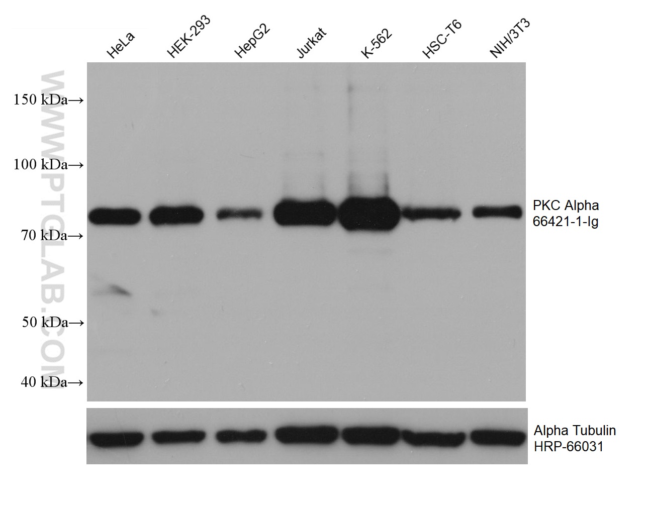
Various lysates were subjected to SDS PAGE followed by western blot with 66421-1-Ig (PKC Alpha antibody) at dilution of 1:10000 incubated at room temperature for 1.5 hours.
Various lysates were subjected to SDS PAGE followed by western blot with 66421-1-Ig (PKC Alpha antibody) at dilution of 1:10000 incubated at room temperature for 1.5 hours.
WB analysis of HeLa using 66421-1-Ig
HeLa cells were subjected to SDS PAGE followed by western blot with 66421-1-Ig (PKC alpha antibody at dilution of 1:10000 incubated at room temperature for 1.5 hours.
HeLa cells were subjected to SDS PAGE followed by western blot with 66421-1-Ig (PKC alpha antibody at dilution of 1:10000 incubated at room temperature for 1.5 hours.
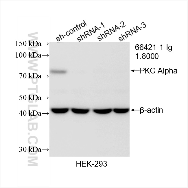
WB analysis of HEK-293 using 66421-1-Ig
WB result of PKC Alpha antibody (66421-1-Ig; 1:8000; incubated at room temperature for 1.5 hours) with sh-Control and sh-PKC Alpha transfected HEK-293 cells.
WB result of PKC Alpha antibody (66421-1-Ig; 1:8000; incubated at room temperature for 1.5 hours) with sh-Control and sh-PKC Alpha transfected HEK-293 cells.
WB analysis of HEK-293 using 66421-1-Ig
HEK-293 cells were subjected to SDS PAGE followed by western blot with 66421-1-Ig (PKC alpha antibody at dilution of 1:10000 incubated at room temperature for 1.5 hours.
HEK-293 cells were subjected to SDS PAGE followed by western blot with 66421-1-Ig (PKC alpha antibody at dilution of 1:10000 incubated at room temperature for 1.5 hours.
WB analysis of C6 using 66421-1-Ig
C6 cells were subjected to SDS PAGE followed by western blot with 66421-1-Ig (PKC alpha antibody at dilution of 1:10000 incubated at room temperature for 1.5 hours.
C6 cells were subjected to SDS PAGE followed by western blot with 66421-1-Ig (PKC alpha antibody at dilution of 1:10000 incubated at room temperature for 1.5 hours.
WB analysis of RAW 264.7 using 66421-1-Ig
RAW 264.7 cells were subjected to SDS PAGE followed by western blot with 66421-1-Ig (PKC alpha antibody at dilution of 1:10000 incubated at room temperature for 1.5 hours.
RAW 264.7 cells were subjected to SDS PAGE followed by western blot with 66421-1-Ig (PKC alpha antibody at dilution of 1:10000 incubated at room temperature for 1.5 hours.
IHC staining of human lung cancer using 66421-1-Ig
Immunohistochemical analysis of paraffin-embedded human lung cancer tissue slide using 66421-1-Ig (PKC alpha antibody at dilution of 1:200 (under 10x lens). Heat mediated antigen retrieval with Tris-EDTA buffer (pH 9.0).
Immunohistochemical analysis of paraffin-embedded human lung cancer tissue slide using 66421-1-Ig (PKC alpha antibody at dilution of 1:200 (under 10x lens). Heat mediated antigen retrieval with Tris-EDTA buffer (pH 9.0).
IHC staining of human lung cancer using 66421-1-Ig
Immunohistochemical analysis of paraffin-embedded human lung cancer tissue slide using 66421-1-Ig (PKC alpha antibody at dilution of 1:200 (under 40x lens). Heat mediated antigen retrieval with Tris-EDTA buffer (pH 9.0).
Immunohistochemical analysis of paraffin-embedded human lung cancer tissue slide using 66421-1-Ig (PKC alpha antibody at dilution of 1:200 (under 40x lens). Heat mediated antigen retrieval with Tris-EDTA buffer (pH 9.0).
IF Staining of HeLa using 66421-1-Ig
Immunofluorescent analysis of (-20°C Methanol) fixed HeLa cells using PKC Alpha antibody (66421-1-Ig, Clone: 1D11F4 ) at dilution of 1:800 and CoraLite®488-Conjugated Goat Anti-Mouse IgG(H+L).
Immunofluorescent analysis of (-20°C Methanol) fixed HeLa cells using PKC Alpha antibody (66421-1-Ig, Clone: 1D11F4 ) at dilution of 1:800 and CoraLite®488-Conjugated Goat Anti-Mouse IgG(H+L).
IF Staining of HeLa using 66421-1-Ig
Immunofluorescent analysis of (-20°C Ethanol) fixed HeLa cells using PKC Alpha antibody (66421-1-Ig, Clone: 1D11F4 ) at dilution of 1:400 and CoraLite®488-Conjugated Goat Anti-Mouse IgG(H+L).
Immunofluorescent analysis of (-20°C Ethanol) fixed HeLa cells using PKC Alpha antibody (66421-1-Ig, Clone: 1D11F4 ) at dilution of 1:400 and CoraLite®488-Conjugated Goat Anti-Mouse IgG(H+L).
IF Staining of HeLa using 66421-1-Ig
Immunofluorescent analysis of (-20℃ Ethanol ) fixed HeLa cells using 66421-1-Ig(PKC alpha antibody) at dilution of 1:100 and Alexa Fluor 488-conjugated Goat Anti-Mouse IgG(H+L).
Immunofluorescent analysis of (-20℃ Ethanol ) fixed HeLa cells using 66421-1-Ig(PKC alpha antibody) at dilution of 1:100 and Alexa Fluor 488-conjugated Goat Anti-Mouse IgG(H+L).
FC experiment of HeLa using 66421-1-Ig
1X10^6 HeLa cells were intracellularly stained with 0.4 ug Anti-Human PKC Alpha (66421-1-Ig, Clone:1D11F4) and CoraLite®488-Conjugated Goat Anti-Mouse IgG(H+L) at dilution 1:1000 (red), or 0.4 ug Mouse IgG1 Isotype Control (MOPC-21) (65124-1-Ig, Clone: MOPC-21) (blue). Cells were fixed with 4% PFA and permeabilized with Flow Cytometry Perm Buffer (PF00011-C).
1X10^6 HeLa cells were intracellularly stained with 0.4 ug Anti-Human PKC Alpha (66421-1-Ig, Clone:1D11F4) and CoraLite®488-Conjugated Goat Anti-Mouse IgG(H+L) at dilution 1:1000 (red), or 0.4 ug Mouse IgG1 Isotype Control (MOPC-21) (65124-1-Ig, Clone: MOPC-21) (blue). Cells were fixed with 4% PFA and permeabilized with Flow Cytometry Perm Buffer (PF00011-C).
The Proteintech guarantee covers Proteintech antibodies in any species and any application, including those not listed on the datasheet. If the antibody doesn’t perform, you can receive a hassle-free refund or credit note.
HeLa cells, HEK-293 cells, C6 cells, RAW 264.7 cells, HepG2 cells, Jurkat cells, K-562 cells, HSC-T6 cells, NIH/3T3 cells
Positive IHC detected in
human lung cancer tissue Note: suggested antigen retrieval with TE buffer pH 9.0; (*) Alternatively, antigen retrieval may be performed with citrate buffer pH 6.0
Positive IF/ICC detected in
HeLa cells
Positive FC (Intra) detected in
HeLa cells
Recommended dilution
Application
Dilution
Western Blot (WB)
WB : 1:5000-1:50000
Immunohistochemistry (IHC)
IHC : 1:50-1:500
Immunofluorescence (IF)/ICC
IF/ICC : 1:400-1:1600
Flow Cytometry (FC) (INTRA)
FC (INTRA) : 0.40 ug per 10^6 cells in a 100 µl suspension
It is recommended that this reagent should be titrated in each testing system to obtain optimal results.
Sample-dependent, Check data in validation data gallery.
PBS with 0.02% sodium azide and 50% glycerol pH 7.3.
Storage Conditions
Store at -20°C. Stable for one year after shipment. Aliquoting is unnecessary for -20oC storage. 20ul sizes contain 0.1% BSA.
Background Information
Protein kinase C (PKC) is a family of serine- and threonine-specific protein kinases that can be activated by calcium and the second messenger diacylglycerol. PKC family members phosphorylate a wide variety of protein targets and are known to be involved in diverse cellular signaling pathways. PKC family members also serve as major receptors for phorbol esters, a class of tumor promoters. Each member of the PKC family has a specific expression profile and is believed to play a distinct role in cells. PRKCA is one of the PKC family members. This kinase has been reported to play roles in many different cellular processes, such as cell adhesion, cell transformation, cell cycle checkpoint, and cell volume control. Knockout studies in mice suggest that this kinase may be a fundamental regulator of cardiac contractility and Ca(2+) handling in myocytes.
Various lysates were subjected to SDS PAGE followed by western blot with 66421-1-Ig (PKC Alpha antibody) at dilution of 1:10000 incubated at room temperature for 1.5 hours.
WB analysis of HeLa using 66421-1-Ig
HeLa cells were subjected to SDS PAGE followed by western blot with 66421-1-Ig (PKC alpha antibody at dilution of 1:10000 incubated at room temperature for 1.5 hours.
WB analysis of HEK-293 using 66421-1-Ig
WB result of PKC Alpha antibody (66421-1-Ig; 1:8000; incubated at room temperature for 1.5 hours) with sh-Control and sh-PKC Alpha transfected HEK-293 cells.
WB analysis of HEK-293 using 66421-1-Ig
HEK-293 cells were subjected to SDS PAGE followed by western blot with 66421-1-Ig (PKC alpha antibody at dilution of 1:10000 incubated at room temperature for 1.5 hours.
WB analysis of C6 using 66421-1-Ig
C6 cells were subjected to SDS PAGE followed by western blot with 66421-1-Ig (PKC alpha antibody at dilution of 1:10000 incubated at room temperature for 1.5 hours.
WB analysis of RAW 264.7 using 66421-1-Ig
RAW 264.7 cells were subjected to SDS PAGE followed by western blot with 66421-1-Ig (PKC alpha antibody at dilution of 1:10000 incubated at room temperature for 1.5 hours.
IHC Figures
IHC staining of human lung cancer using 66421-1-Ig
Immunohistochemical analysis of paraffin-embedded human lung cancer tissue slide using 66421-1-Ig (PKC alpha antibody at dilution of 1:200 (under 10x lens). Heat mediated antigen retrieval with Tris-EDTA buffer (pH 9.0).
IHC staining of human lung cancer using 66421-1-Ig
Immunohistochemical analysis of paraffin-embedded human lung cancer tissue slide using 66421-1-Ig (PKC alpha antibody at dilution of 1:200 (under 40x lens). Heat mediated antigen retrieval with Tris-EDTA buffer (pH 9.0).
IF/ICC Figures
IF Staining of HeLa using 66421-1-Ig
Immunofluorescent analysis of (-20°C Methanol) fixed HeLa cells using PKC Alpha antibody (66421-1-Ig, Clone: 1D11F4 ) at dilution of 1:800 and CoraLite®488-Conjugated Goat Anti-Mouse IgG(H+L).
IF Staining of HeLa using 66421-1-Ig
Immunofluorescent analysis of (-20°C Ethanol) fixed HeLa cells using PKC Alpha antibody (66421-1-Ig, Clone: 1D11F4 ) at dilution of 1:400 and CoraLite®488-Conjugated Goat Anti-Mouse IgG(H+L).
IF Staining of HeLa using 66421-1-Ig
Immunofluorescent analysis of (-20℃ Ethanol ) fixed HeLa cells using 66421-1-Ig(PKC alpha antibody) at dilution of 1:100 and Alexa Fluor 488-conjugated Goat Anti-Mouse IgG(H+L).
FC (INTRA) Figures
FC experiment of HeLa using 66421-1-Ig
1X10^6 HeLa cells were intracellularly stained with 0.4 ug Anti-Human PKC Alpha (66421-1-Ig, Clone:1D11F4) and CoraLite®488-Conjugated Goat Anti-Mouse IgG(H+L) at dilution 1:1000 (red), or 0.4 ug Mouse IgG1 Isotype Control (MOPC-21) (65124-1-Ig, Clone: MOPC-21) (blue). Cells were fixed with 4% PFA and permeabilized with Flow Cytometry Perm Buffer (PF00011-C).
The species listed in Tested Reactivity are in-house verified and applicable species. For unlisted species, please refer to the homology analysis of the immunogen sequence and related species. For rabbit polyclonal antibodies, homology >70% is recommended. For mouse monoclonal antibodies and rabbit recombinant antibodies, homology >90% is recommended. Generally, the higher the homology, the greater the applicability. However, there will be certain differences in protein expression in different species, tissues or cells. Therefore, the homology analysis results are for reference only and do not serve as a guarantee.
At Proteintech, we pride ourselves on our antibody quality, customer service and transparency. As such, we are comparing our antibodies with other vendors, enabling easy identification and comparisons of key data to help you choose the suitable antibody for your needs.
We have selected the top cited antibodies from these vendors for you to compare.
Proteintech
KD/KO VALIDATED
PKC Alpha Monoclonal antibody
Catalog Number
66421-1-Ig
Citations
1
Dilutions
WB : 1:5000-1:50000 IHC : 1:50-1:500 IF/ICC : 1:400-1:1600 FC (INTRA) : 0.40 ug per 10^6 cells in a 100 µl suspension
Applications
WB, IHC, IF/ICC, FC (Intra), ELISA
Reactivity
human, mouse, rat
Product Guarantee
Covers any species including not listed on datasheet
Covers any applications including not listed on datasheet
 at dilution of 1:10000 incubated at room temperature for 1.5 hours.")

 with sh-Control and sh-PKC Alpha transfected HEK-293 cells.")


. Heat mediated antigen retrieval with Tris-EDTA buffer (pH 9.0).")
. Heat mediated antigen retrieval with Tris-EDTA buffer (pH 9.0).")
 fixed HeLa cells using PKC Alpha antibody (66421-1-Ig, Clone: 1D11F4 ) at dilution of 1:800 and CoraLite®488-Conjugated Goat Anti-Mouse IgG(H+L).")
 fixed HeLa cells using PKC Alpha antibody (66421-1-Ig, Clone: 1D11F4 ) at dilution of 1:400 and CoraLite®488-Conjugated Goat Anti-Mouse IgG(H+L).")
 fixed HeLa cells using 66421-1-Ig(PKC alpha antibody) at dilution of 1:100 and Alexa Fluor 488-conjugated Goat Anti-Mouse IgG(H+L).")
 and CoraLite®488-Conjugated Goat Anti-Mouse IgG(H+L) at dilution 1:1000 (red), or 0.4 ug Mouse IgG1 Isotype Control (MOPC-21) (65124-1-Ig, Clone: MOPC-21) (blue). Cells were fixed with 4% PFA and permeabilized with Flow Cytometry Perm Buffer (PF00011-C).")