Various lysates were subjected to SDS PAGE followed by western blot with 55103-1-AP (CDK3 antibody) at dilution of 1:2000 incubated at room temperature for 1.5 hours.
Various lysates were subjected to SDS PAGE followed by western blot with 55103-1-AP (CDK3 antibody) at dilution of 1:2000 incubated at room temperature for 1.5 hours.
WB analysis of Jurkat using 55103-1-AP
Jurkat cells were subjected to SDS PAGE followed by western blot with 55103-1-AP (CDK3 antibody) at dilution of 1:800 incubated at room temperature for 1.5 hours.
Jurkat cells were subjected to SDS PAGE followed by western blot with 55103-1-AP (CDK3 antibody) at dilution of 1:800 incubated at room temperature for 1.5 hours.
WB analysis of MCF-7 using 55103-1-AP
MCF7 cells were subjected to SDS PAGE followed by western blot with 55103-1-AP (CDK3 antibody) at dilution of 1:800 incubated at room temperature for 1.5 hours.
MCF7 cells were subjected to SDS PAGE followed by western blot with 55103-1-AP (CDK3 antibody) at dilution of 1:800 incubated at room temperature for 1.5 hours.
WB analysis of MCF-7 using 55103-1-AP
MCF-7 cells were subjected to SDS PAGE followed by western blot with 55103-1-AP (CDK3 antibody) at dilution of 1:600 incubated at room temperature for 1.5 hours.
MCF-7 cells were subjected to SDS PAGE followed by western blot with 55103-1-AP (CDK3 antibody) at dilution of 1:600 incubated at room temperature for 1.5 hours.
IHC staining of human breast cancer using 55103-1-AP
Immunohistochemical analysis of paraffin-embedded human breast cancer tissue slide using 55103-1-AP (CDK3 Antibody) at dilution of 1:200 (under 10x lens). Heat mediated antigen retrieval with Tris-EDTA buffer (pH 9.0).
Immunohistochemical analysis of paraffin-embedded human breast cancer tissue slide using 55103-1-AP (CDK3 Antibody) at dilution of 1:200 (under 10x lens). Heat mediated antigen retrieval with Tris-EDTA buffer (pH 9.0).
IHC staining of human breast cancer using 55103-1-AP
Immunohistochemical analysis of paraffin-embedded human breast cancer tissue slide using 55103-1-AP (CDK3 Antibody) at dilution of 1:200 (under 40x lens). Heat mediated antigen retrieval with Tris-EDTA buffer (pH 9.0).
Immunohistochemical analysis of paraffin-embedded human breast cancer tissue slide using 55103-1-AP (CDK3 Antibody) at dilution of 1:200 (under 40x lens). Heat mediated antigen retrieval with Tris-EDTA buffer (pH 9.0).
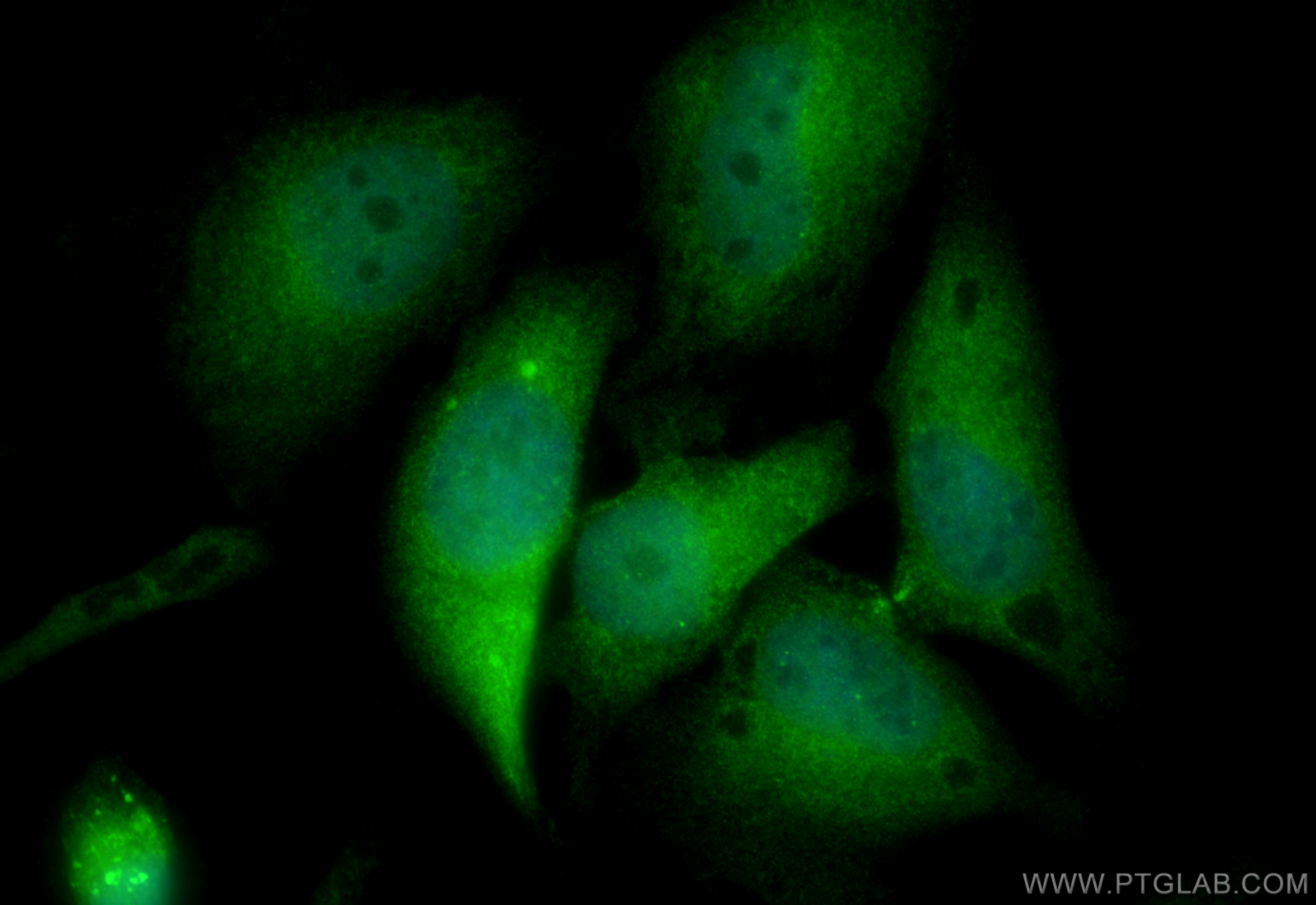
IF Staining of HeLa using 55103-1-AP
Immunofluorescent analysis of (4% PFA) fixed HeLa cells using CDK3 antibody (55103-1-AP) at dilution of 1:400 and CoraLite®488-Conjugated Goat Anti-Rabbit IgG(H+L) (SA00013-2).
Immunofluorescent analysis of (4% PFA) fixed HeLa cells using CDK3 antibody (55103-1-AP) at dilution of 1:400 and CoraLite®488-Conjugated Goat Anti-Rabbit IgG(H+L) (SA00013-2).
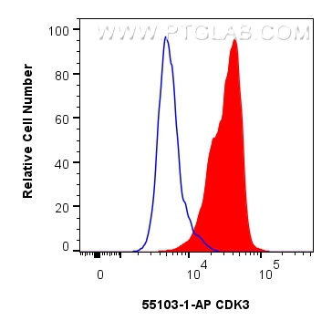
FC experiment of HeLa using 55103-1-AP
1x10^6 HeLa cells were intracellularly stained with 0.25 ug CDK3 Polyclonal antibody (55103-1-AP) and CoraLite®488-Conjugated Goat Anti-Rabbit IgG(H+L) (SA00013-2)(red), or 0.25 ug Rabbit IgG control Rabbit PolyAb (30000-0-AP) (blue). Cells were fixed and permeabilized with Transcription Factor Staining Buffer Kit (PF00011).
1x10^6 HeLa cells were intracellularly stained with 0.25 ug CDK3 Polyclonal antibody (55103-1-AP) and CoraLite®488-Conjugated Goat Anti-Rabbit IgG(H+L) (SA00013-2)(red), or 0.25 ug Rabbit IgG control Rabbit PolyAb (30000-0-AP) (blue). Cells were fixed and permeabilized with Transcription Factor Staining Buffer Kit (PF00011).
The Proteintech guarantee covers Proteintech antibodies in any species and any application, including those not listed on the datasheet. If the antibody doesn’t perform, you can receive a hassle-free refund or credit note.
mouse brain tissue, MCF-7 cells, Jurkat cells, rat brain tissue
Positive IHC detected in
human breast cancer tissue Note: suggested antigen retrieval with TE buffer pH 9.0; (*) Alternatively, antigen retrieval may be performed with citrate buffer pH 6.0
Positive IF/ICC detected in
HeLa cells
Positive FC (Intra) detected in
HeLa cells
Recommended dilution
Application
Dilution
Western Blot (WB)
WB : 1:1000-1:4000
Immunohistochemistry (IHC)
IHC : 1:100-1:400
Immunofluorescence (IF)/ICC
IF/ICC : 1:200-1:800
Flow Cytometry (FC) (INTRA)
FC (INTRA) : 0.25 ug per 10^6 cells in a 100 µl suspension
It is recommended that this reagent should be titrated in each testing system to obtain optimal results.
Sample-dependent, Check data in validation data gallery.
PBS with 0.02% sodium azide and 50% glycerol , pH 7.3
Storage Conditions
Store at -20°C. Stable for one year after shipment. Aliquoting is unnecessary for -20oC storage. 20ul sizes contain 0.1% BSA.
Background Information
CDK3 belongs to the protein kinase superfamily, CMGC Ser/Thr protein kinase family and CDC2/CDKX subfamily. It is probably involved in the control of the cell cycle. CDK3 interacts with a yet unknown type of cyclin. It can phosphorylate histone H1. Endogenous Cyclin C/Cdk3 complexes are critical to promote exit from quiescence in an Rb-dependent manner. Cyclin C/Cdk3 complexes is a key regulators of cell cycle reentry in human cells. (PMID: 15130482 )The antibody is specific to CDK3.
Various lysates were subjected to SDS PAGE followed by western blot with 55103-1-AP (CDK3 antibody) at dilution of 1:2000 incubated at room temperature for 1.5 hours.
WB analysis of Jurkat using 55103-1-AP
Jurkat cells were subjected to SDS PAGE followed by western blot with 55103-1-AP (CDK3 antibody) at dilution of 1:800 incubated at room temperature for 1.5 hours.
WB analysis of MCF-7 using 55103-1-AP
MCF7 cells were subjected to SDS PAGE followed by western blot with 55103-1-AP (CDK3 antibody) at dilution of 1:800 incubated at room temperature for 1.5 hours.
WB analysis of MCF-7 using 55103-1-AP
MCF-7 cells were subjected to SDS PAGE followed by western blot with 55103-1-AP (CDK3 antibody) at dilution of 1:600 incubated at room temperature for 1.5 hours.
IHC Figures
IHC staining of human breast cancer using 55103-1-AP
Immunohistochemical analysis of paraffin-embedded human breast cancer tissue slide using 55103-1-AP (CDK3 Antibody) at dilution of 1:200 (under 10x lens). Heat mediated antigen retrieval with Tris-EDTA buffer (pH 9.0).
IHC staining of human breast cancer using 55103-1-AP
Immunohistochemical analysis of paraffin-embedded human breast cancer tissue slide using 55103-1-AP (CDK3 Antibody) at dilution of 1:200 (under 40x lens). Heat mediated antigen retrieval with Tris-EDTA buffer (pH 9.0).
IF/ICC Figures
IF Staining of HeLa using 55103-1-AP
Immunofluorescent analysis of (4% PFA) fixed HeLa cells using CDK3 antibody (55103-1-AP) at dilution of 1:400 and CoraLite®488-Conjugated Goat Anti-Rabbit IgG(H+L) (SA00013-2).
FC (INTRA) Figures
FC experiment of HeLa using 55103-1-AP
1x10^6 HeLa cells were intracellularly stained with 0.25 ug CDK3 Polyclonal antibody (55103-1-AP) and CoraLite®488-Conjugated Goat Anti-Rabbit IgG(H+L) (SA00013-2)(red), or 0.25 ug Rabbit IgG control Rabbit PolyAb (30000-0-AP) (blue). Cells were fixed and permeabilized with Transcription Factor Staining Buffer Kit (PF00011).
The species listed in Tested Reactivity are in-house verified and applicable species. For unlisted species, please refer to the homology analysis of the immunogen sequence and related species. For rabbit polyclonal antibodies, homology >70% is recommended. For mouse monoclonal antibodies and rabbit recombinant antibodies, homology >90% is recommended. Generally, the higher the homology, the greater the applicability. However, there will be certain differences in protein expression in different species, tissues or cells. Therefore, the homology analysis results are for reference only and do not serve as a guarantee.
At Proteintech, we pride ourselves on our antibody quality, customer service and transparency. As such, we are comparing our antibodies with other vendors, enabling easy identification and comparisons of key data to help you choose the suitable antibody for your needs.
We have selected the top cited antibodies from these vendors for you to compare.
Proteintech
CDK3 Polyclonal antibody
Catalog Number
55103-1-AP
Citations
4
Dilutions
WB : 1:1000-1:4000 IHC : 1:100-1:400 IF/ICC : 1:200-1:800 FC (INTRA) : 0.25 ug per 10^6 cells in a 100 µl suspension
Applications
WB, IHC, IF/ICC, FC (Intra), ELISA
Reactivity
human, mouse, rat
Product Guarantee
Covers any species including not listed on datasheet
Covers any applications including not listed on datasheet
 at dilution of 1:2000 incubated at room temperature for 1.5 hours.")
 at dilution of 1:800 incubated at room temperature for 1.5 hours.")
 at dilution of 1:800 incubated at room temperature for 1.5 hours.")
 at dilution of 1:600 incubated at room temperature for 1.5 hours.")
 at dilution of 1:200 (under 10x lens). Heat mediated antigen retrieval with Tris-EDTA buffer (pH 9.0).")
 at dilution of 1:200 (under 40x lens). Heat mediated antigen retrieval with Tris-EDTA buffer (pH 9.0).")
 fixed HeLa cells using CDK3 antibody (55103-1-AP) at dilution of 1:400 and CoraLite®488-Conjugated Goat Anti-Rabbit IgG(H+L) (SA00013-2).")
 and CoraLite®488-Conjugated Goat Anti-Rabbit IgG(H+L) (SA00013-2)(red), or 0.25 ug Rabbit IgG control Rabbit PolyAb (30000-0-AP) (blue). Cells were fixed and permeabilized with Transcription Factor Staining Buffer Kit (PF00011).")