IF Visualization
Learn the difference between direct and indirect visualization and when to use each method.
IF Visualization
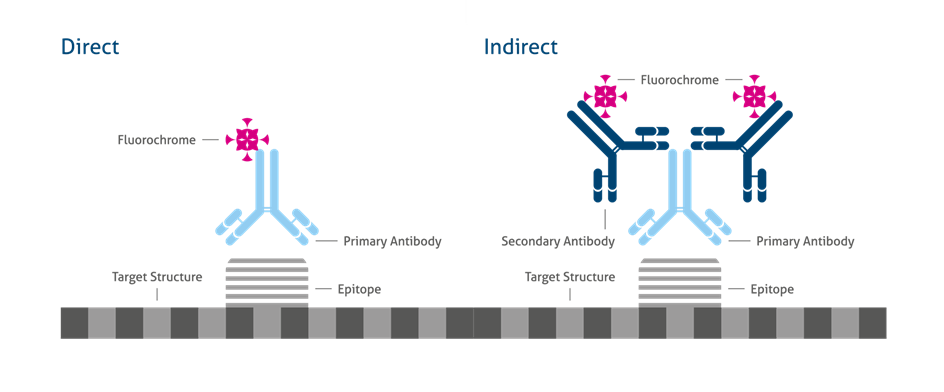
Depending on the target of interest, direct or indirect visualization is recommended.
Direct visualization
-
Direct visualization requires the use of a labeled primary antibody:
-
Short protocol, fast analysis, easy to handle.
-
Expensive, a labeled primary antibody for each target is needed.
-
A highly specific primary antibody is needed.
-
No signal amplification, not suitable for low expressed targets.
Indirect visualization
-
Indirect visualization requires the use of a labeled secondary antibody:
-
Longer protocol.
-
Less expensive, a labeled secondary antibody can be used for different primary antibodies.
-
Higher background noise.
-
Signal amplification, suitable for medium to high expressed targets.
Direct Vs. Indirect Immunofluorescence
Most Commonly Used Labels For IF Staining
Mounting Media
Since a fluorescent signal fades away during time, mounting media is added at the end of the staining protocol to maintain the sample. It is also needed to optimize the refractive index for imaging. Mounting media are a solution of glycerol in a special buffer. This helps to maintain the fluorescence signal and slow down photobleaching. Mounting media are optimized for different conditions and dyes, and some questions to consider when selecting the right one are:
-
Fixed samples for long-term conservation?
-
Fixed samples for immediate imaging?
-
Which kind of dye/fluorophore?
-
Multistaining?