Tested Applications
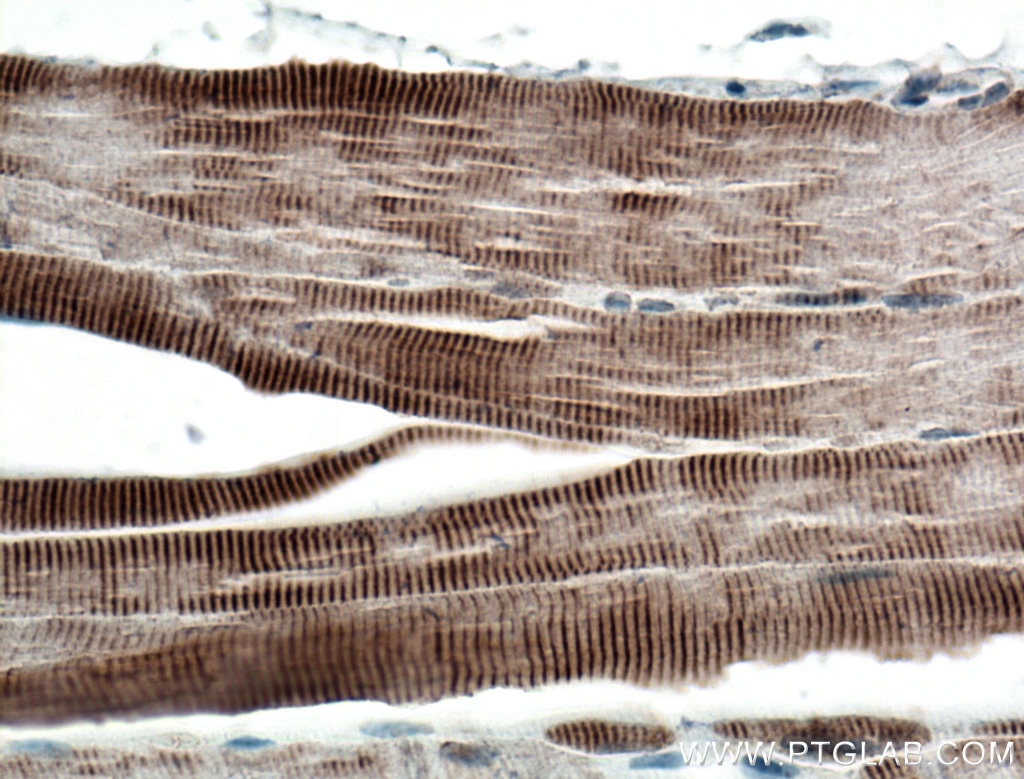
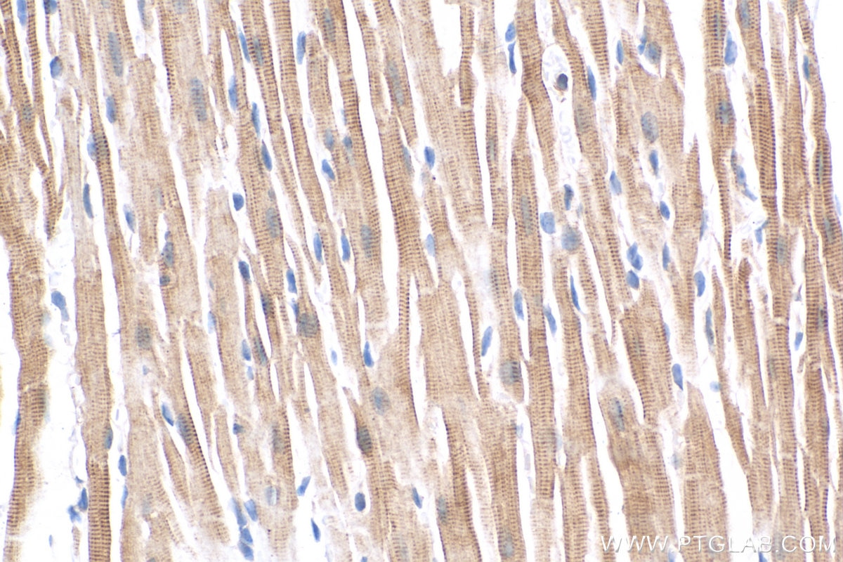
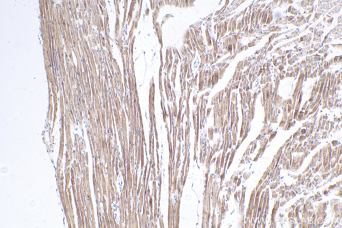
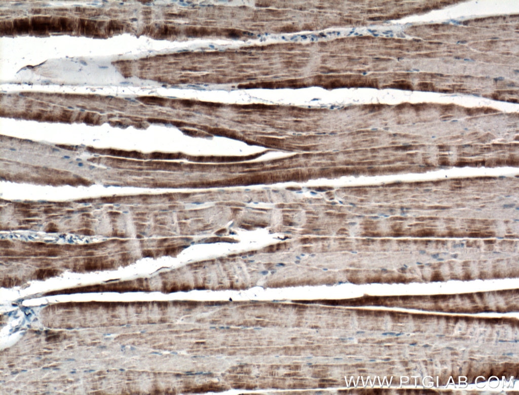
| Positive IHC detected in | mouse heart tissue, mouse skeletal muscle tissue Note: suggested antigen retrieval with TE buffer pH 9.0; (*) Alternatively, antigen retrieval may be performed with citrate buffer pH 6.0 |
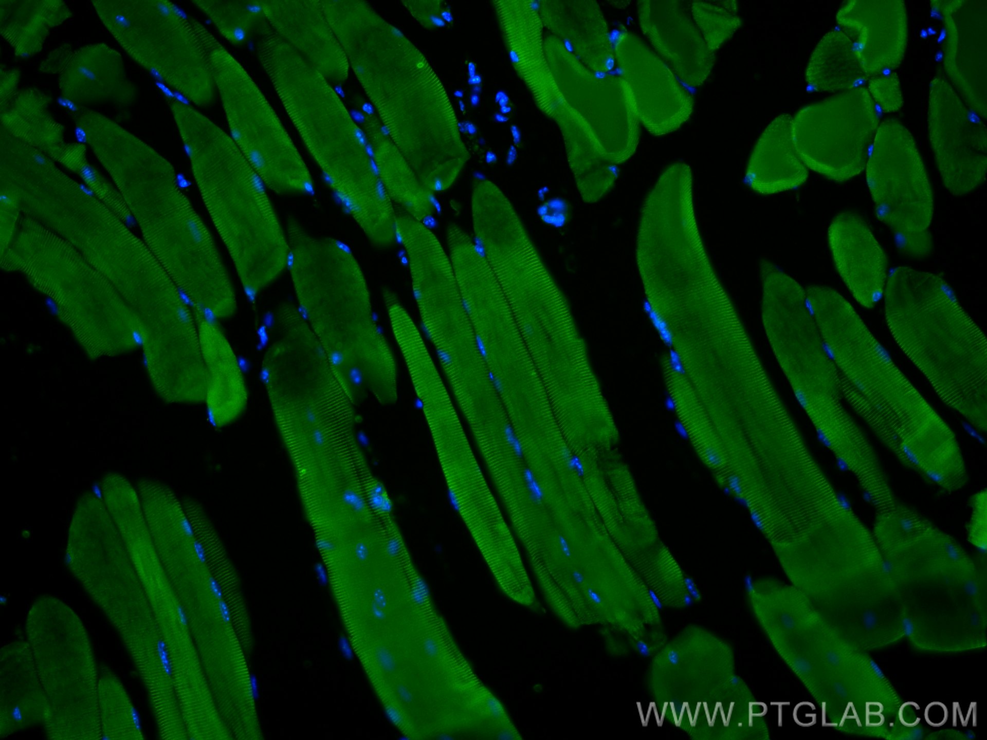
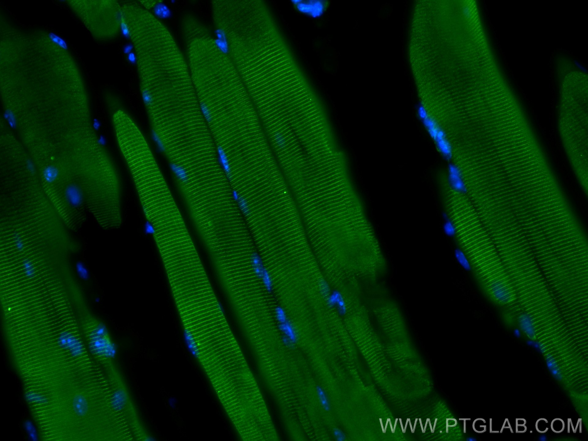
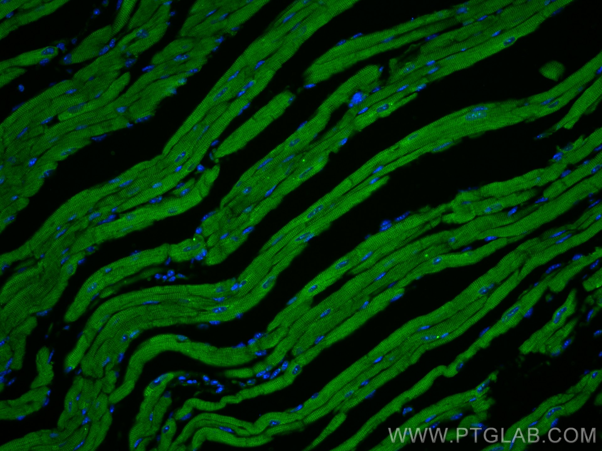
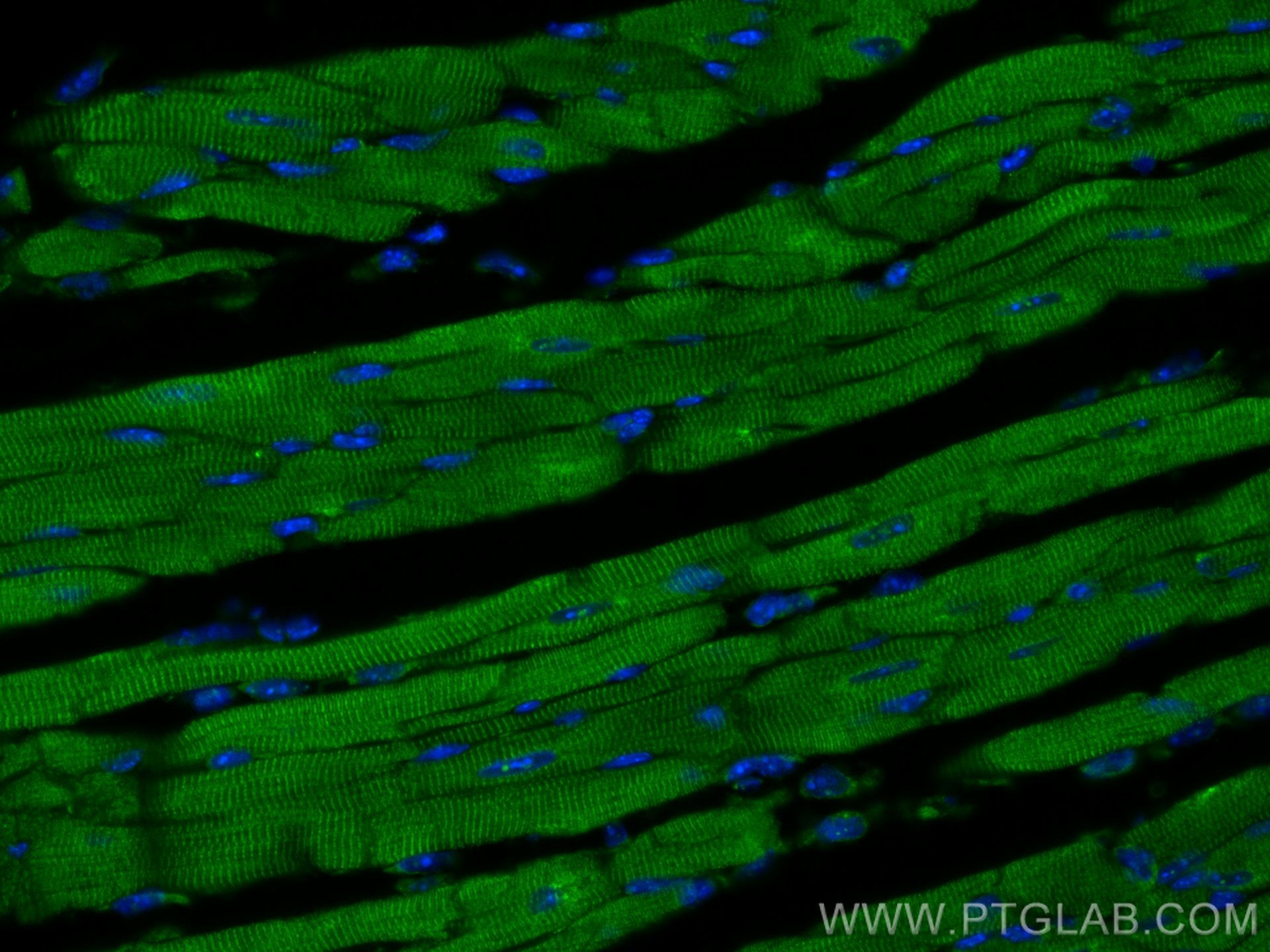
| Positive IF-P detected in | mouse skeletal muscle tissue, mouse heart tissue |
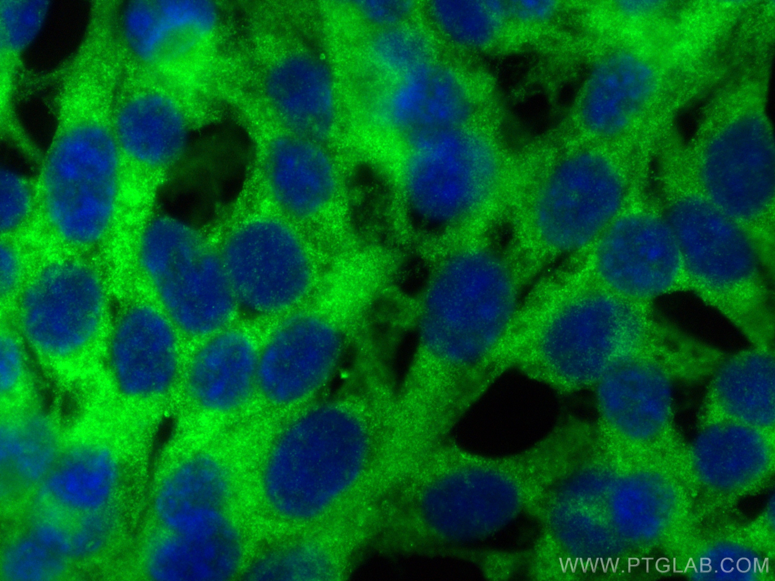
| Positive IF/ICC detected in | NIH/3T3 cells |
Recommended dilution
| Application | Dilution |
|---|---|
| Immunohistochemistry (IHC) | IHC : 1:200-1:800 |
| Immunofluorescence (IF)-P | IF-P : 1:50-1:500 |
| Immunofluorescence (IF)/ICC | IF/ICC : 1:200-1:800 |
| It is recommended that this reagent should be titrated in each testing system to obtain optimal results. | |
| Sample-dependent, Check data in validation data gallery. | |
Published Applications
| IF | See 6 publications below |
Product Information
27867-1-AP targets Titin in IHC, IF/ICC, IF-P, ELISA applications and shows reactivity with human, mouse samples.
| Tested Reactivity | human, mouse |
| Cited Reactivity | human, mouse |
| Host / Isotype | Rabbit / IgG |
| Class | Polyclonal |
| Type | Antibody |
| Immunogen |
CatNo: Ag27496 Product name: Recombinant human TTN protein Source: e coli.-derived, PGEX-4T Tag: GST Domain: 33738-33969 aa of NM_001256850 Sequence: MFKSIHEKVSKISETKKSDQKTTESTVTRKTEPKAPEPISSKPVIVTGLQDTTVSSDSVAKFAVKATGEPRPTAIWTKDGKAITQGGKYKLSEDKGGFFLEIHKTDTSDSGLYTCTVKNSAGSVSSSCKLTIKAIKDTEAQKVSTQKTSEITPQKKAVVQEEISQKALRSEEIKMSEAKSQEKLALKEEASKVLISEEVKKSAATSLEKSIVHEEITKTSQASEEVRTHAEIK Predict reactive species |
| Full Name | titin |
| Calculated Molecular Weight | 3816 kDa |
| GenBank Accession Number | NM_001256850 |
| Gene Symbol | Titin |
| Gene ID (NCBI) | 7273 |
| RRID | AB_2880998 |
| Conjugate | Unconjugated |
| Form | Liquid |
| Purification Method | Antigen affinity purification |
| UNIPROT ID | Q8WZ42 |
| Storage Buffer | PBS with 0.02% sodium azide and 50% glycerol, pH 7.3. |
| Storage Conditions | Store at -20°C. Stable for one year after shipment. Aliquoting is unnecessary for -20oC storage. 20ul sizes contain 0.1% BSA. |
Background Information
Titin, or connectin, is a giant muscle protein expressed in the cardiac and skeletal muscles that spans half of the sarcomere from Z line to M line. It plays a key role in muscle contraction and relaxation, maintaining the structural integrity of the myotome(PMID: 11846417). Mutations in Titin are associated with familial hypertrophic cardiomyopathy 9, and autoantibodies to titin are produced in patients with the autoimmune disease scleroderma(PMID: 15802564).
Protocols
| Product Specific Protocols | |
|---|---|
| IF protocol for Titin antibody 27867-1-AP | Download protocol |
| IHC protocol for Titin antibody 27867-1-AP | Download protocol |
| Standard Protocols | |
|---|---|
| Click here to view our Standard Protocols |
Publications
| Species | Application | Title |
|---|---|---|
Elife A transcriptome atlas of the mouse iris at single-cell resolution defines cell types and the genomic response to pupil dilation. | ||
Dev Biol Aberrant differentiation of second heart field mesoderm prefigures cellular defects in the outflow tract in response to loss of FGF8 | ||
Cell Stem Cell Generation of self-organized neuromusculoskeletal tri-tissue organoids from human pluripotent stem cells | ||
Physiol Rep Germline deletion of Rgs2 and/or Rgs5 in male mice does not exacerbate left ventricular remodeling induced by subchronic isoproterenol infusion | ||
Stem Cell Res Generation of a human induced pluripotent stem cell line YCMi004-A from a patient with dilated cardiomyopathy carrying a protein-truncating mutation of the Titin gene and its differentiation towards cardiomyocytes |
Reviews
The reviews below have been submitted by verified Proteintech customers who received an incentive for providing their feedback.
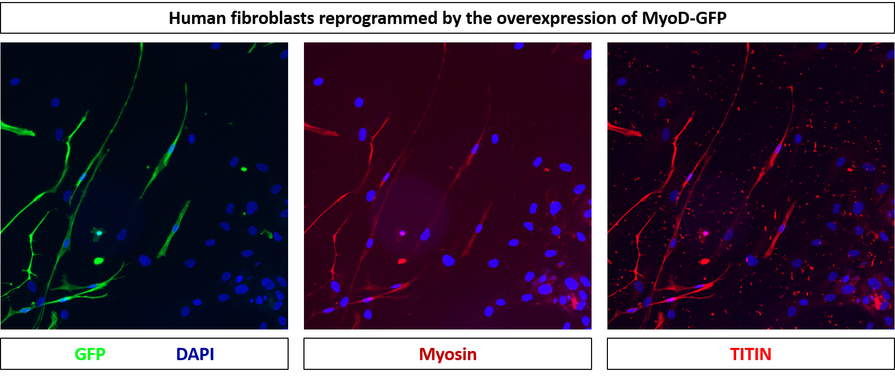
FH Víctor (Verified Customer) (01-26-2023) | The antibody is a bit dirty but specific for muscle cells. I'm trying to evaluate specific sarcomeric structures with higher magnifications, and they seem to be marked. So the antibody is OK.
 |
FH Joseph (Verified Customer) (01-14-2020) | I tested the Titin antibody using IHC on 16uM mouse skeletal muscle sections (Tibialis anterior and Soleus) using 1:100 and 1:300 dilutions. Both dilutions worked very well.
|

