at dilution of 1:20000 incubated at room temperature for 1.5 hours.")
 at dilution of 1:20000 incubated at room temperature for 1.5 hours.")
 at dilution of 1:20000 incubated at room temperature for 1.5 hours.")
 at dilution of 1:20000 incubated at room temperature for 1.5 hours.")
 at dilution of 1:20000 incubated at room temperature for 1.5 hours.")
 at dilution of 1:4000 (under 10x lens). Heat mediated antigen retrieval with Tris-EDTA buffer (pH 9.0).")
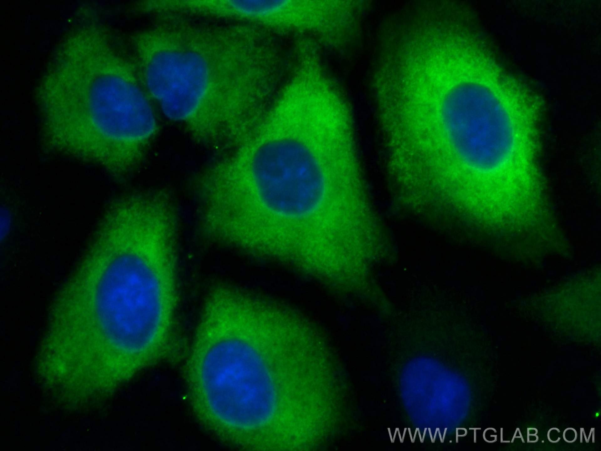
 fixed A549 cells using TGM2 antibody (68006-1-Ig, Clone: 2D4C11 ) at dilution of 1:800 and Multi-rAb CoraLite ® Plus 488-Goat Anti-Mouse Recombinant Secondary Antibody (H+L) (RGAM002).")
 and CoraLite®488-Conjugated AffiniPure Goat Anti-Mouse IgG(H+L) at dilution 1:1000 (red), or 0.4 ug Mouse IgG2a Isotype Control (C1.18.4) (65208-1-Ig, Clone: C1.18.4) (blue). Cells were fixed with 4% PFA and permeabilized with Flow Cytometry Perm Buffer.")
Tested Applications
| Positive WB detected in | HeLa cells, human placenta tissue, HUVEC cells, HepG2 cells, K-562 cells |
| Positive IHC detected in | human liver cancer tissue Note: suggested antigen retrieval with TE buffer pH 9.0; (*) Alternatively, antigen retrieval may be performed with citrate buffer pH 6.0 |
| Positive IF/ICC detected in | A549 cells |
| Positive FC (Intra) detected in | HeLa cells |
Recommended dilution
| Application | Dilution |
|---|---|
| Western Blot (WB) | WB : 1:5000-1:50000 |
| Immunohistochemistry (IHC) | IHC : 1:2000-1:8000 |
| Immunofluorescence (IF)/ICC | IF/ICC : 1:400-1:1600 |
| Flow Cytometry (FC) (INTRA) | FC (INTRA) : 0.40 ug per 10^6 cells in a 100 µl suspension |
| It is recommended that this reagent should be titrated in each testing system to obtain optimal results. | |
| Sample-dependent, Check data in validation data gallery. | |
Published Applications
| KD/KO | See 2 publications below |
| WB | See 3 publications below |
| IHC | See 2 publications below |
| IF | See 2 publications below |
| CoIP | See 1 publications below |
Product Information
68006-1-Ig targets TGM2 in WB, IHC, IF/ICC, FC (Intra), CoIP, ELISA applications and shows reactivity with human samples.
| Tested Reactivity | human |
| Cited Reactivity | human, mouse, hamster |
| Host / Isotype | Mouse / IgG2a |
| Class | Monoclonal |
| Type | Antibody |
| Immunogen |
CatNo: Ag7462 Product name: Recombinant human TGM2 protein Source: e coli.-derived, PET28a Tag: 6*His Domain: 1-349 aa of BC003551 Sequence: MAEELVLERCDLELETNGRDHHTADLCREKLVVRRGQPFWLTLHFEGRNYEASVDSLTFSVVTGPAPSQEAGTKARFPLRDAVEEGDWTATVVDQQDCTLSLQLTTPANAPIGLYRLSLEASTGYQGSSFVLGHFILLFNAWCPADAVYLDSEEERQEYVLTQQGFIYQGSAKFIKNIPWNFGQFEDGILDICLILLDVNPKFLKNAGRDCSRRSSPVYVGRVVSGMVNCNDDQGVLLGRWDNNYGDGVSPMSWIGSVDILRRWKNHGCQRVKYGQCWVFAAVACTVLRCLGIPTRVVTNYNSAHDQNSNLLIEYFRNEFGEIQGDKSEMIWNFHCWVESWMTRPDLQP Predict reactive species |
| Full Name | transglutaminase 2 (C polypeptide, protein-glutamine-gamma-glutamyltransferase) |
| Calculated Molecular Weight | 77 kDa |
| Observed Molecular Weight | 80 kDa |
| GenBank Accession Number | BC003551 |
| Gene Symbol | TGM2 |
| Gene ID (NCBI) | 7052 |
| ENSEMBL Gene ID | ENSG00000198959 |
| RRID | AB_2918753 |
| Conjugate | Unconjugated |
| Form | Liquid |
| Purification Method | Protein A purification |
| UNIPROT ID | P21980 |
| Storage Buffer | PBS with 0.02% sodium azide and 50% glycerol, pH 7.3. |
| Storage Conditions | Store at -20°C. Stable for one year after shipment. Aliquoting is unnecessary for -20oC storage. 20ul sizes contain 0.1% BSA. |
Background Information
Transglutaminase 2 (TGM2) is a ubiquitous and multifunctional calcium-dependent enzyme belonging to the transglutaminase family. It is best known for its canonical activity of catalyzing the cross-linking of proteins by forming stable ε-(γ-glutamyl)lysine isopeptide bonds, which contributes to extracellular matrix stabilization and wound healing. Beyond this, TGM2 exhibits GTPase activity, allowing it to function as a signaling G-protein in intracellular processes. It is implicated in a wide range of physiological functions, including cell adhesion, proliferation, and apoptosis, as well as pathological conditions such as celiac disease, fibrosis, neurodegenerative disorders, and cancer metastasis, where its dysregulated expression often contributes to disease progression.
Protocols
| Product Specific Protocols | |
|---|---|
| IF protocol for TGM2 antibody 68006-1-Ig | Download protocol |
| IHC protocol for TGM2 antibody 68006-1-Ig | Download protocol |
| WB protocol for TGM2 antibody 68006-1-Ig | Download protocol |
| Standard Protocols | |
|---|---|
| Click here to view our Standard Protocols |
Publications
| Species | Application | Title |
|---|---|---|
Biomaterials Tumor-specific nitric oxide generator to amplify peroxynitrite based on highly penetrable nanoparticles for metastasis inhibition and enhanced cancer therapy. | ||
Transl Res Counteracting TGM2 by a Fibroin peptide ameliorated Adriamycin-induced nephropathy via regulation of lipid metabolism through PANX1-PPAR α/PANK1 pathway | ||
F1000Res A guide to selecting high-performing antibodies for Protein-glutamine gamma-glutamyltransferase 2 (TGM2) for use in western blot, immunoprecipitation and immunofluorescence | ||
ACS Synth Biol Overexpression of Tgm2 in Chinese Hamster Ovary Cells Enhances Recombinant Monoclonal Antibody Expression and Promotes Cell Proliferation through Reduction of Apoptosis
| ||
Discov Oncol Transglutaminase 2 nuclear localization enhances glioblastoma radiation resistance
|