tissue using 60019-2-Ig")


Tested Applications
| Positive WB detected in | LNCaP cells, HeLa cells, HEK-293 cells, HepG2 cells, Jurkat cells, K-562 cells |
| Positive IP detected in | K-562 cells |
| Positive IHC detected in | human gliomas tissue, human pancreas cancer tissue, human brain(FTLD-U) tissue Note: suggested antigen retrieval with TE buffer pH 9.0; (*) Alternatively, antigen retrieval may be performed with citrate buffer pH 6.0 |
Recommended dilution
| Application | Dilution |
|---|---|
| Western Blot (WB) | WB : 1:5000-1:50000 |
| Immunoprecipitation (IP) | IP : 0.5-4.0 ug for 1.0-3.0 mg of total protein lysate |
| Immunohistochemistry (IHC) | IHC : 1:5000-1:20000 |
| It is recommended that this reagent should be titrated in each testing system to obtain optimal results. | |
| Sample-dependent, Check data in validation data gallery. | |
Published Applications
| KD/KO | See 1 publications below |
| WB | See 60 publications below |
| IHC | See 31 publications below |
| IP | See 4 publications below |
| CoIP | See 2 publications below |
| RIP | See 1 publications below |
Product Information
60019-2-Ig targets TDP-43 (human specific) in WB, IHC, IP, CoIP, RIP, ELISA applications and shows reactivity with human samples.
| Tested Reactivity | human |
| Cited Reactivity | human, rabbit, yeast |
| Host / Isotype | Mouse / IgG1 |
| Class | Monoclonal |
| Type | Antibody |
| Immunogen |
Recombinant protein Predict reactive species |
| Full Name | TAR DNA binding protein |
| Calculated Molecular Weight | 43 kDa |
| Observed Molecular Weight | 43 kDa |
| GenBank Accession Number | BC001487 |
| Gene Symbol | TDP-43 |
| Gene ID (NCBI) | 23435 |
| RRID | AB_2200520 |
| Conjugate | Unconjugated |
| Form | Liquid |
| Purification Method | Protein G purification |
| UNIPROT ID | Q13148 |
| Storage Buffer | PBS with 0.02% sodium azide and 50% glycerol, pH 7.3. |
| Storage Conditions | Store at -20°C. Stable for one year after shipment. Aliquoting is unnecessary for -20oC storage. 20ul sizes contain 0.1% BSA. |
Background Information
Transactivation response (TAR) DNA-binding protein of 43 kDa (also known as TARDBP or TDP-43) was first isolated as a transcriptional inactivator binding to the TAR DNA element of the HIV-1 virus. Neumann et al. (2006) found that a hyperphosphorylated, ubiquitinated, and cleaved form of TARDBP, known as pathologic TDP-43, is the major component of the tau-negative and ubiquitin-positive inclusions that characterize amyotrophic lateral sclerosis (ALS) and the most common pathological subtype of frontotemporal lobar degeneration (FTLD-U). Various forms of TDP-43 exist, including 18-35 kDa of cleaved C-terminal fragments, 45-50 kDa phospho-protein, 55 kDa glycosylated form, 75 kDa hyperphosphorylated form, and 90-300 kDa cross-linked form. (PMID: 17023659,19823856, 21666678, 22193176). 60019-2-Ig is a mouse monoclonal antibody recognizing the cleavage product of 20-30 kDa in addition to the native and phosphorylated forms of TDP-43. Immunohistochemical analyses of TDP-43 using this antibody detect both normal diffuse nuclear staining and insoluble inclusions in pathologic tissues. Notably this antibody only recognizes human TDP-43 but not reacts with mouse or rat TDP-43.
Protocols
| Product Specific Protocols | |
|---|---|
| IHC protocol for TDP-43 (human specific) antibody 60019-2-Ig | Download protocol |
| IP protocol for TDP-43 (human specific) antibody 60019-2-Ig | Download protocol |
| WB protocol for TDP-43 (human specific) antibody 60019-2-Ig | Download protocol |
| Standard Protocols | |
|---|---|
| Click here to view our Standard Protocols |
Publications
| Species | Application | Title |
|---|---|---|
Nature Mutations in UBQLN2 cause dominant X-linked juvenile and adult-onset ALS and ALS/dementia. | ||
Nat Med The inhibition of TDP-43 mitochondrial localization blocks its neuronal toxicity. | ||
Nat Neurosci TREM2 interacts with TDP-43 and mediates microglial neuroprotection against TDP-43-related neurodegeneration. | ||
Nat Neurosci TDP-43 extracted from frontotemporal lobar degeneration subject brains displays distinct aggregate assemblies and neurotoxic effects reflecting disease progression rates. |
Reviews
The reviews below have been submitted by verified Proteintech customers who received an incentive for providing their feedback.
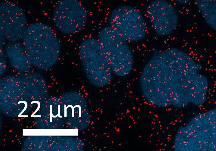
FH Lidiya (Verified Customer) (05-26-2022) | The antibody works great! Antibody was used for WB in Neuroblastoma cell lines (SK-N-BE(2)C and IMR32) with HRP-conjugated secondary. Antibody was also used for a Proximity Ligation Assay to assess interaction with another protein in SK-N-BE(2)C cells. 1:50 of antibody was used for this assay.
 |
FH David (Verified Customer) (01-13-2020) | Great antibody for several applications. Works nicely for immunoblot and immunofluourescence. Can also be used for RNA immunoprecipitation.
|