Various lysates were subjected to SDS PAGE followed by western blot with 68155-1-Ig (STIP1 antibody) at dilution of 1:10000 incubated at room temperature for 1.5 hours. The membrane was stripped and reblotted with HRP-conjugated GAPDH Monoclonal antibody (HRP-60004) as loading control.
Various lysates were subjected to SDS PAGE followed by western blot with 68155-1-Ig (STIP1 antibody) at dilution of 1:10000 incubated at room temperature for 1.5 hours. The membrane was stripped and reblotted with HRP-conjugated GAPDH Monoclonal antibody (HRP-60004) as loading control.
IP experiment of Jurkat using 68155-1-Ig
IP result of anti-STIP1 (IP:68155-1-Ig, 5ug; Detection:68155-1-Ig 1:1000) with Jurkat cells lysate 2400 ug.
Immunohistochemical analysis of paraffin-embedded mouse liver tissue slide using 68155-1-Ig (STIP1 antibody) at dilution of 1:1000 (under 40x lens). Heat mediated antigen retrieval with Tris-EDTA buffer (pH 9.0).
IF Staining of HeLa using 68155-1-Ig
Immunofluorescent analysis of (-20°C Methanol) fixed HeLa cells using STIP1 antibody (68155-1-Ig, Clone: 1B5H2 ) at dilution of 1:800 and CoraLite®488-Conjugated AffiniPure Goat Anti-Mouse IgG(H+L).
Immunofluorescent analysis of (-20°C Methanol) fixed HeLa cells using STIP1 antibody (68155-1-Ig, Clone: 1B5H2 ) at dilution of 1:800 and CoraLite®488-Conjugated AffiniPure Goat Anti-Mouse IgG(H+L).
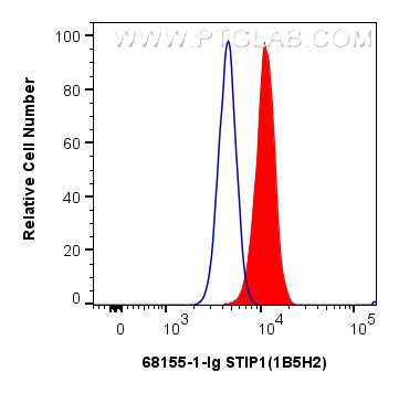
FC experiment of MCF-7 using 68155-1-Ig
1x10^6 MCF-7 cells were intracellularly stained with 0.4 ug STIP1 Monoclonal antibody (68155-1-Ig, Clone:1B5H2) and CoraLite®488-Conjugated Goat Anti-Mouse IgG(H+L) (SA00013-1)(red), or 0.4 ug Mouse IgG1 isotype control Mouse McAb (66360-1-Ig, Clone: 1F8D3) (blue). Cells were fixed with 4% PFA and permeabilized with Flow Cytometry Perm Buffer (PF00011-C).
1x10^6 MCF-7 cells were intracellularly stained with 0.4 ug STIP1 Monoclonal antibody (68155-1-Ig, Clone:1B5H2) and CoraLite®488-Conjugated Goat Anti-Mouse IgG(H+L) (SA00013-1)(red), or 0.4 ug Mouse IgG1 isotype control Mouse McAb (66360-1-Ig, Clone: 1F8D3) (blue). Cells were fixed with 4% PFA and permeabilized with Flow Cytometry Perm Buffer (PF00011-C).
The Proteintech guarantee covers Proteintech antibodies in any species and any application, including those not listed on the datasheet. If the antibody doesn’t perform, you can receive a hassle-free refund or credit note.
mouse liver tissue Note: suggested antigen retrieval with TE buffer pH 9.0; (*) Alternatively, antigen retrieval may be performed with citrate buffer pH 6.0
Positive IF/ICC detected in
HeLa cells
Positive FC (Intra) detected in
MCF-7 cells
Recommended dilution
Application
Dilution
Western Blot (WB)
WB : 1:5000-1:50000
Immunoprecipitation (IP)
IP : 0.5-4.0 ug for 1.0-3.0 mg of total protein lysate
Immunohistochemistry (IHC)
IHC : 1:500-1:2000
Immunofluorescence (IF)/ICC
IF/ICC : 1:400-1:1600
Flow Cytometry (FC) (INTRA)
FC (INTRA) : 0.40 ug per 10^6 cells in a 100 µl suspension
It is recommended that this reagent should be titrated in each testing system to obtain optimal results.
Sample-dependent, Check data in validation data gallery.
Product Information
68155-1-Ig targets STIP1 in WB, IHC, IF/ICC, FC (Intra), IP, ELISA applications and shows reactivity with human, mouse, rat samples.
PBS with 0.02% sodium azide and 50% glycerol , pH 7.3
Storage Conditions
Store at -20°C. Stable for one year after shipment. Aliquoting is unnecessary for -20oC storage. 20ul sizes contain 0.1% BSA.
Background Information
STIP1, also named as Hop and NY-REN-11. Its expression is low in the hypothalamus. STIP1 has some isoforms with MW 62 kDa, 68 kDa and 59 kDa. Acts as a co-chaperone for HSP90AA1.Mediates the association of the molecular chaperones HSPA8/HSC70 and HSP90, Probably forms a complex composed of chaperones HSP90 and HSP70, co-chaperones STIP1/HOP, CDC37, PPP5C, PTGES3/p23, TSC1 and client protein TSC2. STIP1 interacts with PACRG, EEF1AKMT3, FLCN, FNIP1 and FNIP2 (PMID:24880080).
Various lysates were subjected to SDS PAGE followed by western blot with 68155-1-Ig (STIP1 antibody) at dilution of 1:10000 incubated at room temperature for 1.5 hours. The membrane was stripped and reblotted with HRP-conjugated GAPDH Monoclonal antibody (HRP-60004) as loading control.
IHC Figures
IHC staining of mouse liver using 68155-1-Ig
Immunohistochemical analysis of paraffin-embedded mouse liver tissue slide using 68155-1-Ig (STIP1 antibody) at dilution of 1:1000 (under 10x lens). Heat mediated antigen retrieval with Tris-EDTA buffer (pH 9.0).
IHC staining of mouse liver using 68155-1-Ig
Immunohistochemical analysis of paraffin-embedded mouse liver tissue slide using 68155-1-Ig (STIP1 antibody) at dilution of 1:1000 (under 40x lens). Heat mediated antigen retrieval with Tris-EDTA buffer (pH 9.0).
IP Figures
IP experiment of Jurkat using 68155-1-Ig
IP result of anti-STIP1 (IP:68155-1-Ig, 5ug; Detection:68155-1-Ig 1:1000) with Jurkat cells lysate 2400 ug.
IP experiment of HeLa using 68155-1-Ig
IP result of anti-STIP1 (IP:68155-1-Ig, 5ug; Detection:68155-1-Ig 1:1000) with HeLa cells lysate 1880 ug.
IF/ICC Figures
IF Staining of HeLa using 68155-1-Ig
Immunofluorescent analysis of (-20°C Methanol) fixed HeLa cells using STIP1 antibody (68155-1-Ig, Clone: 1B5H2 ) at dilution of 1:800 and CoraLite®488-Conjugated AffiniPure Goat Anti-Mouse IgG(H+L).
FC (INTRA) Figures
FC experiment of MCF-7 using 68155-1-Ig
1x10^6 MCF-7 cells were intracellularly stained with 0.4 ug STIP1 Monoclonal antibody (68155-1-Ig, Clone:1B5H2) and CoraLite®488-Conjugated Goat Anti-Mouse IgG(H+L) (SA00013-1)(red), or 0.4 ug Mouse IgG1 isotype control Mouse McAb (66360-1-Ig, Clone: 1F8D3) (blue). Cells were fixed with 4% PFA and permeabilized with Flow Cytometry Perm Buffer (PF00011-C).
The species listed in Tested Reactivity are in-house verified and applicable species. For unlisted species, please refer to the homology analysis of the immunogen sequence and related species. For rabbit polyclonal antibodies, homology >70% is recommended. For mouse monoclonal antibodies and rabbit recombinant antibodies, homology >90% is recommended. Generally, the higher the homology, the greater the applicability. However, there will be certain differences in protein expression in different species, tissues or cells. Therefore, the homology analysis results are for reference only and do not serve as a guarantee.
At Proteintech, we pride ourselves on our antibody quality, customer service and transparency. As such, we are comparing our antibodies with other vendors, enabling easy identification and comparisons of key data to help you choose the suitable antibody for your needs.
We have selected the top cited antibodies from these vendors for you to compare.
Proteintech
STIP1 Monoclonal antibody
Catalog Number
68155-1-Ig
Citations
-
Dilutions
WB : 1:5000-1:50000 IP : 0.5-4.0 ug for IP and 0.5-4.0 ug for 1.0-3.0 mg of total protein lysate for WB IHC : 1:500-1:2000 IF/ICC : 1:400-1:1600 FC (INTRA) : 0.40 ug per 10^6 cells in a 100 µl suspension
Applications
WB, IHC, IF/ICC, FC (Intra), IP, ELISA
Reactivity
human, mouse, rat
Product Guarantee
Covers any species including not listed on datasheet
Covers any applications including not listed on datasheet
 at dilution of 1:10000 incubated at room temperature for 1.5 hours. The membrane was stripped and reblotted with HRP-conjugated GAPDH Monoclonal antibody (HRP-60004) as loading control.")
 with Jurkat cells lysate 2400 ug.")
 with HeLa cells lysate 1880 ug.")
 at dilution of 1:1000 (under 10x lens). Heat mediated antigen retrieval with Tris-EDTA buffer (pH 9.0).")
 at dilution of 1:1000 (under 40x lens). Heat mediated antigen retrieval with Tris-EDTA buffer (pH 9.0).")
 fixed HeLa cells using STIP1 antibody (68155-1-Ig, Clone: 1B5H2 ) at dilution of 1:800 and CoraLite®488-Conjugated AffiniPure Goat Anti-Mouse IgG(H+L).")
 and CoraLite®488-Conjugated Goat Anti-Mouse IgG(H+L) (SA00013-1)(red), or 0.4 ug Mouse IgG1 isotype control Mouse McAb (66360-1-Ig, Clone: 1F8D3) (blue). Cells were fixed with 4% PFA and permeabilized with Flow Cytometry Perm Buffer (PF00011-C).")