Tested Applications
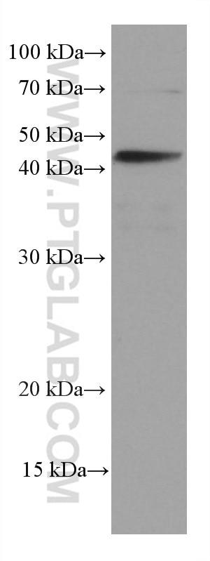
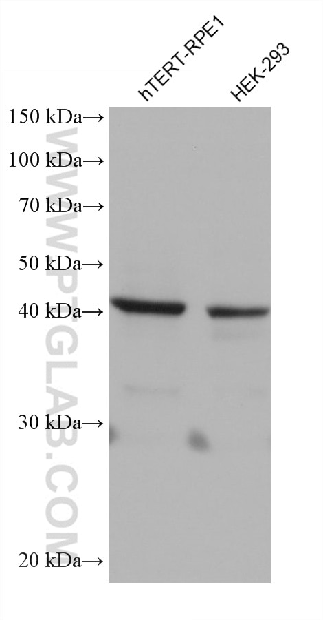
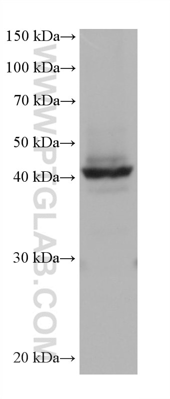
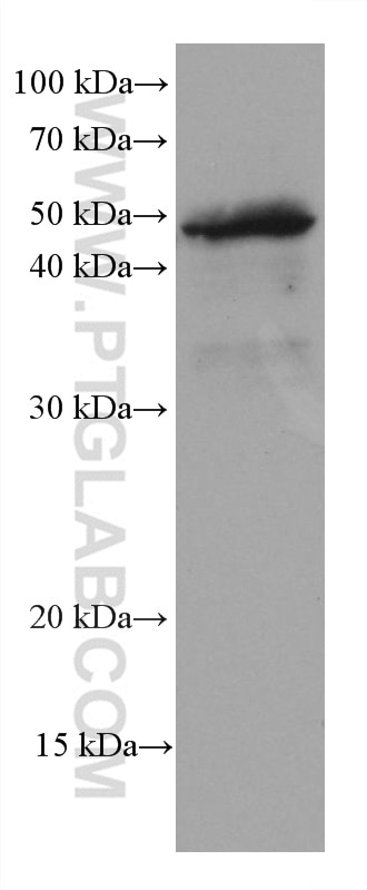
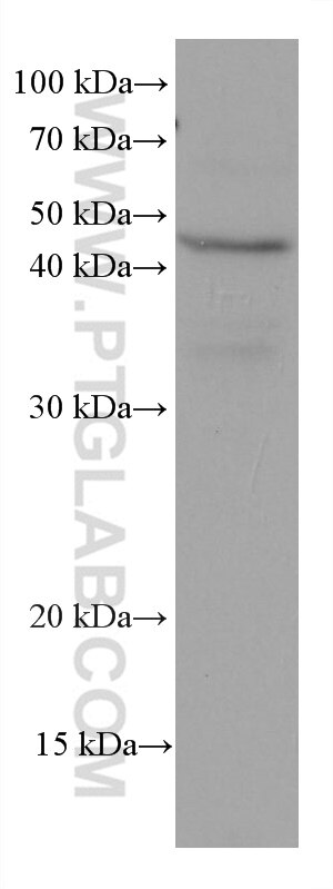
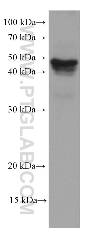
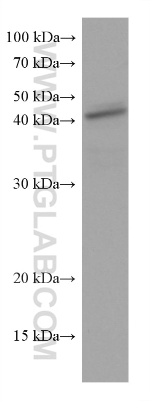
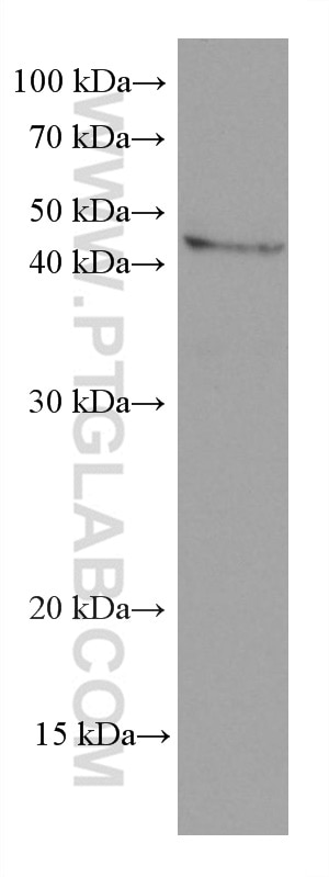
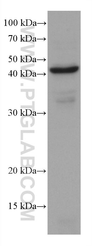
| Positive WB detected in | A549 cells, hTERT-RPE1 cells, T-47D cells, HEK-293 cells, HT-29 cells, HCT 116 cells, HepG2 cells, PC-3 cells, pig brain tissue |
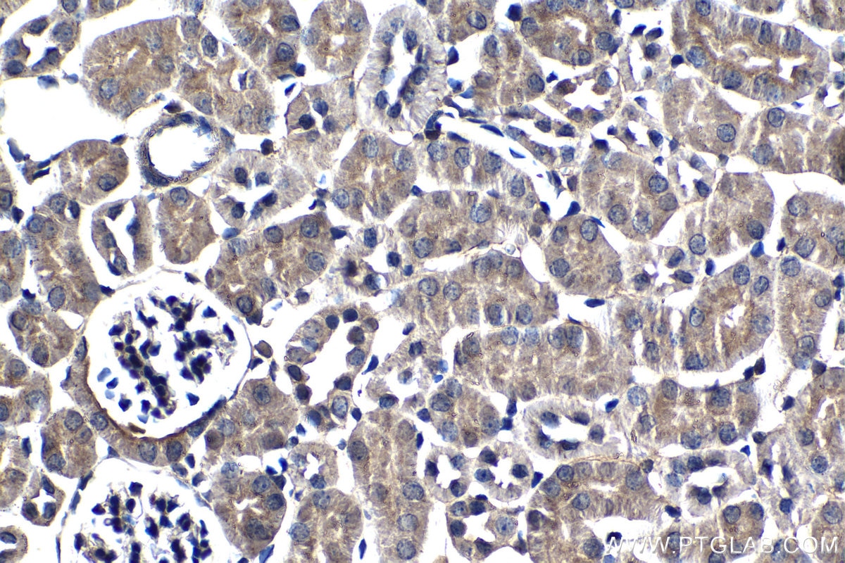
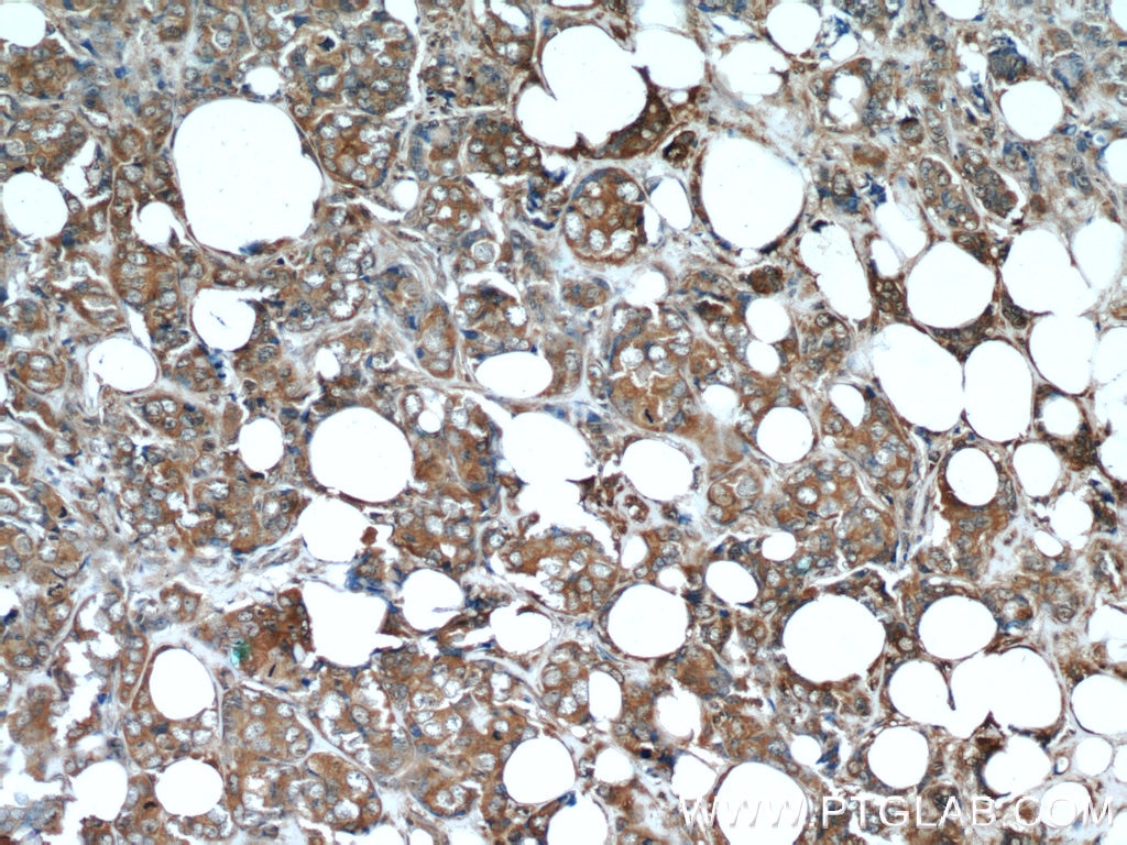
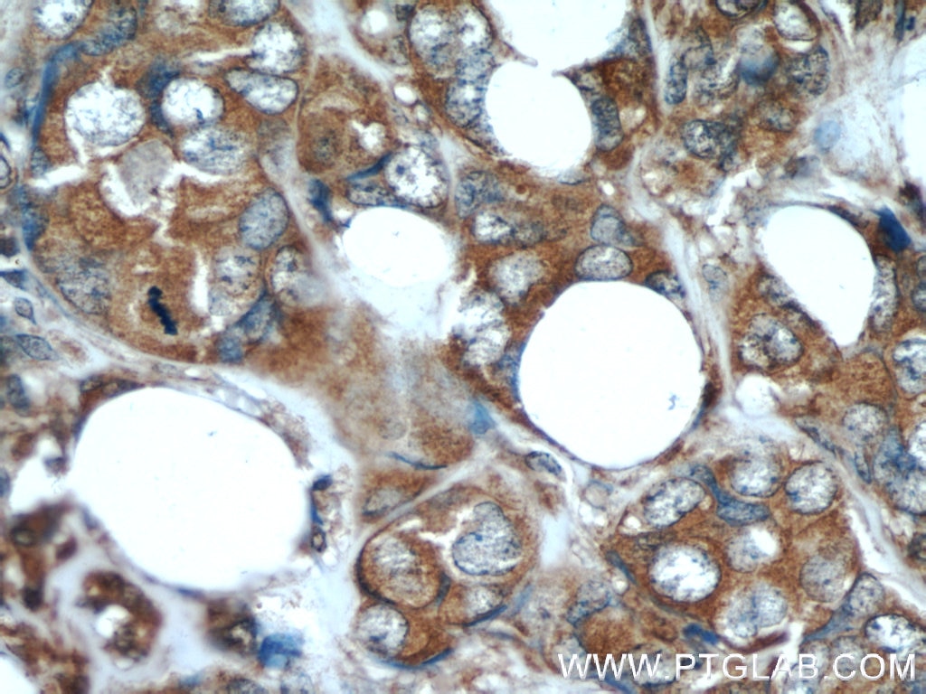
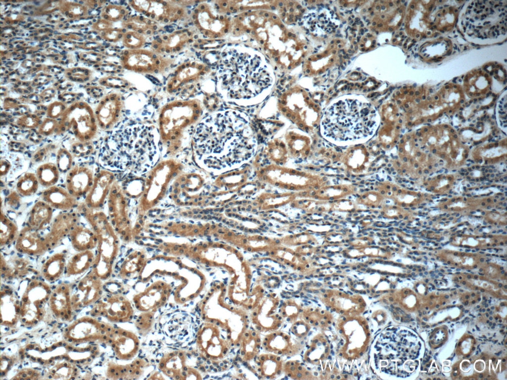
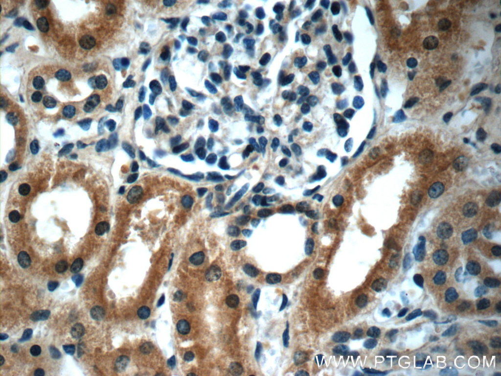
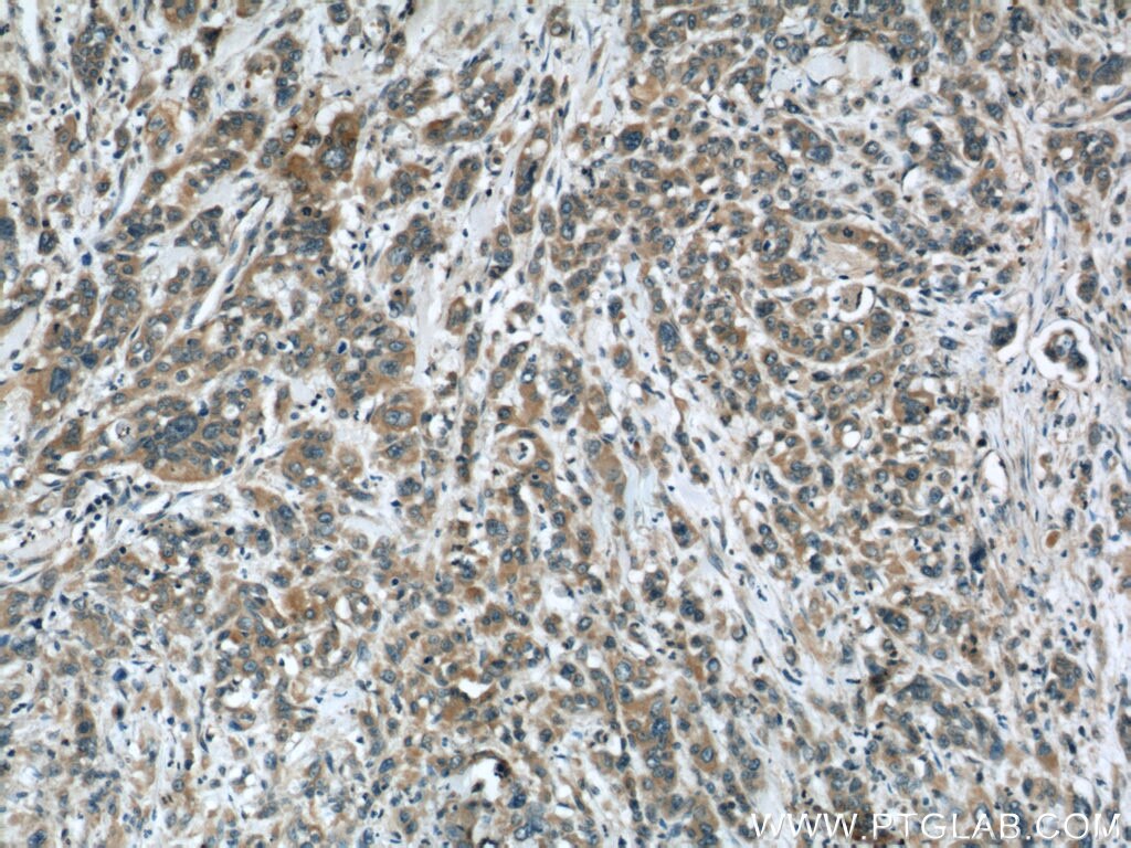
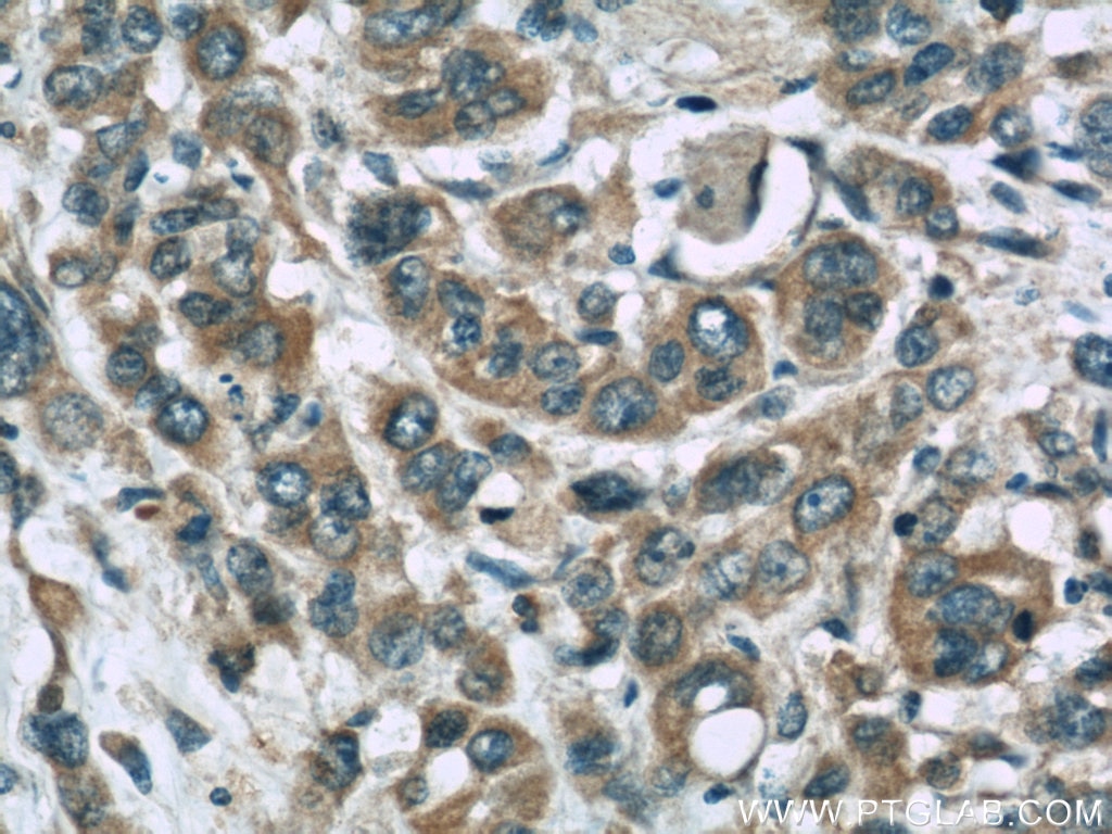
| Positive IHC detected in | mouse kidney tissue, human breast cancer tissue, human kidney tissue, human stomach cancer tissue Note: suggested antigen retrieval with TE buffer pH 9.0; (*) Alternatively, antigen retrieval may be performed with citrate buffer pH 6.0 |
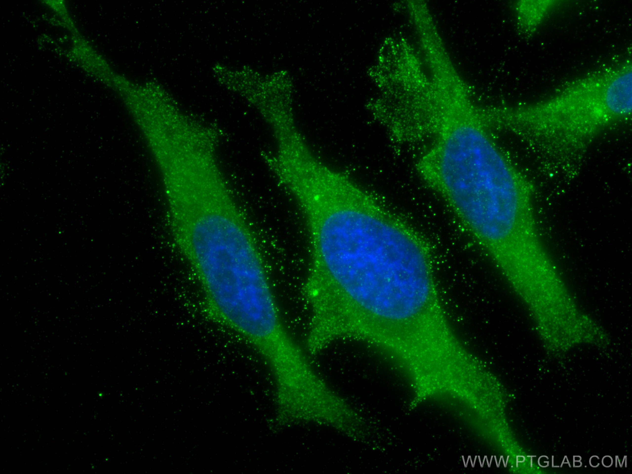
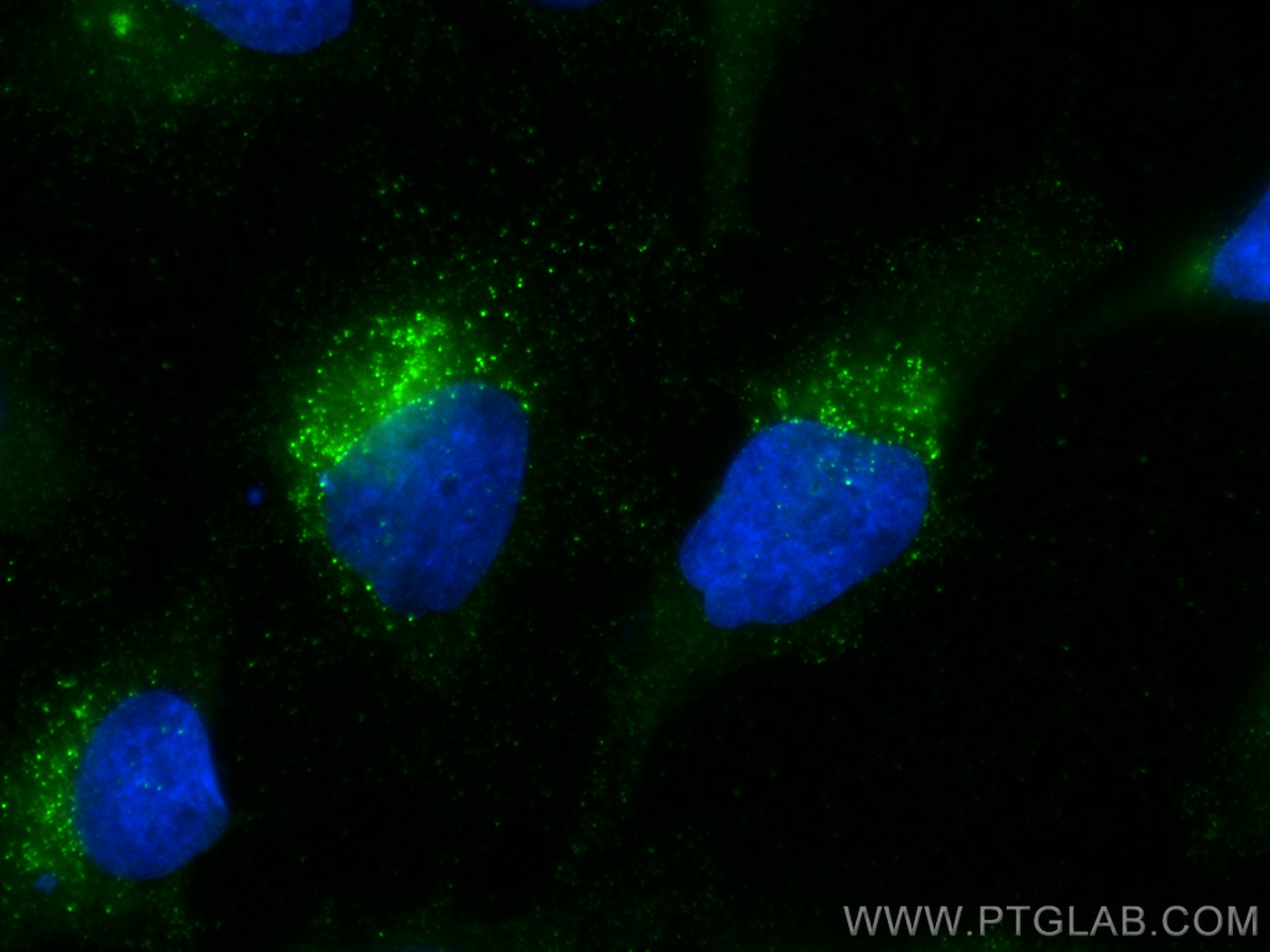
| Positive IF/ICC detected in | HeLa cells |
Recommended dilution
| Application | Dilution |
|---|---|
| Western Blot (WB) | WB : 1:1000-1:6000 |
| Immunohistochemistry (IHC) | IHC : 1:500-1:2000 |
| Immunofluorescence (IF)/ICC | IF/ICC : 1:400-1:1600 |
| It is recommended that this reagent should be titrated in each testing system to obtain optimal results. | |
| Sample-dependent, Check data in validation data gallery. | |
Published Applications
| KD/KO | See 3 publications below |
| WB | See 6 publications below |
| IHC | See 3 publications below |
Product Information
60063-1-Ig targets Stanniocalcin 2 in WB, IHC, IF/ICC, ELISA applications and shows reactivity with human, mouse, pig samples.
| Tested Reactivity | human, mouse, pig |
| Cited Reactivity | human, mouse |
| Host / Isotype | Mouse / IgG1 |
| Class | Monoclonal |
| Type | Antibody |
| Immunogen | Stanniocalcin 2 fusion protein Ag0359 Predict reactive species |
| Full Name | stanniocalcin 2 |
| Calculated Molecular Weight | 33 kDa |
| Observed Molecular Weight | 44 kDa |
| GenBank Accession Number | BC000658 |
| Gene Symbol | Stanniocalcin 2 |
| Gene ID (NCBI) | 8614 |
| RRID | AB_2197544 |
| Conjugate | Unconjugated |
| Form | Liquid |
| Purification Method | Protein G purification |
| UNIPROT ID | O76061 |
| Storage Buffer | PBS with 0.02% sodium azide and 50% glycerol, pH 7.3. |
| Storage Conditions | Store at -20°C. Stable for one year after shipment. Aliquoting is unnecessary for -20oC storage. 20ul sizes contain 0.1% BSA. |
Background Information
Stanniocalcin (STC) is a family of secreted glycoprotein hormones that originally discovered in the corpuscles of Stannius, an endocrine gland of fish. STC1 and STC2, two homologues of STC family, are reported to involve in calcium and phosphate homeostasis. It is expressed in a wide variety of tissues such as kidney, spleen, heart, and pancreas. The protein may play a role in the regulation of renal and intestinal calcium and phosphate transport, cell metabolism, or cellular calcium/phosphate homeostasis. STC2 overexpression could promote tumor cell proliferation, invasion and metastasis in prostate cancer, ovarian cancer or neuroblastoma. STC2 is also vital for cytoprotective properties when exposed to ER stress and hypoxia.
Protocols
| Product Specific Protocols | |
|---|---|
| WB protocol for Stanniocalcin 2 antibody 60063-1-Ig | Download protocol |
| IHC protocol for Stanniocalcin 2 antibody 60063-1-Ig | Download protocol |
| IF protocol for Stanniocalcin 2 antibody 60063-1-Ig | Download protocol |
| Standard Protocols | |
|---|---|
| Click here to view our Standard Protocols |
Publications
| Species | Application | Title |
|---|---|---|
J Hepatocell Carcinoma The Oncogenic and Diagnostic Potential of Stanniocalcin 2 in Hepatocellular Carcinoma. | ||
Biochem Biophys Res Commun The novel prognostic risk factor STC2 can regulate the occurrence and progression of osteosarcoma via the glycolytic pathway.
| ||
Oncotarget STC2 promotes head and neck squamous cell carcinoma metastasis through modulating the PI3K/AKT/Snail signaling.
| ||
Biomed Res Int STC2 Is a Potential Prognostic Biomarker for Pancreatic Cancer and Promotes Migration and Invasion by Inducing Epithelial-Mesenchymal Transition.
| ||
BMC Med Genomics Osteosarcoma transcriptome data exploration reveals STC2 as a novel risk indicator in disease progression | ||
Arch Biochem Biophys STC2 knockdown inhibits cell proliferation and glycolysis in hepatocellular carcinoma through promoting autophagy by PI3K/Akt/mTOR pathway |
