HeLa cells were subjected to SDS PAGE followed by western blot with 12273-1-AP (SSSCA1 antibody) at dilution of 1:500 incubated at room temperature for 1.5 hours.
HeLa cells were subjected to SDS PAGE followed by western blot with 12273-1-AP (SSSCA1 antibody) at dilution of 1:500 incubated at room temperature for 1.5 hours.
WB analysis of HeLa using 12273-1-AP
HeLa cells were subjected to SDS PAGE followed by western blot with 12273-1-AP (SSSCA1 antibody) at dilution of 1:1000 incubated at room temperature for 1.5 hours.
HeLa cells were subjected to SDS PAGE followed by western blot with 12273-1-AP (SSSCA1 antibody) at dilution of 1:1000 incubated at room temperature for 1.5 hours.
WB analysis of NIH/3T3 using 12273-1-AP
NIH/3T3 cells were subjected to SDS PAGE followed by western blot with 12273-1-AP (SSSCA1 antibody) at dilution of 1:300 incubated at room temperature for 1.5 hours.
NIH/3T3 cells were subjected to SDS PAGE followed by western blot with 12273-1-AP (SSSCA1 antibody) at dilution of 1:300 incubated at room temperature for 1.5 hours.
WB analysis of A549 using 12273-1-AP
A549 cells were subjected to SDS PAGE followed by western blot with 12273-1-AP (SSSCA1 antibody) at dilution of 1:500 incubated at room temperature for 1.5 hours.
A549 cells were subjected to SDS PAGE followed by western blot with 12273-1-AP (SSSCA1 antibody) at dilution of 1:500 incubated at room temperature for 1.5 hours.
IP experiment of HeLa using 12273-1-AP
IP result of anti-SSSCA1 (IP:12273-1-AP, 4ug; Detection:12273-1-AP 1:1000) with HeLa cells lysate 2000ug.
Immunohistochemical analysis of paraffin-embedded mouse testis tissue slide using 12273-1-AP (SSSCA1 antibody) at dilution of 1:100 (under 40x lens. Heat mediated antigen retrieval with Tris-EDTA buffer (pH 9.0).
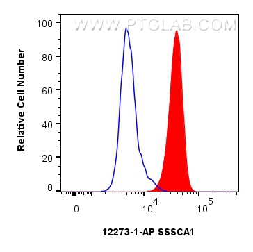
FC experiment of HeLa using 12273-1-AP
1x10^6 HeLa cells were intracellularly stained with 0.25 ug SSSCA1 Polyclonal antibody (12273-1-AP) and CoraLite®488-Conjugated Goat Anti-Rabbit IgG(H+L) (SA00013-2)(red), or 0.25 ug Rabbit IgG control Rabbit PolyAb (30000-0-AP) (blue). Cells were fixed and permeabilized with Transcription Factor Staining Buffer Kit (PF00011).
1x10^6 HeLa cells were intracellularly stained with 0.25 ug SSSCA1 Polyclonal antibody (12273-1-AP) and CoraLite®488-Conjugated Goat Anti-Rabbit IgG(H+L) (SA00013-2)(red), or 0.25 ug Rabbit IgG control Rabbit PolyAb (30000-0-AP) (blue). Cells were fixed and permeabilized with Transcription Factor Staining Buffer Kit (PF00011).
The Proteintech guarantee covers Proteintech antibodies in any species and any application, including those not listed on the datasheet. If the antibody doesn’t perform, you can receive a hassle-free refund or credit note.
mouse testis tissue Note: suggested antigen retrieval with TE buffer pH 9.0; (*) Alternatively, antigen retrieval may be performed with citrate buffer pH 6.0
Positive FC (Intra) detected in
HeLa cells
Recommended dilution
Application
Dilution
Western Blot (WB)
WB : 1:500-1:1000
Immunoprecipitation (IP)
IP : 0.5-4.0 ug for 1.0-3.0 mg of total protein lysate
Immunohistochemistry (IHC)
IHC : 1:50-1:500
Flow Cytometry (FC) (INTRA)
FC (INTRA) : 0.25 ug per 10^6 cells in a 100 µl suspension
It is recommended that this reagent should be titrated in each testing system to obtain optimal results.
Sample-dependent, Check data in validation data gallery.
PBS with 0.02% sodium azide and 50% glycerol, pH 7.3.
Storage Conditions
Store at -20°C. Stable for one year after shipment. Aliquoting is unnecessary for -20oC storage. 20ul sizes contain 0.1% BSA.
Background Information
SSSCA1, also named as Autoantigen p27, is centromere-associated protein. It may induce anti-centromere antibodies. SSSCA1 plays a role in mitosis. It's involved in Sjogren syndrome/scleroderma. The calculated MW of SSSCA1 is 21.5 kDa (native form), with modification the MW is about23- 27 kDa.
Centipeda minima extracts and the active sesquiterpene lactones have therapeutic efficacy in non-small cell lung cancer by suppressing Skp2/p27 signaling pathway
HeLa cells were subjected to SDS PAGE followed by western blot with 12273-1-AP (SSSCA1 antibody) at dilution of 1:500 incubated at room temperature for 1.5 hours.
WB analysis of HeLa using 12273-1-AP
HeLa cells were subjected to SDS PAGE followed by western blot with 12273-1-AP (SSSCA1 antibody) at dilution of 1:1000 incubated at room temperature for 1.5 hours.
WB analysis of NIH/3T3 using 12273-1-AP
NIH/3T3 cells were subjected to SDS PAGE followed by western blot with 12273-1-AP (SSSCA1 antibody) at dilution of 1:300 incubated at room temperature for 1.5 hours.
WB analysis of A549 using 12273-1-AP
A549 cells were subjected to SDS PAGE followed by western blot with 12273-1-AP (SSSCA1 antibody) at dilution of 1:500 incubated at room temperature for 1.5 hours.
IHC Figures
IHC staining of mouse testis using 12273-1-AP
Immunohistochemical analysis of paraffin-embedded mouse testis tissue slide using 12273-1-AP (SSSCA1 antibody) at dilution of 1:100 (under 10x lens. Heat mediated antigen retrieval with Tris-EDTA buffer (pH 9.0).
IHC staining of mouse testis using 12273-1-AP
Immunohistochemical analysis of paraffin-embedded mouse testis tissue slide using 12273-1-AP (SSSCA1 antibody) at dilution of 1:100 (under 40x lens. Heat mediated antigen retrieval with Tris-EDTA buffer (pH 9.0).
IP Figures
IP experiment of HeLa using 12273-1-AP
IP result of anti-SSSCA1 (IP:12273-1-AP, 4ug; Detection:12273-1-AP 1:1000) with HeLa cells lysate 2000ug.
FC (INTRA) Figures
FC experiment of HeLa using 12273-1-AP
1x10^6 HeLa cells were intracellularly stained with 0.25 ug SSSCA1 Polyclonal antibody (12273-1-AP) and CoraLite®488-Conjugated Goat Anti-Rabbit IgG(H+L) (SA00013-2)(red), or 0.25 ug Rabbit IgG control Rabbit PolyAb (30000-0-AP) (blue). Cells were fixed and permeabilized with Transcription Factor Staining Buffer Kit (PF00011).
The species listed in Tested Reactivity are in-house verified and applicable species. For unlisted species, please refer to the homology analysis of the immunogen sequence and related species. For rabbit polyclonal antibodies, homology >70% is recommended. For mouse monoclonal antibodies and rabbit recombinant antibodies, homology >90% is recommended. Generally, the higher the homology, the greater the applicability. However, there will be certain differences in protein expression in different species, tissues or cells. Therefore, the homology analysis results are for reference only and do not serve as a guarantee.
At Proteintech, we pride ourselves on our antibody quality, customer service and transparency. As such, we are comparing our antibodies with other vendors, enabling easy identification and comparisons of key data to help you choose the suitable antibody for your needs.
We have selected the top cited antibodies from these vendors for you to compare.
Proteintech
KD/KO VALIDATED
SSSCA1 Polyclonal antibody
Catalog Number
12273-1-AP
Citations
2
Dilutions
WB : 1:500-1:1000 IP : 0.5-4.0 ug for IP and 0.5-4.0 ug for 1.0-3.0 mg of total protein lysate for WB IHC : 1:50-1:500 FC (INTRA) : 0.25 ug per 10^6 cells in a 100 µl suspension
Applications
WB, IHC, FC (Intra), IP, ELISA
Reactivity
human, mouse, rat
Product Guarantee
Covers any species including not listed on datasheet
Covers any applications including not listed on datasheet
 at dilution of 1:500 incubated at room temperature for 1.5 hours.")
 at dilution of 1:1000 incubated at room temperature for 1.5 hours.")
 at dilution of 1:300 incubated at room temperature for 1.5 hours.")
 at dilution of 1:500 incubated at room temperature for 1.5 hours.")
 with HeLa cells lysate 2000ug.")
 at dilution of 1:100 (under 10x lens. Heat mediated antigen retrieval with Tris-EDTA buffer (pH 9.0).")
 at dilution of 1:100 (under 40x lens. Heat mediated antigen retrieval with Tris-EDTA buffer (pH 9.0).")
 and CoraLite®488-Conjugated Goat Anti-Rabbit IgG(H+L) (SA00013-2)(red), or 0.25 ug Rabbit IgG control Rabbit PolyAb (30000-0-AP) (blue). Cells were fixed and permeabilized with Transcription Factor Staining Buffer Kit (PF00011).")