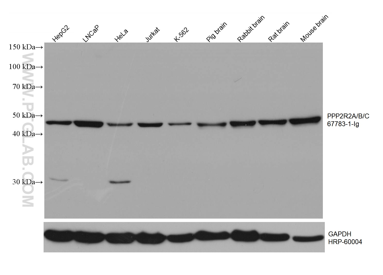
Various lysates were subjected to SDS PAGE followed by western blot with 67783-1-Ig (PPP2R2A/B/C antibody) at dilution of 1:3000 incubated at room temperature for 1.5 hours. The membrane was stripped and reblotted with HRP-conjugated GAPDH Monoclonal antibody (HRP-60004) as loading control.
Various lysates were subjected to SDS PAGE followed by western blot with 67783-1-Ig (PPP2R2A/B/C antibody) at dilution of 1:3000 incubated at room temperature for 1.5 hours. The membrane was stripped and reblotted with HRP-conjugated GAPDH Monoclonal antibody (HRP-60004) as loading control.
WB analysis using 67783-1-Ig
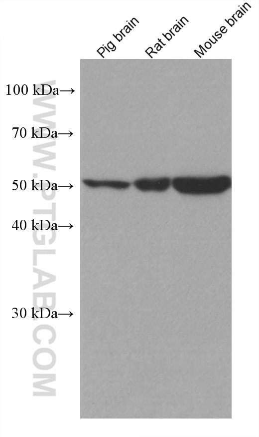
Various lysates were subjected to SDS PAGE followed by western blot with 67783-1-Ig (PPP2R2A/B/C antibody) at dilution of 1:2000 incubated at room temperature for 1.5 hours.
Various lysates were subjected to SDS PAGE followed by western blot with 67783-1-Ig (PPP2R2A/B/C antibody) at dilution of 1:2000 incubated at room temperature for 1.5 hours.
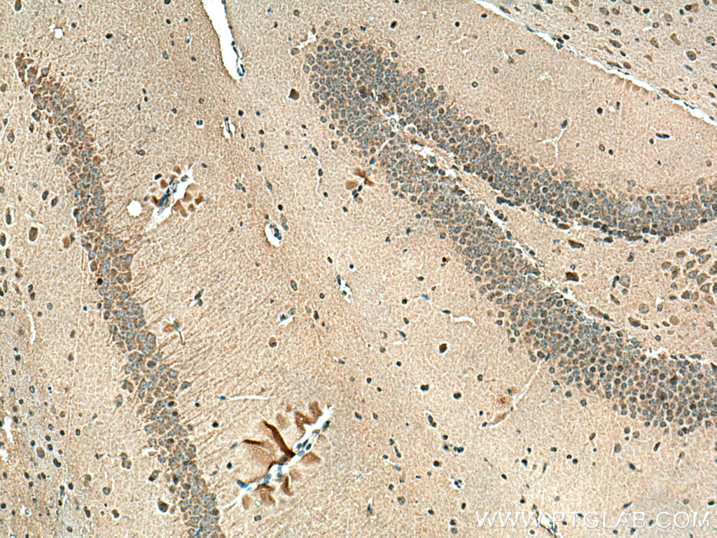
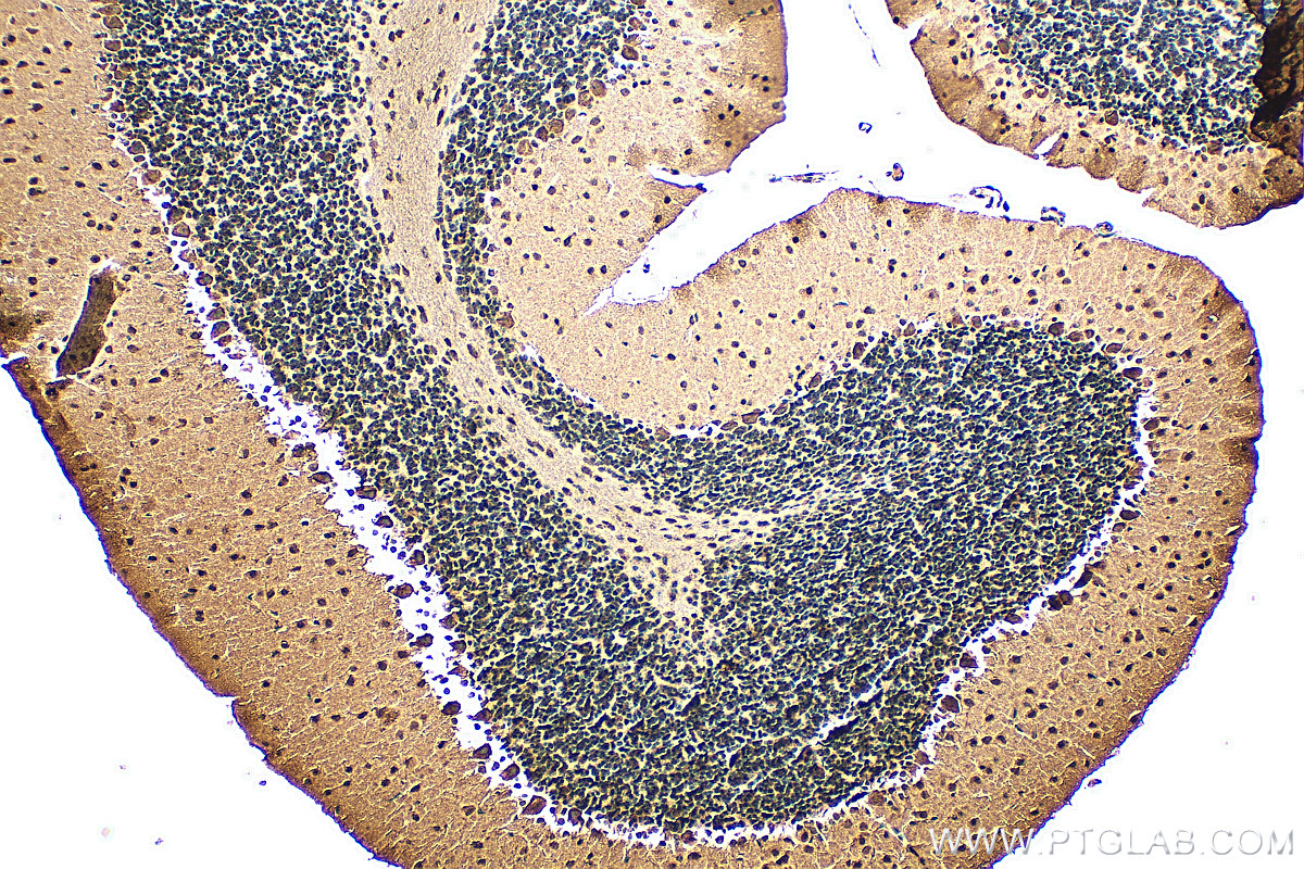
IHC staining of mouse brain using 67783-1-Ig
Immunohistochemical analysis of paraffin-embedded mouse brain tissue slide using 67783-1-Ig (PPP2R2A/B/C antibody) at dilution of 1:200 (under 10x lens). Heat mediated antigen retrieval with Tris-EDTA buffer (pH 9.0).
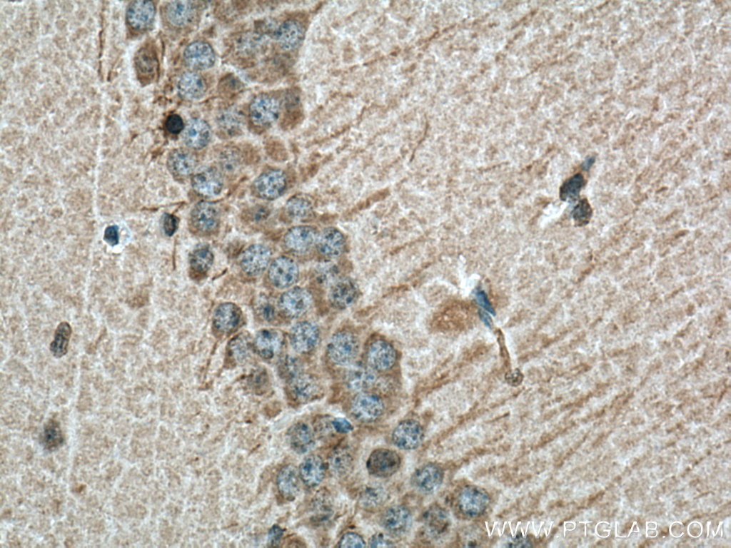
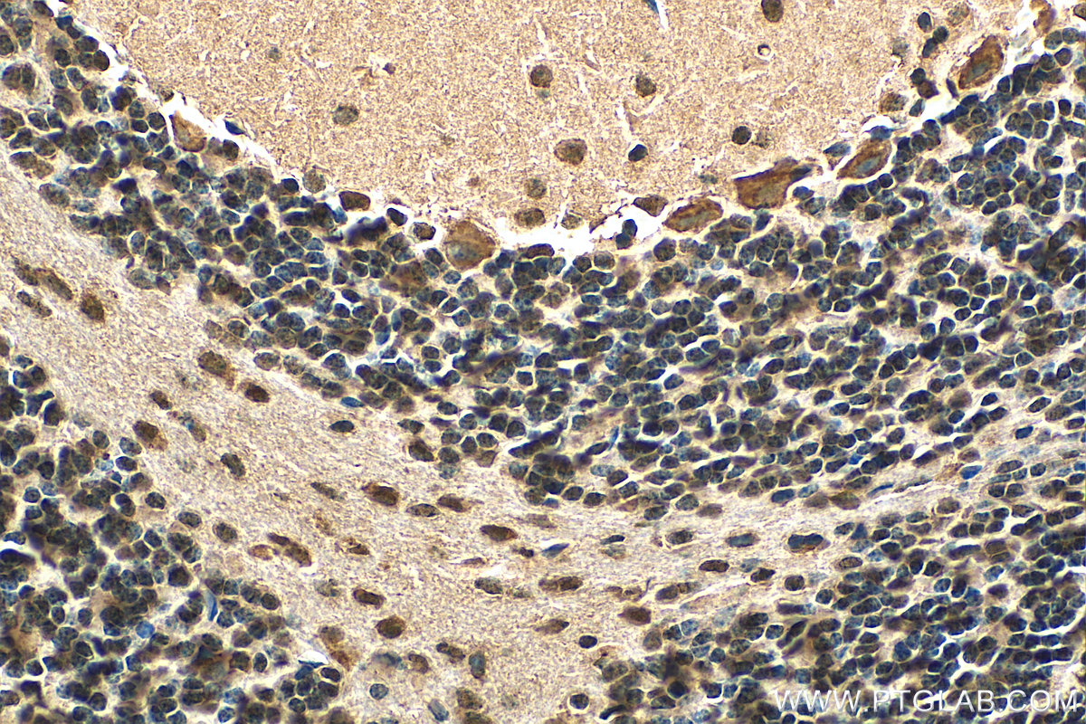
Immunohistochemical analysis of paraffin-embedded mouse cerebellum tissue slide using 67783-1-Ig (PPP2R2A/B/C antibody) at dilution of 1:300 (under 40x lens). Heat mediated antigen retrieval with Tris-EDTA buffer (pH 9.0).
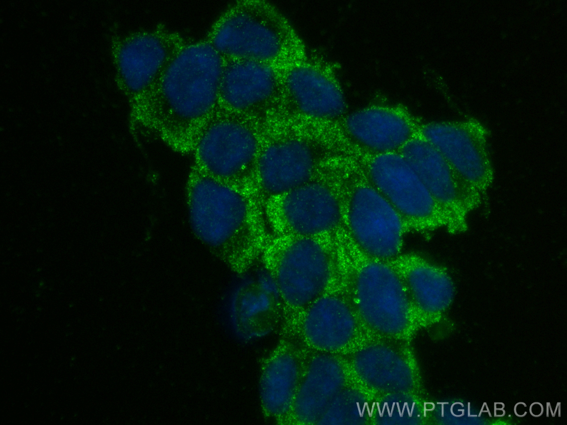
IF Staining of SH-SY5Y using 67783-1-Ig
Immunofluorescent analysis of (-20°C Ethanol) fixed SH-SY5Y cells using PPP2R2A/B/C antibody (67783-1-Ig, Clone: 2E1D5 ) at dilution of 1:400 and CoraLite®488-Conjugated Goat Anti-Mouse IgG(H+L) (SA00013-1).
Immunofluorescent analysis of (-20°C Ethanol) fixed SH-SY5Y cells using PPP2R2A/B/C antibody (67783-1-Ig, Clone: 2E1D5 ) at dilution of 1:400 and CoraLite®488-Conjugated Goat Anti-Mouse IgG(H+L) (SA00013-1).
The Proteintech guarantee covers Proteintech antibodies in any species and any application, including those not listed on the datasheet. If the antibody doesn’t perform, you can receive a hassle-free refund or credit note.
mouse brain tissue, mouse cerebellum tissue Note: suggested antigen retrieval with TE buffer pH 9.0; (*) Alternatively, antigen retrieval may be performed with citrate buffer pH 6.0
Positive IF/ICC detected in
SH-SY5Y cells
Recommended dilution
Application
Dilution
Western Blot (WB)
WB : 1:1000-1:6000
Immunohistochemistry (IHC)
IHC : 1:50-1:500
Immunofluorescence (IF)/ICC
IF/ICC : 1:200-1:800
It is recommended that this reagent should be titrated in each testing system to obtain optimal results.
Sample-dependent, Check data in validation data gallery.
PBS with 0.02% sodium azide and 50% glycerol , pH 7.3
Storage Conditions
Store at -20°C. Stable for one year after shipment. Aliquoting is unnecessary for -20oC storage. 20ul sizes contain 0.1% BSA.
Background Information
The PPP2R2B gene encodes a deduced 443 amino acid protein of approximately 52 kDa, which is a brain-specific regulatory subunit B of protein phosphatase 2. PPP2R2B is a Subunit of PP2A, a highly conserved constitutive enzyme(PMID:11719278). PPP2R2B (Bβ) is an important regulator of protein phosphatase 2A (PP2A) activity in the brain. Through differential promoter usage and alternative splicing, two major isoforms Bβ1 and Bβ2 with divergent sub-cellular targeting N termini are produced. Bβ plays an important role in neuronal survival. It has 5 isoforms produced by alternative splicing. This antibody can recognize PPP2R2A and PPP2R2C due to the immunogen of PPP2R2B having high homology with PPP2R2A and PPP2R2C.
Various lysates were subjected to SDS PAGE followed by western blot with 67783-1-Ig (PPP2R2A/B/C antibody) at dilution of 1:3000 incubated at room temperature for 1.5 hours. The membrane was stripped and reblotted with HRP-conjugated GAPDH Monoclonal antibody (HRP-60004) as loading control.
WB analysis using 67783-1-Ig
Various lysates were subjected to SDS PAGE followed by western blot with 67783-1-Ig (PPP2R2A/B/C antibody) at dilution of 1:2000 incubated at room temperature for 1.5 hours.
IHC Figures
IHC staining of mouse brain using 67783-1-Ig
Immunohistochemical analysis of paraffin-embedded mouse brain tissue slide using 67783-1-Ig (PPP2R2A/B/C antibody) at dilution of 1:200 (under 10x lens). Heat mediated antigen retrieval with Tris-EDTA buffer (pH 9.0).
IHC staining of mouse brain using 67783-1-Ig
Immunohistochemical analysis of paraffin-embedded mouse brain tissue slide using 67783-1-Ig (PPP2R2A/B/C antibody) at dilution of 1:200 (under 40x lens). Heat mediated antigen retrieval with Tris-EDTA buffer (pH 9.0).
IHC staining of mouse cerebellum using 67783-1-Ig
Immunohistochemical analysis of paraffin-embedded mouse cerebellum tissue slide using 67783-1-Ig (PPP2R2A/B/C antibody) at dilution of 1:300 (under 10x lens). Heat mediated antigen retrieval with Tris-EDTA buffer (pH 9.0).
IHC staining of mouse cerebellum using 67783-1-Ig
Immunohistochemical analysis of paraffin-embedded mouse cerebellum tissue slide using 67783-1-Ig (PPP2R2A/B/C antibody) at dilution of 1:300 (under 40x lens). Heat mediated antigen retrieval with Tris-EDTA buffer (pH 9.0).
IF/ICC Figures
IF Staining of SH-SY5Y using 67783-1-Ig
Immunofluorescent analysis of (-20°C Ethanol) fixed SH-SY5Y cells using PPP2R2A/B/C antibody (67783-1-Ig, Clone: 2E1D5 ) at dilution of 1:400 and CoraLite®488-Conjugated Goat Anti-Mouse IgG(H+L) (SA00013-1).
The species listed in Tested Reactivity are in-house verified and applicable species. For unlisted species, please refer to the homology analysis of the immunogen sequence and related species. For rabbit polyclonal antibodies, homology >70% is recommended. For mouse monoclonal antibodies and rabbit recombinant antibodies, homology >90% is recommended. Generally, the higher the homology, the greater the applicability. However, there will be certain differences in protein expression in different species, tissues or cells. Therefore, the homology analysis results are for reference only and do not serve as a guarantee.
At Proteintech, we pride ourselves on our antibody quality, customer service and transparency. As such, we are comparing our antibodies with other vendors, enabling easy identification and comparisons of key data to help you choose the suitable antibody for your needs.
We have selected the top cited antibodies from these vendors for you to compare.
 at dilution of 1:3000 incubated at room temperature for 1.5 hours. The membrane was stripped and reblotted with HRP-conjugated GAPDH Monoclonal antibody (HRP-60004) as loading control.")
 at dilution of 1:2000 incubated at room temperature for 1.5 hours.")
 at dilution of 1:200 (under 10x lens). Heat mediated antigen retrieval with Tris-EDTA buffer (pH 9.0).")
 at dilution of 1:200 (under 40x lens). Heat mediated antigen retrieval with Tris-EDTA buffer (pH 9.0).")
 at dilution of 1:300 (under 10x lens). Heat mediated antigen retrieval with Tris-EDTA buffer (pH 9.0).")
 at dilution of 1:300 (under 40x lens). Heat mediated antigen retrieval with Tris-EDTA buffer (pH 9.0).")
 fixed SH-SY5Y cells using PPP2R2A/B/C antibody (67783-1-Ig, Clone: 2E1D5 ) at dilution of 1:400 and CoraLite®488-Conjugated Goat Anti-Mouse IgG(H+L) (SA00013-1).")