Filter:
 at dilution of 1:10000 incubated at room temperature for 1.5 hours. The membrane was stripped and reblotted with HRP-conjugated Alpha Tubulin Monoclonal antibody (HRP-66031) as loading control.")
 at dilution of 1:300 incubated at room temperature for 1.5 hours.")
 with si-Control and si-PLOD3 transfected HepG2 cells.")
 at dilution of 1:10000 incubated at room temperature for 1.5 hours.")
 at dilution of 1:300 incubated at room temperature for 1.5 hours.")
 with HeLa cells lysate 1400ug.")
 at dilution of 1:100 (under 10x lens).")
 at dilution of 1:100 (under 40x lens).")
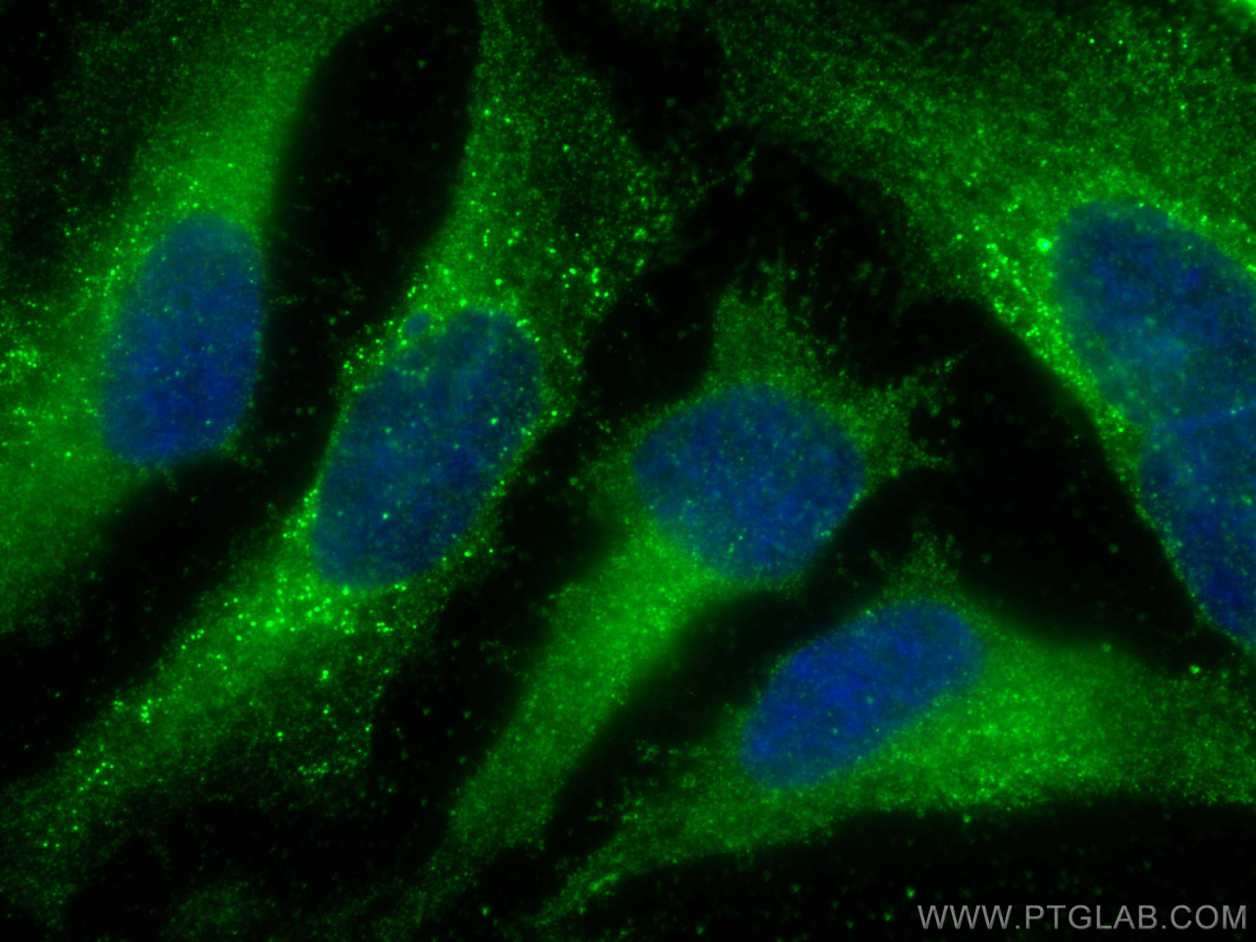
 fixed HeLa cells using PLOD3 antibody (60058-1-Ig, Clone: 2G12B8 ) at dilution of 1:400 and CoraLite®488-Conjugated AffiniPure Goat Anti-Mouse IgG(H+L).")
 at dilution of 1:25 and Rhodamine-Goat anti-Mouse IgG.")
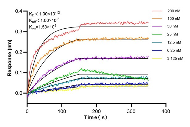
 kinetic assays of 60058-1-Ig against Human PLOD3 were performed. The Affinity Constant is below 1 pM.")
Tested Applications
| Positive WB detected in | A549 cells, HepG2 cells, human placenta tissue, PC-3 cells, K-562 cells, LNCaP cells, HeLa cells, HSC-T6 cells, NIH/3T3 cells, 4T1 cells |
| Positive IP detected in | HeLa cells |
| Positive IHC detected in | human pancreas cancer tissue Note: suggested antigen retrieval with TE buffer pH 9.0; (*) Alternatively, antigen retrieval may be performed with citrate buffer pH 6.0 |
| Positive IF/ICC detected in | HeLa cells |
Recommended dilution
| Application | Dilution |
|---|---|
| Western Blot (WB) | WB : 1:5000-1:50000 |
| Immunoprecipitation (IP) | IP : 0.5-4.0 ug for 1.0-3.0 mg of total protein lysate |
| Immunohistochemistry (IHC) | IHC : 1:20-1:200 |
| Immunofluorescence (IF)/ICC | IF/ICC : 1:200-1:800 |
| It is recommended that this reagent should be titrated in each testing system to obtain optimal results. | |
| Sample-dependent, Check data in validation data gallery. | |
Published Applications
| WB | See 3 publications below |
Product Information
60058-1-Ig targets PLOD3 in WB, IHC, IF/ICC, IP, ELISA applications and shows reactivity with human, mouse, rat samples.
| Tested Reactivity | human, mouse, rat |
| Cited Reactivity | human, mouse |
| Host / Isotype | Mouse / IgG2a |
| Class | Monoclonal |
| Type | Antibody |
| Immunogen | PLOD3 fusion protein Ag1480 Predict reactive species |
| Full Name | procollagen-lysine, 2-oxoglutarate 5-dioxygenase 3 |
| Calculated Molecular Weight | 738 aa, 85 kDa |
| Observed Molecular Weight | 80-85 kDa |
| GenBank Accession Number | BC011674 |
| Gene Symbol | PLOD3 |
| Gene ID (NCBI) | 8985 |
| RRID | AB_2165779 |
| Conjugate | Unconjugated |
| Form | Liquid |
| Purification Method | Protein A purification |
| UNIPROT ID | O60568 |
| Storage Buffer | PBS with 0.02% sodium azide and 50% glycerol, pH 7.3. |
| Storage Conditions | Store at -20°C. Stable for one year after shipment. Aliquoting is unnecessary for -20oC storage. 20ul sizes contain 0.1% BSA. |
Background Information
PLOD3, also named as LH3, forms hydroxylysine residues in -Xaa-Lys-Gly- sequences in collagens. These hydroxylysines serve as sites of attachment for carbohydrate units and are essential for the stability of the intermolecular collagen cross-links. The major function of PLOD3 in osteoblasts is to glucosylate galactosylhydroxylysine residues in type I collagen.
Protocols
| Product Specific Protocols | |
|---|---|
| WB protocol for PLOD3 antibody 60058-1-Ig | Download protocol |
| IHC protocol for PLOD3 antibody 60058-1-Ig | Download protocol |
| IF protocol for PLOD3 antibody 60058-1-Ig | Download protocol |
| IP protocol for PLOD3 antibody 60058-1-Ig | Download protocol |
| Standard Protocols | |
|---|---|
| Click here to view our Standard Protocols |
Publications
| Species | Application | Title |
|---|---|---|
PLoS One Lysyl Hydroxylase 3 Localizes to Epidermal Basement Membrane and Is Reduced in Patients with Recessive Dystrophic Epidermolysis Bullosa. | ||
Matrix Biol Plus Use of osteoblast-derived matrix to assess the influence of collagen modifications on cancer cells. | ||
JCI Insight Transcriptional control of a collagen deposition and adhesion process that promotes lung adenocarcinoma growth and metastasis. |