
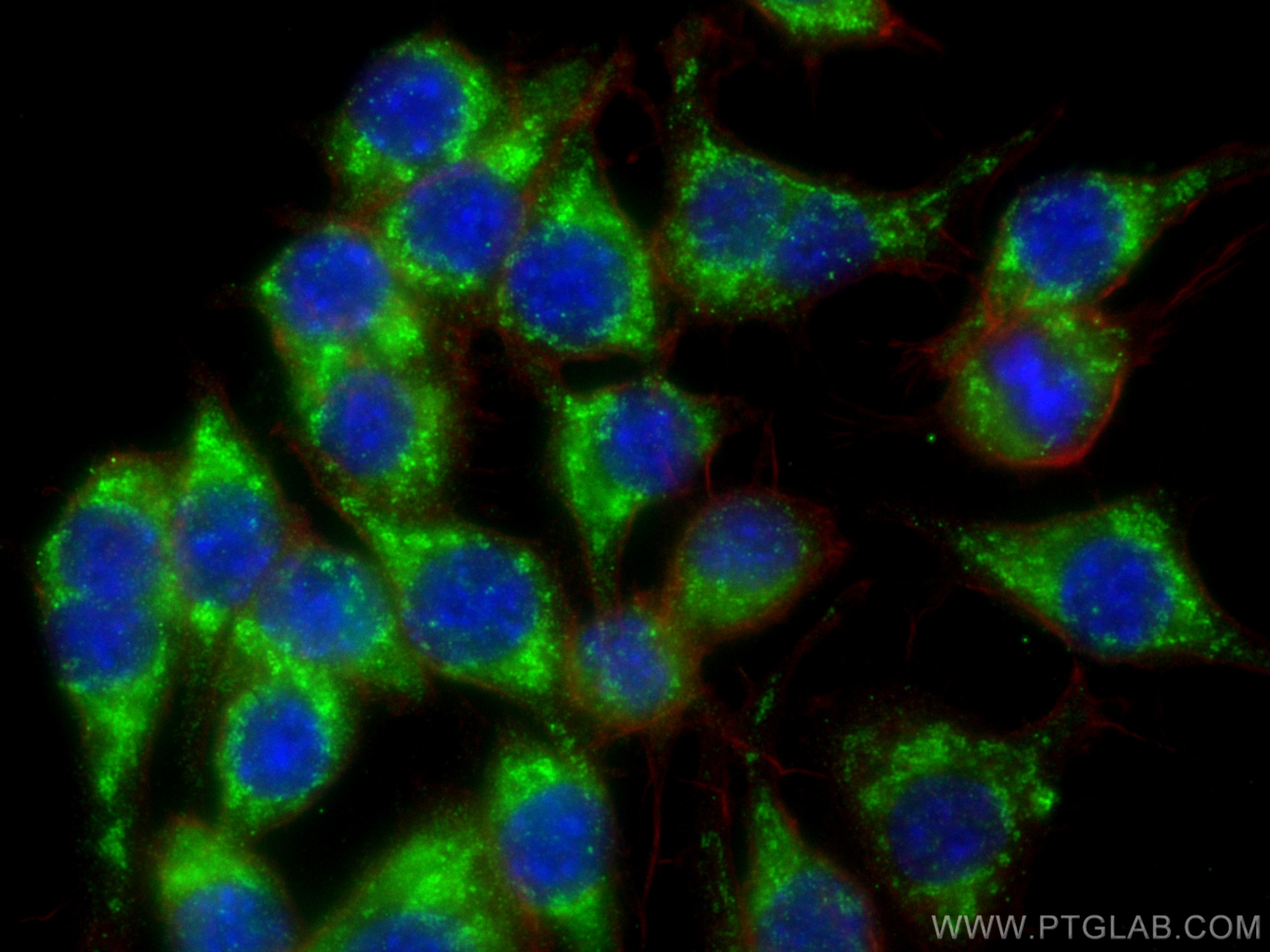

Tested Applications
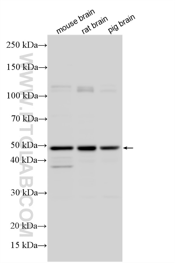
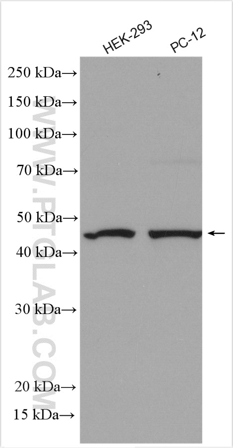
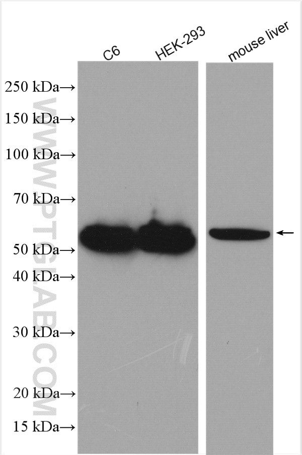
| Positive WB detected in | mouse brain tissue, C6 cells, HEK-293 cells, mouse liver tissue, PC-12 cells, rat brain tissue, pig brain tissue |
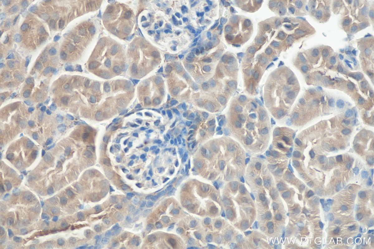
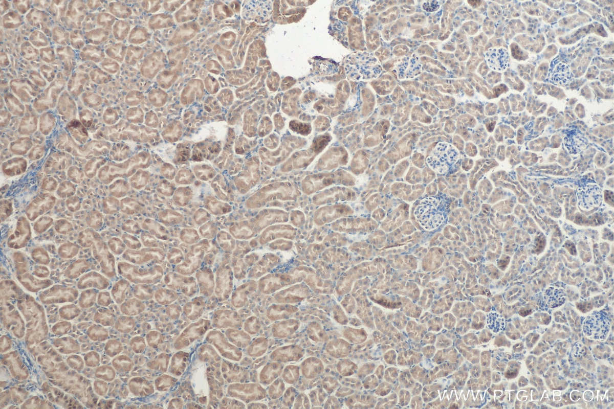
| Positive IHC detected in | mouse kidney tissue Note: suggested antigen retrieval with TE buffer pH 9.0; (*) Alternatively, antigen retrieval may be performed with citrate buffer pH 6.0 |
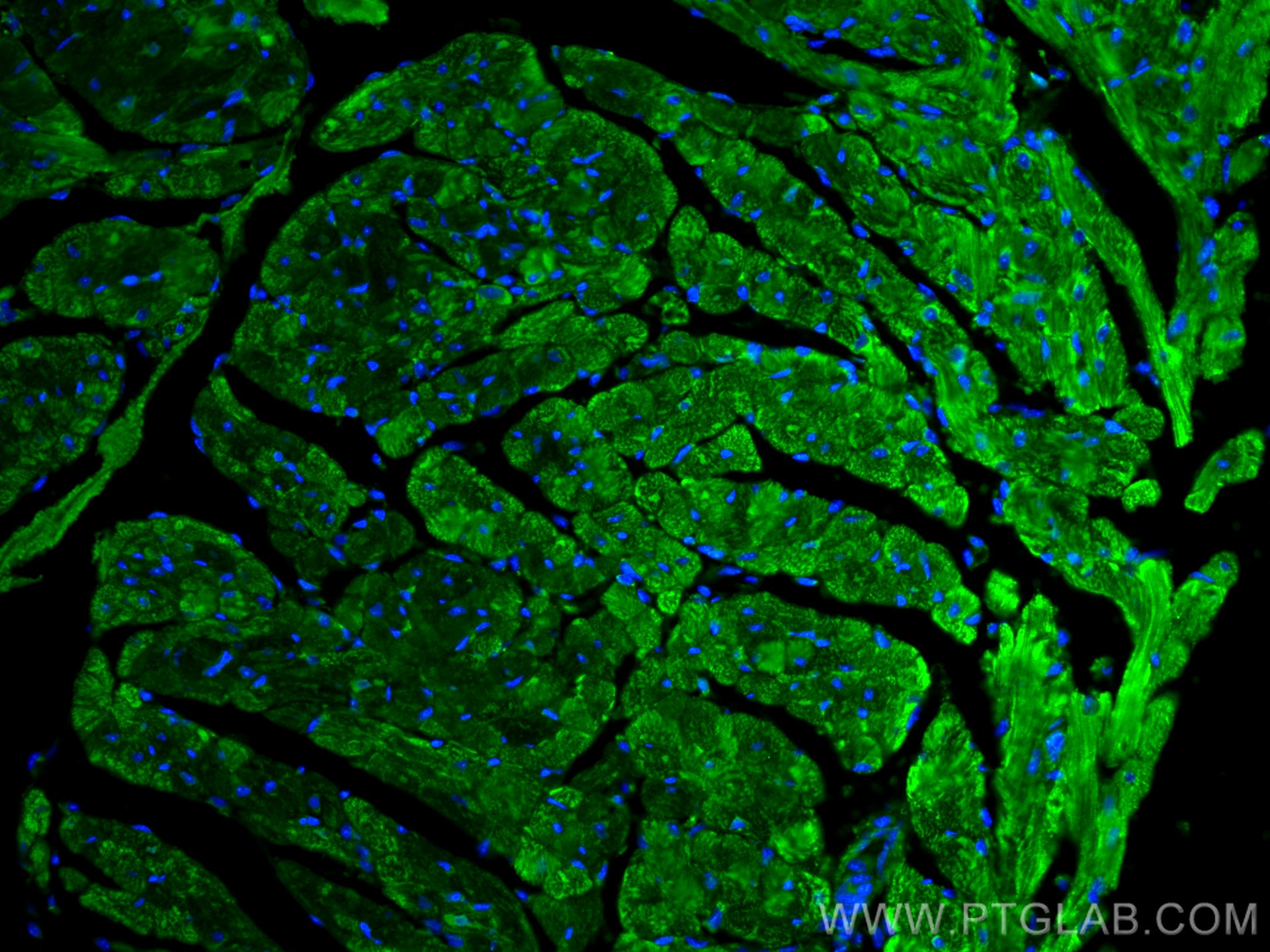
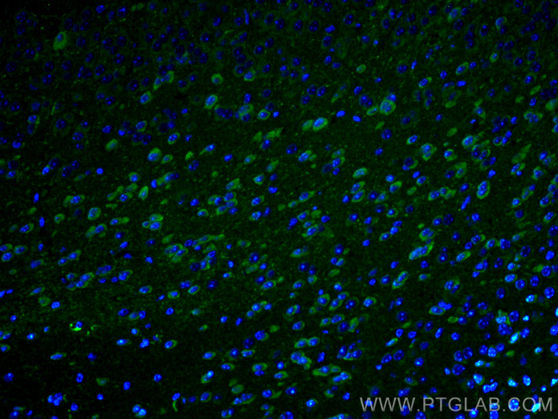
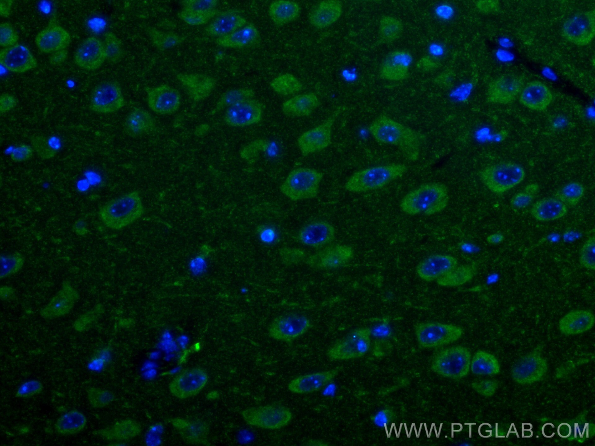
| Positive IF-P detected in | mouse heart tissue, mouse brain tissue |
| Positive IF/ICC detected in | RAW 264.7 cells |
Recommended dilution
| Application | Dilution |
|---|---|
| Western Blot (WB) | WB : 1:1000-1:8000 |
| Immunohistochemistry (IHC) | IHC : 1:50-1:500 |
| Immunofluorescence (IF)-P | IF-P : 1:50-1:500 |
| Immunofluorescence (IF)/ICC | IF/ICC : 1:200-1:800 |
| It is recommended that this reagent should be titrated in each testing system to obtain optimal results. | |
| Sample-dependent, Check data in validation data gallery. | |
Product Information
14060-1-AP targets PARK2/Parkin in WB, IHC, IF/ICC, IF-P, IP, CoIP, ELISA applications and shows reactivity with human, mouse, rat, pig samples.
| Tested Reactivity | human, mouse, rat, pig |
| Cited Reactivity | human, mouse, rat, pig, rabbit, monkey, chicken, cattle, ducks |
| Host / Isotype | Rabbit / IgG |
| Class | Polyclonal |
| Type | Antibody |
| Immunogen |
CatNo: Ag5092 Product name: Recombinant human Parkin protein Source: e coli.-derived, PGEX-4T Tag: GST Domain: 81-387 aa of BC022014 Sequence: NATGGDDPRNAAGGCEREPQSLTRVDLSSSVLPGDSVGLAVILHTDSRKDSPPAGSPAGRSIYNSFYVYCKGPCQRVQPGKLRVQCSTCRQATLTLTQGPSCWDDVLIPNRMSGECQSPHCPGTSAEFFFKCGAHPTSDKETSVALHLIATNSRNITCITCTDVRSPVLVFQCNSRHVICLDCFHLYCVTRLNDRQFVHDPQLGYSLPCVGTGDTVVLRGALGGFRRGVAGCPNSLIKELHHFRILGEEQYNRYQQYGAEECVLQMGGVLCPRPGCGAGLLPEPDQRKVTCEGGNGLGCGYGQRRTK Predict reactive species |
| Full Name | Parkinson disease (autosomal recessive, juvenile) 2, parkin |
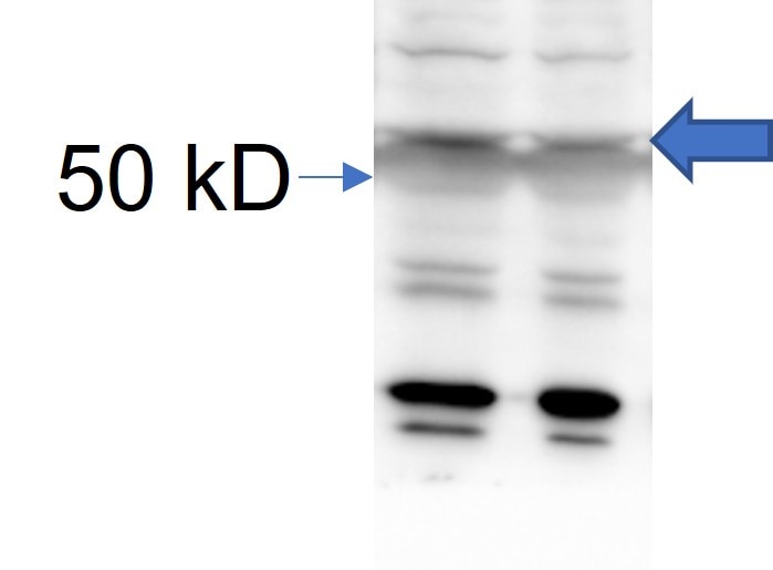
| Calculated Molecular Weight | 52 kDa |
| Observed Molecular Weight | 42-52 kDa |
| GenBank Accession Number | BC022014 |
| Gene Symbol | Parkin |
| Gene ID (NCBI) | 5071 |
| RRID | AB_2878005 |
| Conjugate | Unconjugated |
| Form | Liquid |
| Purification Method | Antigen affinity purification |
| UNIPROT ID | O60260 |
| Storage Buffer | PBS with 0.02% sodium azide and 50% glycerol, pH 7.3. |
| Storage Conditions | Store at -20°C. Stable for one year after shipment. Aliquoting is unnecessary for -20oC storage. 20ul sizes contain 0.1% BSA. |
Background Information
Parkin, a RING-type E3 ubiquitin-protein ligase, is involved in the ubiquitination pathway and contributes to protection from neurotoxicity induced by unfolded protein stresses. Its ubiquitin-protein ligase activity promotes the degradation of a variety of proteins including itself. Mutations in Parkin are implicated in the pathogenesis of autosomal recessive familial Parkinson's disease. It has 8 isoforms produced by alternative splicing with molecular weights of 24, 31, 36 and 42-52 kDa. Sometimes an additional band of 70 kDa or 110 kDa may be detected, which is caused by ubiquitination modification or formation of Parkin complex (PMID: 10976934, PMID: 18190519).
Protocols
| Product Specific Protocols | |
|---|---|
| IF protocol for PARK2/Parkin antibody 14060-1-AP | Download protocol |
| IHC protocol for PARK2/Parkin antibody 14060-1-AP | Download protocol |
| WB protocol for PARK2/Parkin antibody 14060-1-AP | Download protocol |
| Standard Protocols | |
|---|---|
| Click here to view our Standard Protocols |
Publications
What published studies show
Anti-Parkin (Cat# 14060-1-AP) has accumulated 647 citations in journals including Nature Cell Biology, Nature Neuroscience, Nature Communications, Autophagy, Molecular Cell, Science Advances, Cell Reports, Oncogene, Free Radical Biology and Medicine, Advanced Science, and Cell Death & Differentiation. Associated research fields encompass mitophagy, mitochondrial dynamics, cancer biology, neurodegeneration, cardiovascular disease, diabetes, ferroptosis, aging, fibrosis, and infectious disease.
| Species | Application | Title |
|---|---|---|
Bioact Mater Astaxanthin-metformin conjugate nanomicelles-loaded hydrogel reprograms macrophage metabolism via mitochondrion-targeted regulation for diabetic wound healing. | ||
Bioact Mater Copper doped bioactive glass promotes matrix vesicles-mediated biomineralization via osteoblast mitophagy and mitochondrial dynamics during bone regeneration | ||
Bioact Mater NIR-responsive bio-system with sequential antibacterial and immunomodulatory effects for the treatment of periodontitis | ||
Bioact Mater Near infrared enhanced palladium loaded siraitia grosvenorii carbon dots amplify mitophagy for acute lung injury immunotherapy. | ||
Nat Cell Biol Ammonia-induced lysosomal and mitochondrial damage causes cell death of effector CD8+ T cells | ||
Nat Cell Biol Integrated stress response couples mitochondrial fitness with lineage reprogramming to drive cancer evolution. |
Reviews
What customers say
Users consistently rate this antibody positively (average ~4.6/5) for Western Blot at a 1:1000 dilution. Most reviewers describe it as great or perfect for detecting endogenous PARK2 in mouse tissues and cell lines. A few noted minor limitations: one user observed some nonspecific bands alongside the expected ~52 kDa band in mouse brain lysate, and another reported successful detection in only one of three breast cancer cell lines tested, though this may reflect differential expression rather than antibody performance. Overall, users express satisfaction with its reliability for WB applications.
The reviews below have been submitted by verified Proteintech customers who received an incentive for providing their feedback.
MALLIKARJUNA (Verified Customer) (10-17-2025) | GREAT FOR wb
|
Javier (Verified Customer) (09-09-2025) | Perfect antibody for western blotting assay.
|
QX (Verified Customer) (05-08-2025) | It is okay to detect endogenous PARK2 with the right around 52 kd MW although there are some nonspecific bands,
 |
Ajte (Verified Customer) (03-08-2022) | It works well for me
|
Elyssa (Verified Customer) (11-11-2019) | This antibody worked well for me for one cell line (I had tried detecting the protein in 3 cells lines, and only one cell line appeared to have expression. However, I have not looked into this further and cannot say if this has to do with the antibody, or differential expression across lines). Overall, I was happy with the antibody.
|


