WB Figures
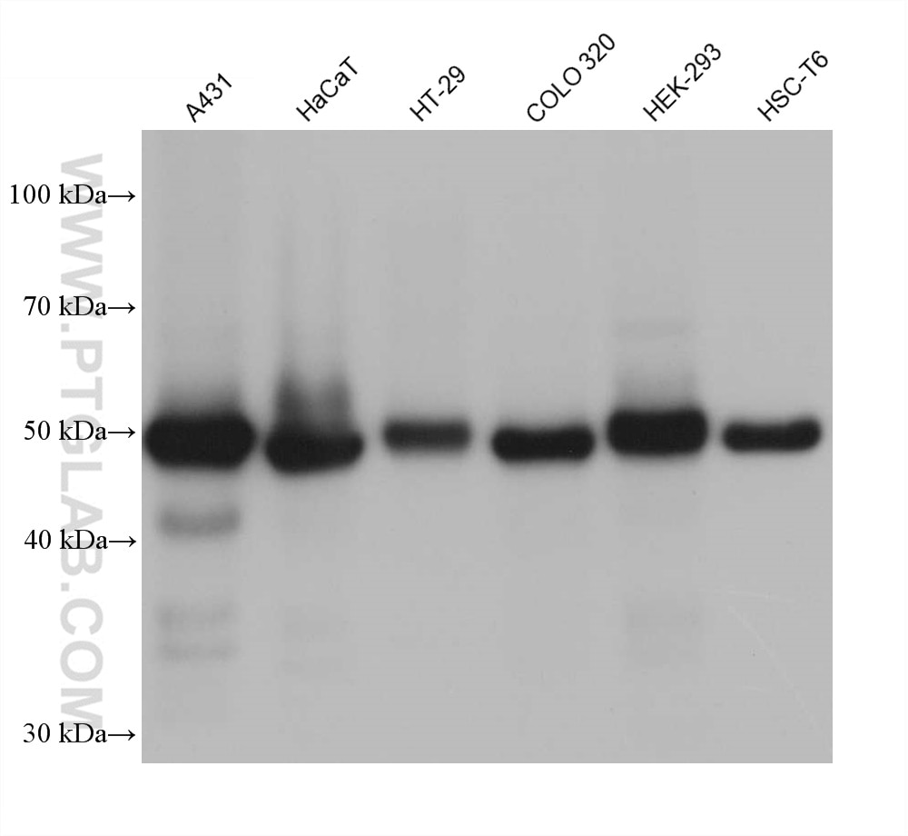
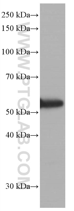
WB analysis using 60283-2-Ig (same clone as 60283-2-PBS)
Various lysates were subjected to SDS PAGE followed by western blot with 60283-2-Ig (P53 antibody) at dilution of 1:20000 incubated at room temperature for 1.5 hours. This data was developed using the same antibody clone with 60283-2-PBS in a different storage buffer formulation.
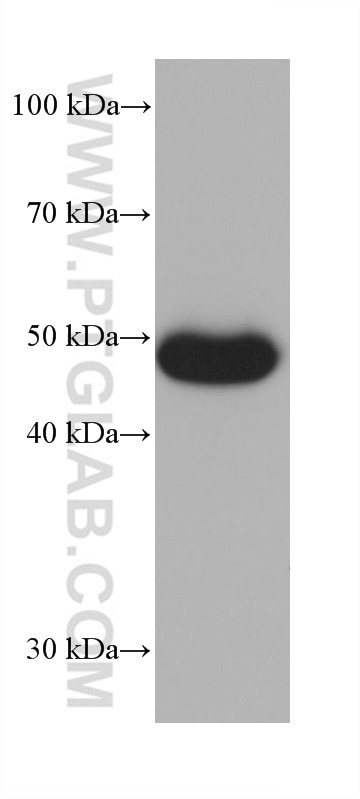
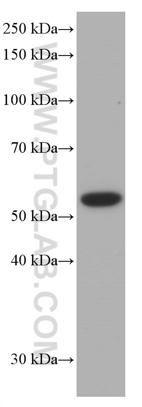
WB analysis of HSC-T6 using 60283-2-Ig (same clone as 60283-2-PBS)
HSC-T6 cells were subjected to SDS PAGE followed by western blot with 60283-2-Ig (P53 antibody) at dilution of 1:20000 incubated at room temperature for 1.5 hours. This data was developed using the same antibody clone with 60283-2-PBS in a different storage buffer formulation.
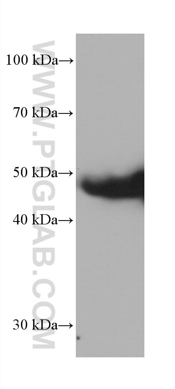
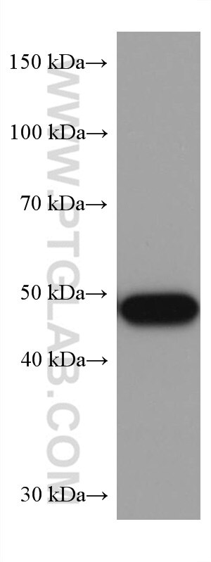
WB analysis of ROS1728 using 60283-2-Ig (same clone as 60283-2-PBS)
ROS1728 cells were subjected to SDS PAGE followed by western blot with 60283-2-Ig (P53 antibody) at dilution of 1:20000 incubated at room temperature for 1.5 hours. This data was developed using the same antibody clone with 60283-2-PBS in a different storage buffer formulation.
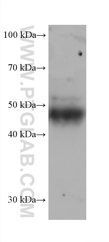
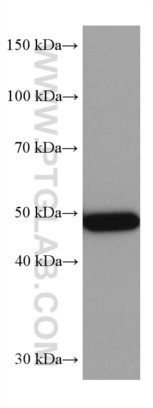
WB analysis of rat thymus using 60283-2-Ig (same clone as 60283-2-PBS)
rat thymus tissue were subjected to SDS PAGE followed by western blot with 60283-2-Ig (P53 antibody) at dilution of 1:20000 incubated at room temperature for 1.5 hours. This data was developed using the same antibody clone with 60283-2-PBS in a different storage buffer formulation.
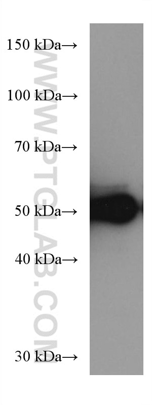
WB analysis of COLO 320 using 60283-2-Ig (same clone as 60283-2-PBS)
COLO 320 cells were subjected to SDS PAGE followed by western blot with 60283-2-Ig (P53 antibody) at dilution of 1:5000 incubated at room temperature for 1.5 hours. This data was developed using the same antibody clone with 60283-2-PBS in a different storage buffer formulation.
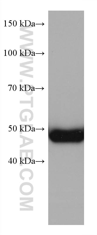
WB analysis of HSC-T6 using 60283-2-Ig (same clone as 60283-2-PBS)
HSC-T6 cells were subjected to SDS PAGE followed by western blot with 60283-2-Ig (P53 antibody) at dilution of 1:5000 incubated at room temperature for 1.5 hours. This data was developed using the same antibody clone with 60283-2-PBS in a different storage buffer formulation.
WB analysis of ROS1728 using 60283-2-Ig (same clone as 60283-2-PBS)
ROS1728 cells were subjected to SDS PAGE followed by western blot with 60283-2-Ig (P53 antibody) at dilution of 1:5000 incubated at room temperature for 1.5 hours. This data was developed using the same antibody clone with 60283-2-PBS in a different storage buffer formulation.
WB analysis of COLO 320 using 60283-2-Ig (same clone as 60283-2-PBS)
COLO 320 cells were subjected to SDS PAGE followed by western blot with 60283-2-Ig (P53 antibody) at dilution of 1:10000 incubated at room temperature for 1.5 hours. This data was developed using the same antibody clone with 60283-2-PBS in a different storage buffer formulation.
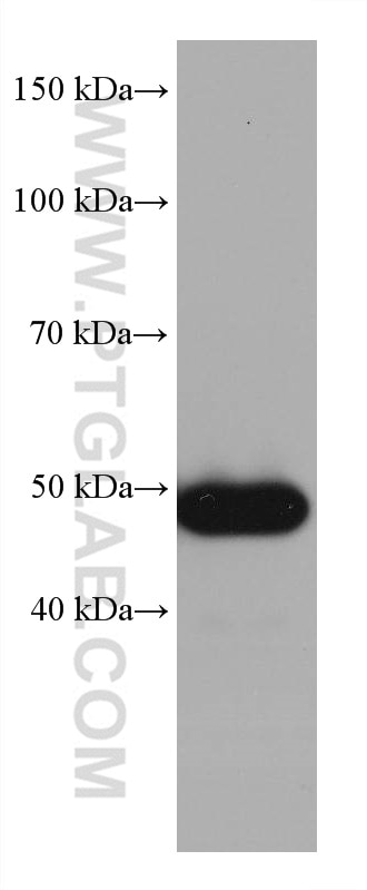
WB analysis of HSC-T6 using 60283-2-Ig (same clone as 60283-2-PBS)
HSC-T6 cells were subjected to SDS PAGE followed by western blot with 60283-2-Ig (P53 antibody) at dilution of 1:10000 incubated at room temperature for 1.5 hours. This data was developed using the same antibody clone with 60283-2-PBS in a different storage buffer formulation.
IHC Figures
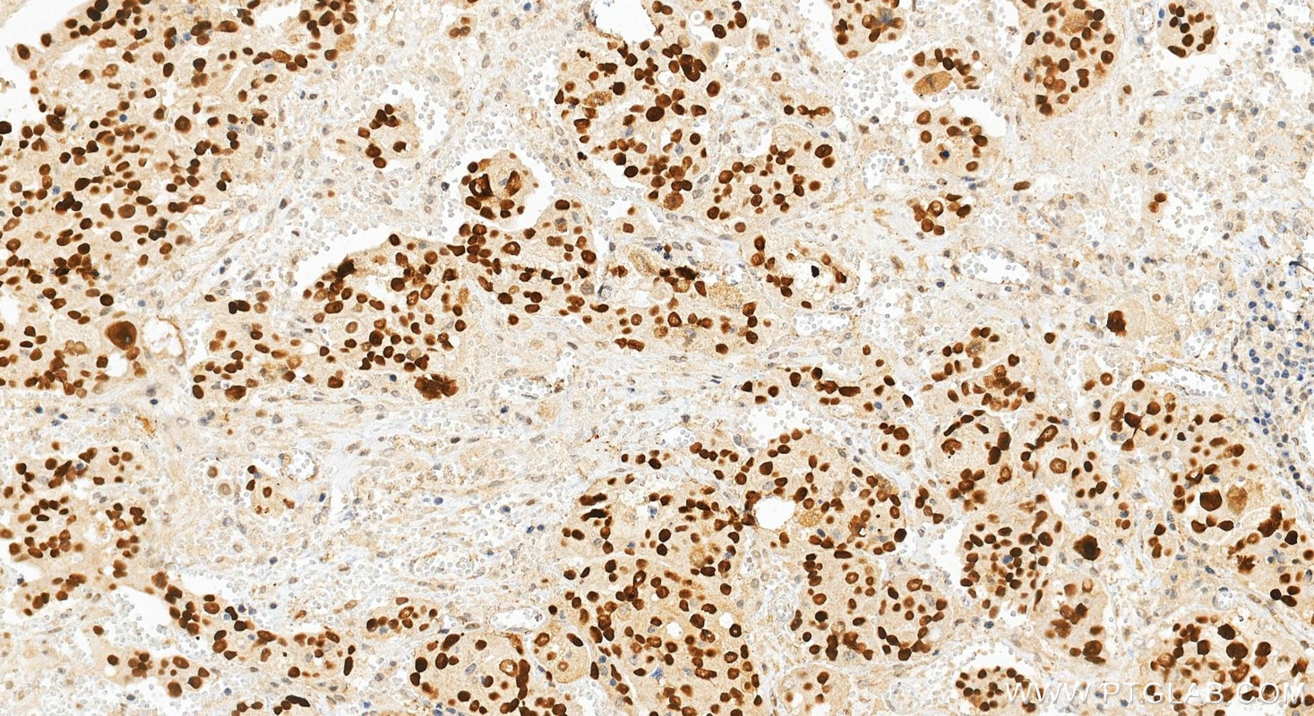
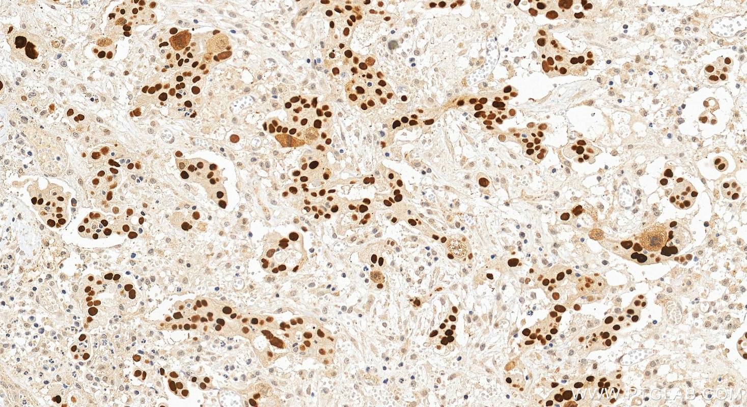
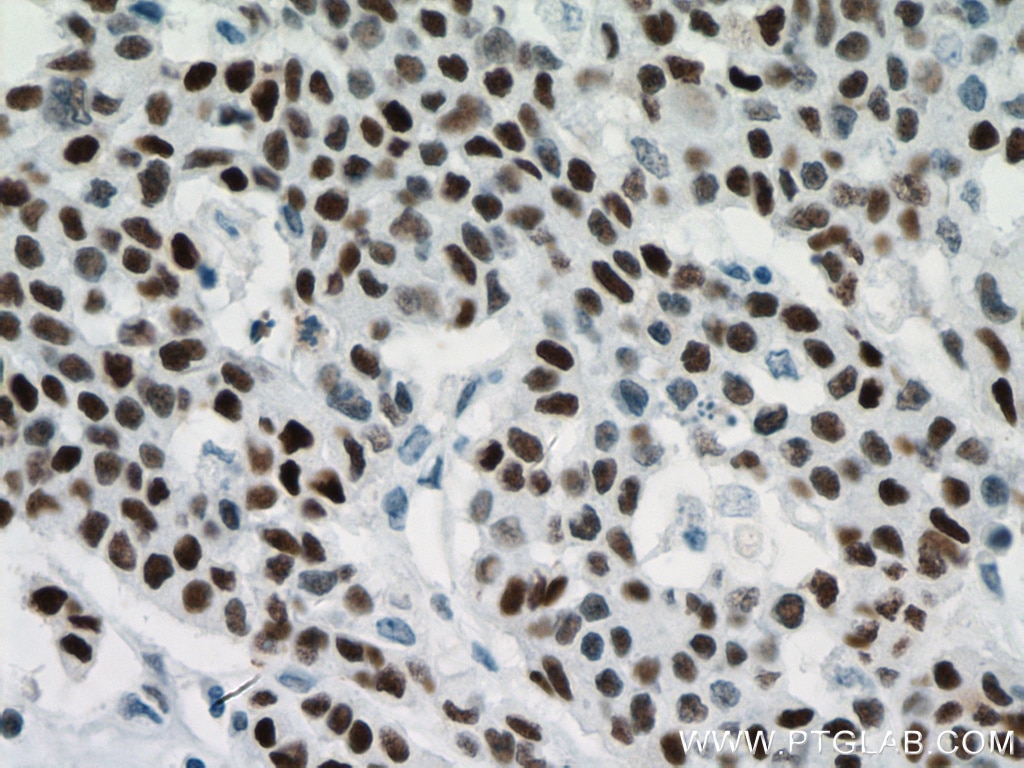
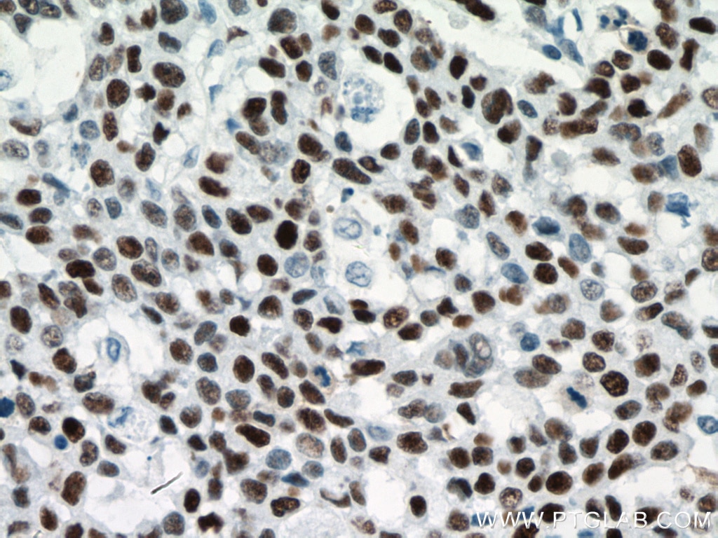
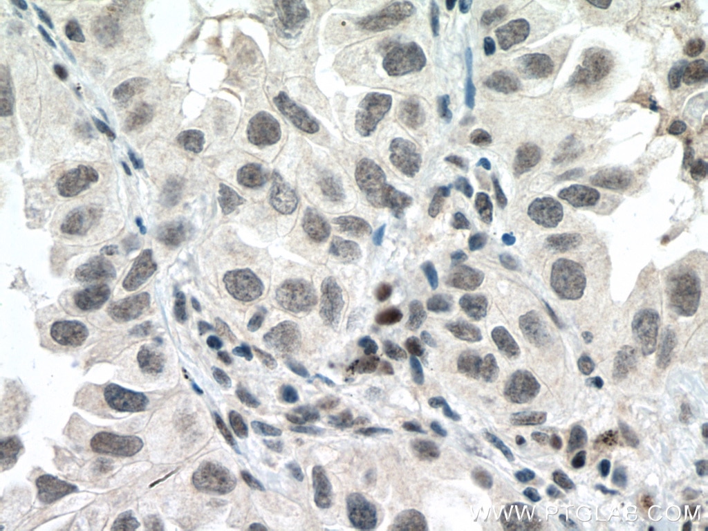
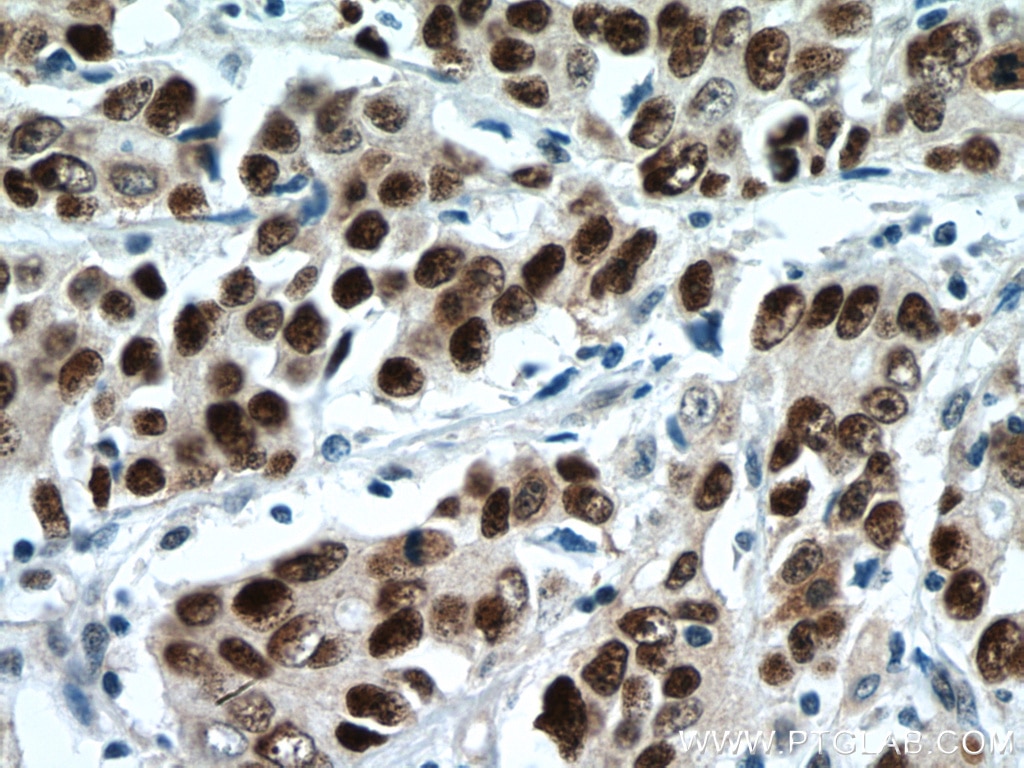
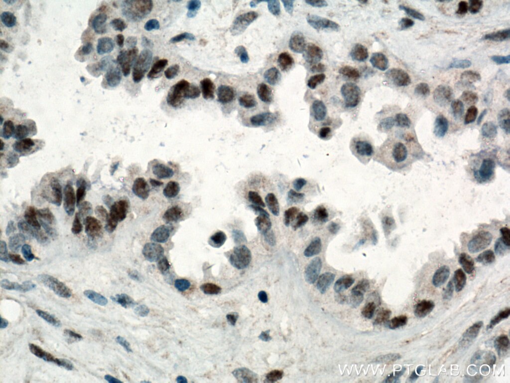
IHC staining of human ovary cancer using 60283-2-Ig (same clone as 60283-2-PBS)
Immunohistochemical analysis of paraffin-embedded human ovary cancer tissue slide using 60283-2-Ig (P53 antibody) at dilution of 1:4000 (under 20x lens). Heat mediated antigen retrieval with Tris-EDTA buffer (pH 9.0). This data was developed using the same antibody clone with 60283-2-PBS in a different storage buffer formulation.
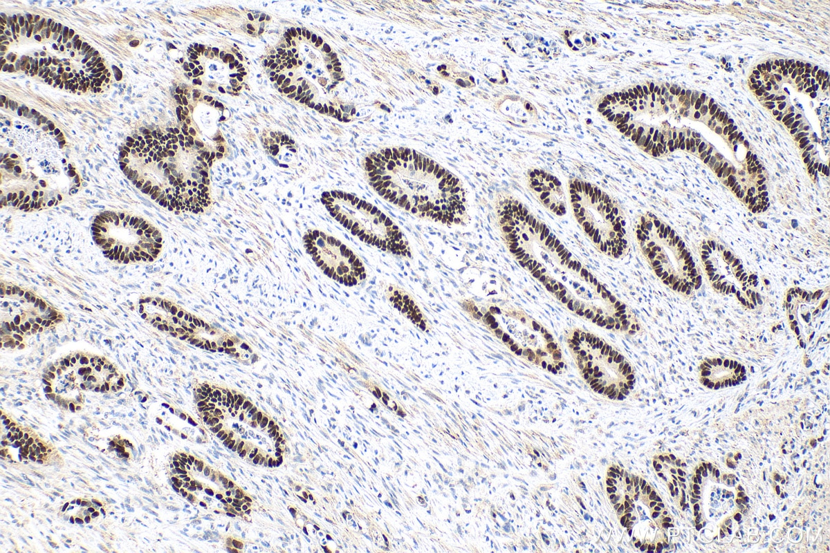
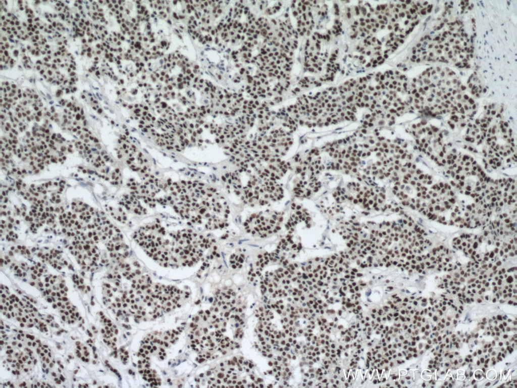
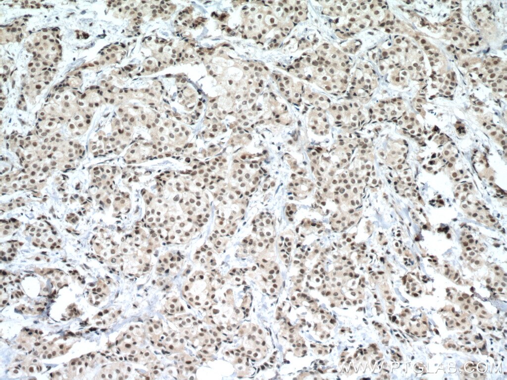
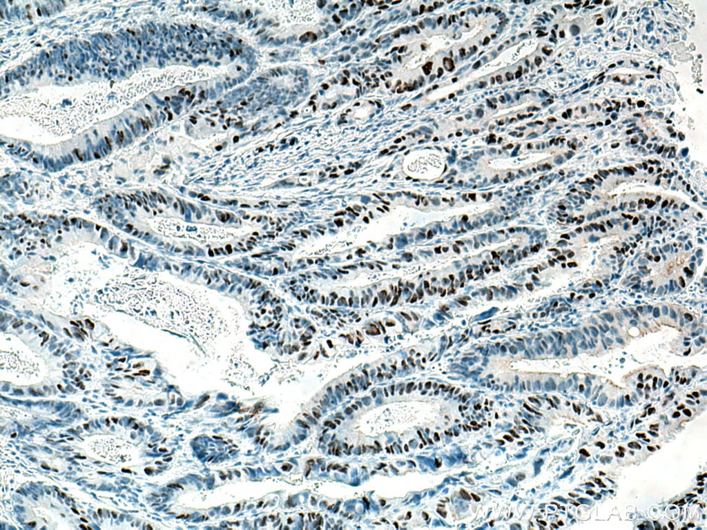
IHC staining of human colon cancer using 60283-2-Ig (same clone as 60283-2-PBS)
Immunohistochemical analysis of paraffin-embedded human colon cancer tissue slide using 60283-2-Ig (P53 antibody) at dilution of 1:3000 (under 10x lens). Heat mediated antigen retrieval with Tris-EDTA buffer (pH 9.0). This data was developed using the same antibody clone with 60283-2-PBS in a different storage buffer formulation.
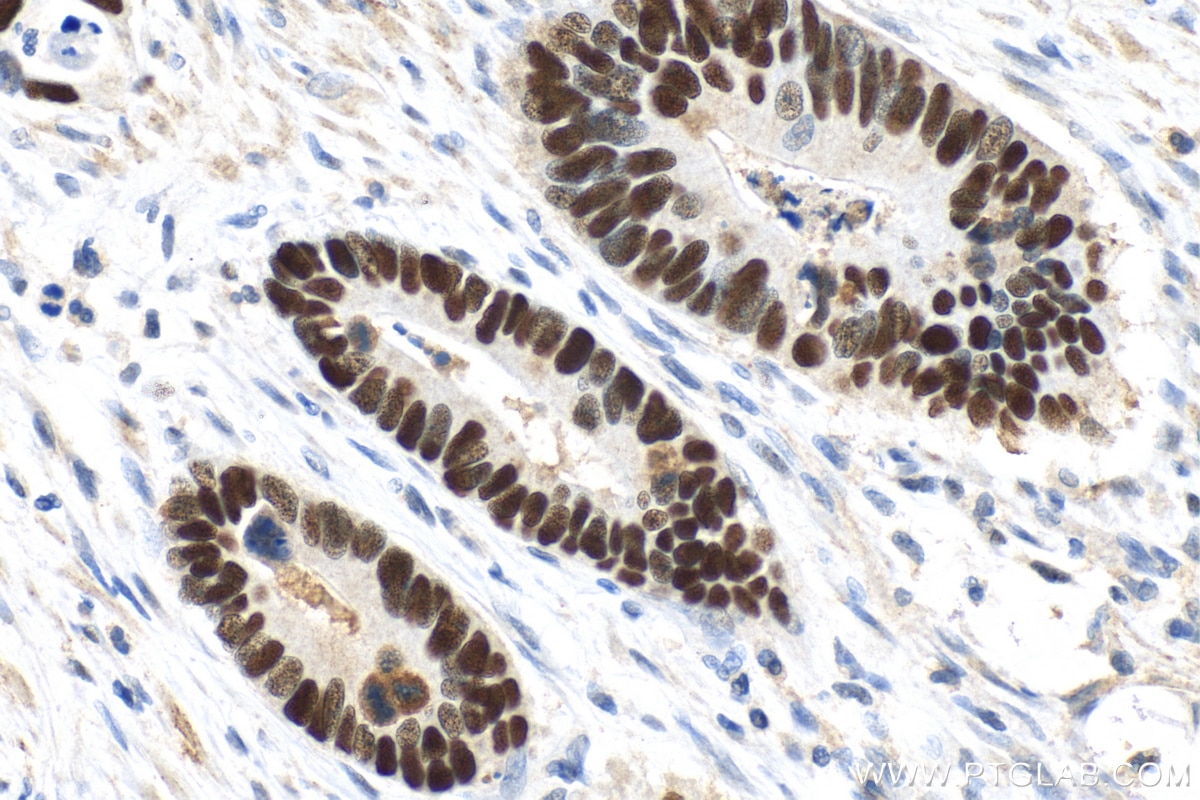
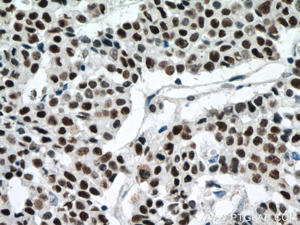
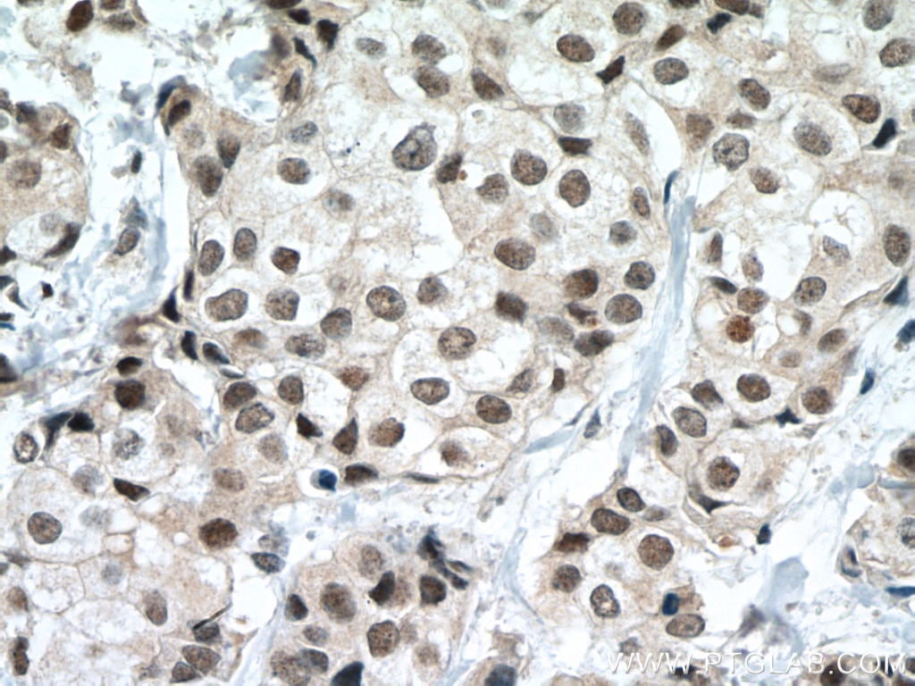
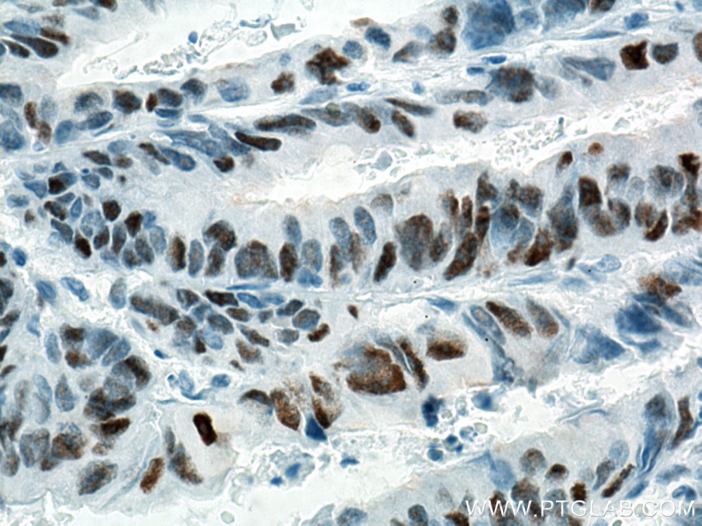
IHC staining of human colon cancer using 60283-2-Ig (same clone as 60283-2-PBS)
Immunohistochemical analysis of paraffin-embedded human colon cancer tissue slide using 60283-2-Ig (P53 antibody) at dilution of 1:3000 (under 40x lens). Heat mediated antigen retrieval with Tris-EDTA buffer (pH 9.0). This data was developed using the same antibody clone with 60283-2-PBS in a different storage buffer formulation.
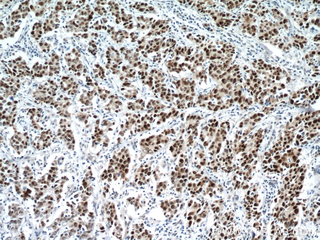
IHC staining of human colon cancer using 60283-2-Ig (same clone as 60283-2-PBS)
Immunohistochemical analysis of paraffin-embedded human colon cancer tissue slide using 60283-2-Ig (P53 antibody) at dilution of 1:400 (under 10x lens. Heat mediated antigen retrieval with Tris-EDTA buffer (pH 9.0). This data was developed using the same antibody clone with 60283-2-PBS in a different storage buffer formulation.
IHC staining of human colon cancer using 60283-2-Ig (same clone as 60283-2-PBS)
Immunohistochemical analysis of paraffin-embedded human colon cancer tissue slide using 60283-2-Ig (P53 antibody) at dilution of 1:400 (under 40x lens. Heat mediated antigen retrieval with Tris-EDTA buffer (pH 9.0). This data was developed using the same antibody clone with 60283-2-PBS in a different storage buffer formulation.
IHC staining of human gliomas using 60283-2-Ig (same clone as 60283-2-PBS)
Immunohistochemical analysis of paraffin-embedded human gliomas tissue slide using 60283-2-Ig (P53 antibody) at dilution of 1:400 (under 10x lens. Heat mediated antigen retrieval with Tris-EDTA buffer (pH 9.0). This data was developed using the same antibody clone with 60283-2-PBS in a different storage buffer formulation.
IHC staining of human gliomas using 60283-2-Ig (same clone as 60283-2-PBS)
Immunohistochemical analysis of paraffin-embedded human gliomas tissue slide using 60283-2-Ig (P53 antibody) at dilution of 1:400 (under 40x lens. Heat mediated antigen retrieval with Tris-EDTA buffer (pH 9.0). This data was developed using the same antibody clone with 60283-2-PBS in a different storage buffer formulation.
IHC staining of human colon cancer using 60283-2-Ig (same clone as 60283-2-PBS)
Immunohistochemical analysis of paraffin-embedded human colon cancer tissue slide using 60283-2-Ig (P53 antibody) at dilution of 1:400 (under 10x lens) Heat mediated antigen retrieved with Sodium Citrate buffer (pH 6.0). This data was developed using the same antibody clone with 60283-2-PBS in a different storage buffer formulation.
IHC staining of human colon cancer using 60283-2-Ig (same clone as 60283-2-PBS)
Immunohistochemical analysis of paraffin-embedded human colon cancer tissue slide using 60283-2-Ig (P53 antibody) at dilution of 1:400 (under 40x lens) Heat mediated antigen retrieved with Sodium Citrate buffer (pH 6.0). This data was developed using the same antibody clone with 60283-2-PBS in a different storage buffer formulation.
IHC staining of human colon cancer using 60283-2-Ig (same clone as 60283-2-PBS)
Immunohistochemical analysis of paraffin-embedded human colon cancer tissue slide using 60283-2-Ig (P53 antibody) at dilution of 1:300 (under 10x lens. Heat mediated antigen retrieval with Tris-EDTA buffer (pH 9.0). This data was developed using the same antibody clone with 60283-2-PBS in a different storage buffer formulation.
IHC staining of human colon cancer using 60283-2-Ig (same clone as 60283-2-PBS)
Immunohistochemical analysis of paraffin-embedded human colon cancer tissue slide using 60283-2-Ig (P53 antibody) at dilution of 1:300 (under 40x lens. Heat mediated antigen retrieval with Tris-EDTA buffer (pH 9.0). This data was developed using the same antibody clone with 60283-2-PBS in a different storage buffer formulation.
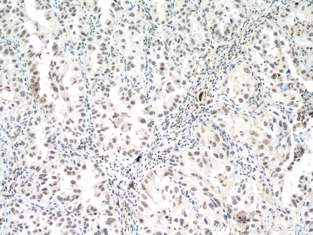
IHC staining of human lung cancer using 60283-2-Ig (same clone as 60283-2-PBS)
Immunohistochemical analysis of paraffin-embedded human lung cancer tissue slide using 60283-2-Ig (P53 antibody) at dilution of 1:300 (under 10x lens. Heat mediated antigen retrieval with Tris-EDTA buffer (pH 9.0). This data was developed using the same antibody clone with 60283-2-PBS in a different storage buffer formulation.
IHC staining of human lung cancer using 60283-2-Ig (same clone as 60283-2-PBS)
Immunohistochemical analysis of paraffin-embedded human lung cancer tissue slide using 60283-2-Ig (P53 antibody) at dilution of 1:300 (under 40x lens. Heat mediated antigen retrieval with Tris-EDTA buffer (pH 9.0). This data was developed using the same antibody clone with 60283-2-PBS in a different storage buffer formulation.
IHC staining of human breast cancer using 60283-2-Ig (same clone as 60283-2-PBS)
Immunohistochemical analysis of paraffin-embedded human breast cancer tissue slide using 60283-2-Ig (P53 antibody) at dilution of 1:300 (under 10x lens. Heat mediated antigen retrieval with Tris-EDTA buffer (pH 9.0). This data was developed using the same antibody clone with 60283-2-PBS in a different storage buffer formulation.
IHC staining of human breast cancer using 60283-2-Ig (same clone as 60283-2-PBS)
Immunohistochemical analysis of paraffin-embedded human breast cancer tissue slide using 60283-2-Ig (P53 antibody) at dilution of 1:300 (under 40x lens. Heat mediated antigen retrieval with Tris-EDTA buffer (pH 9.0). This data was developed using the same antibody clone with 60283-2-PBS in a different storage buffer formulation.
IHC staining of human stomach cancer using 60283-2-Ig (same clone as 60283-2-PBS)
Immunohistochemical analysis of paraffin-embedded human stomach cancer tissue slide using 60283-2-Ig (P53 antibody) at dilution of 1:300 (under 10x lens. Heat mediated antigen retrieval with Tris-EDTA buffer (pH 9.0). This data was developed using the same antibody clone with 60283-2-PBS in a different storage buffer formulation.
IHC staining of human stomach cancer using 60283-2-Ig (same clone as 60283-2-PBS)
Immunohistochemical analysis of paraffin-embedded human stomach cancer tissue slide using 60283-2-Ig (P53 antibody) at dilution of 1:300 (under 40x lens. Heat mediated antigen retrieval with Tris-EDTA buffer (pH 9.0). This data was developed using the same antibody clone with 60283-2-PBS in a different storage buffer formulation.
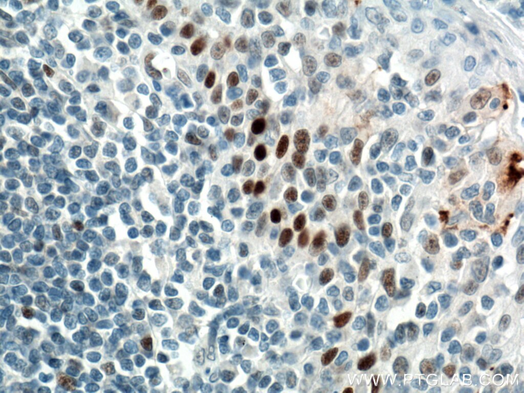
IHC staining of human tonsillitis using 60283-2-Ig (same clone as 60283-2-PBS)
Immunohistochemical analysis of paraffin-embedded human tonsillitis tissue slide using 60283-2-Ig (P53 antibody) at dilution of 1:3200 (under 40x lens. Heat mediated antigen retrieval with Tris-EDTA buffer (pH 9.0). This data was developed using the same antibody clone with 60283-2-PBS in a different storage buffer formulation.
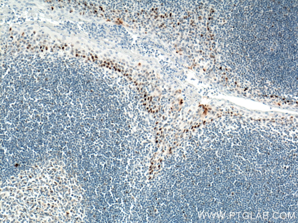
IHC staining of human tonsillitis using 60283-2-Ig (same clone as 60283-2-PBS)
Immunohistochemical analysis of paraffin-embedded human tonsillitis tissue slide using 60283-2-Ig (P53 antibody) at dilution of 1:3200 (under 10x lens. Heat mediated antigen retrieval with Tris-EDTA buffer (pH 9.0). This data was developed using the same antibody clone with 60283-2-PBS in a different storage buffer formulation.
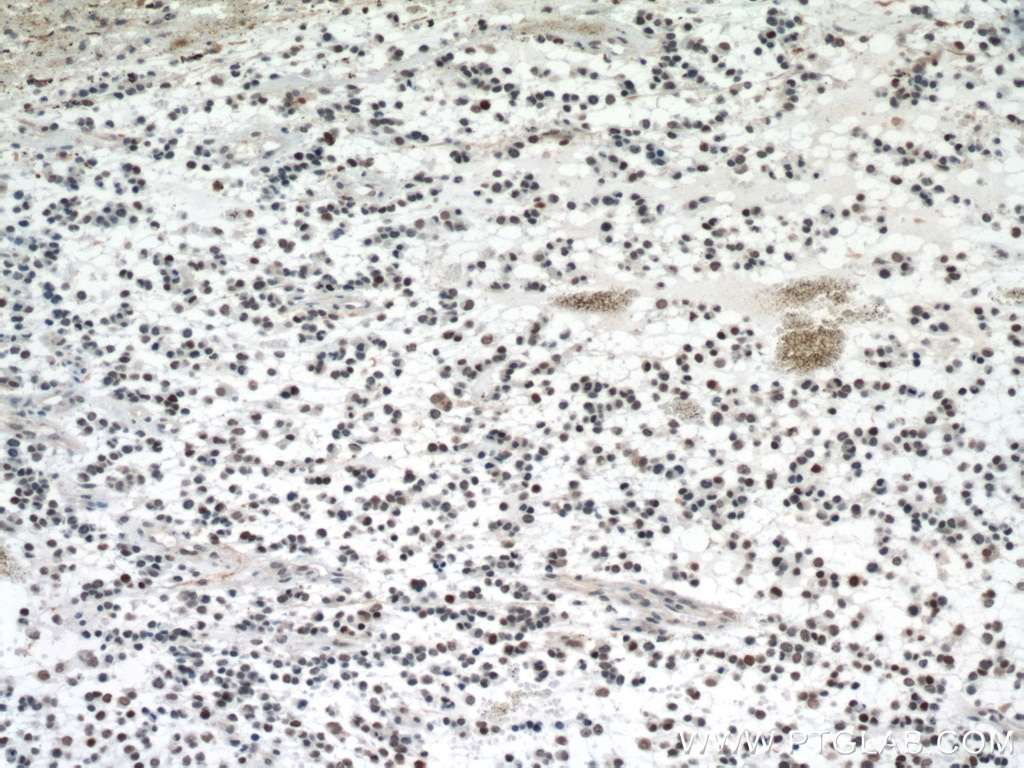
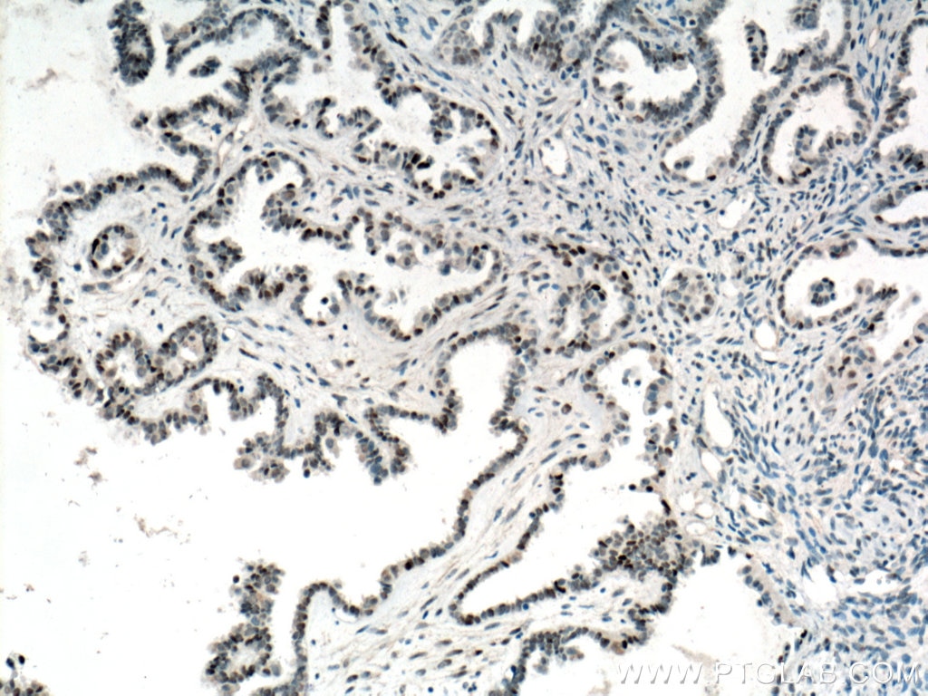
IHC staining of human ovary tumor using 60283-2-Ig (same clone as 60283-2-PBS)
Immunohistochemical analysis of paraffin-embedded human ovary tumor tissue slide using 60283-2-Ig (P53 antibody) at dilution of 1:3200 (under 10x lens. Heat mediated antigen retrieval with Tris-EDTA buffer (pH 9.0). This data was developed using the same antibody clone with 60283-2-PBS in a different storage buffer formulation.
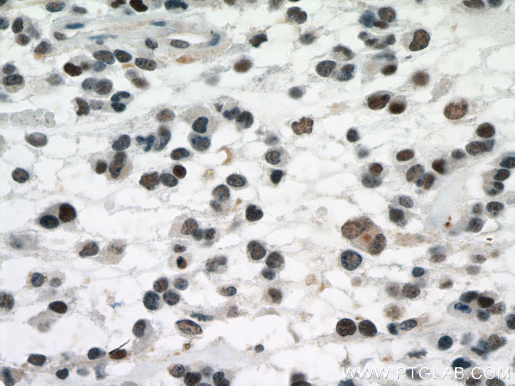
IHC staining of human ovary tumor using 60283-2-Ig (same clone as 60283-2-PBS)
Immunohistochemical analysis of paraffin-embedded human ovary tumor tissue slide using 60283-2-Ig (P53 antibody) at dilution of 1:3200 (under 40x lens. Heat mediated antigen retrieval with Tris-EDTA buffer (pH 9.0). This data was developed using the same antibody clone with 60283-2-PBS in a different storage buffer formulation.
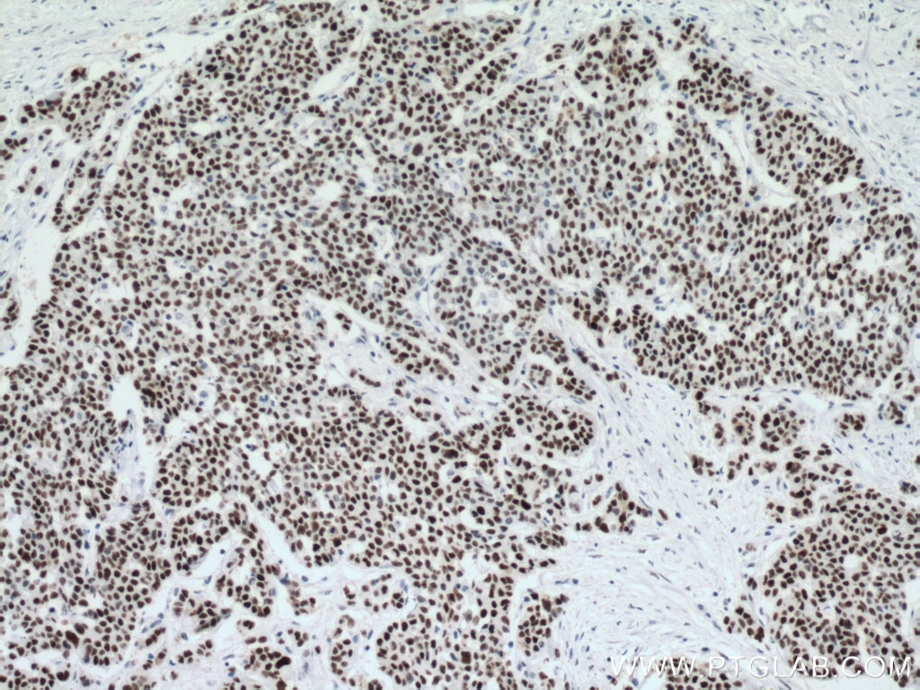
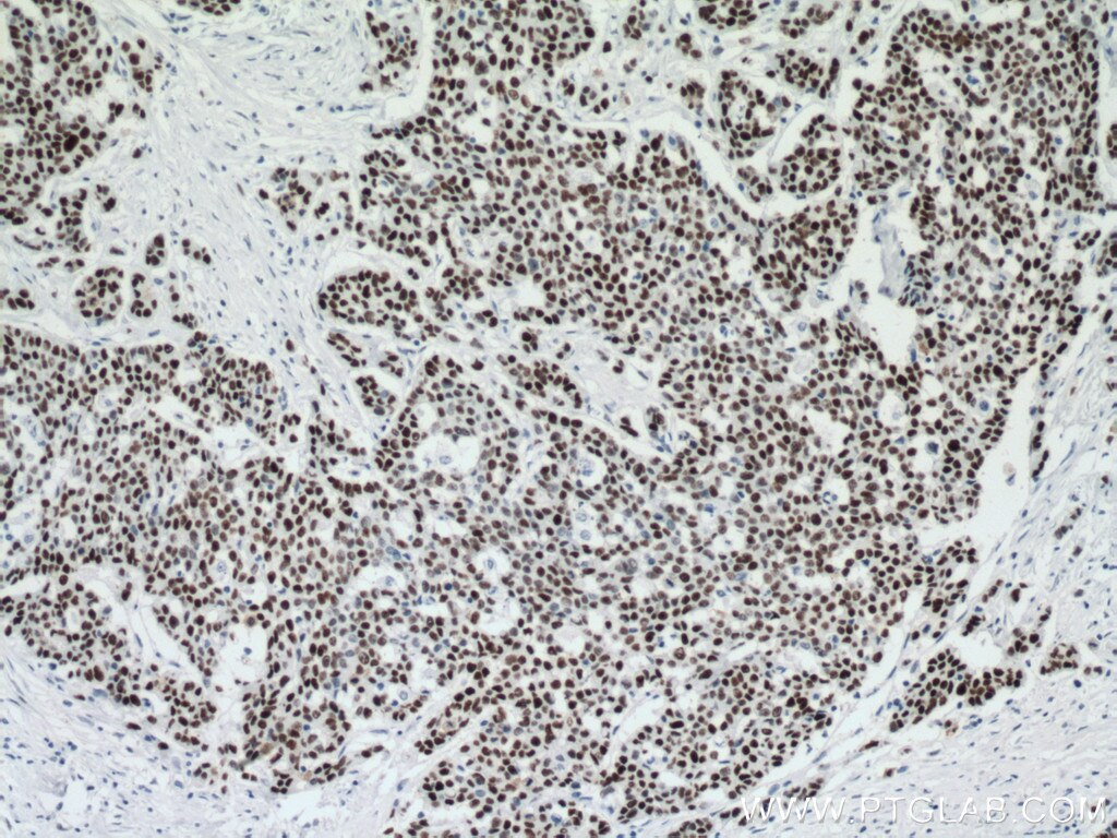
IHC staining of human colon cancer using 60283-2-Ig (same clone as 60283-2-PBS)
Immunohistochemical analysis of paraffin-embedded human colon cancer tissue slide using 60283-2-Ig (P53 antibody) at dilution of 1:1600 (under 10x lens). Heat mediated antigen retrieval with Tris-EDTA buffer (pH 9.0). This data was developed using the same antibody clone with 60283-2-PBS in a different storage buffer formulation.
IHC staining of human colon cancer using 60283-2-Ig (same clone as 60283-2-PBS)
Immunohistochemical analysis of paraffin-embedded human colon cancer tissue slide using 60283-2-Ig (P53 antibody) at dilution of 1:1600 (under 40x lens). Heat mediated antigen retrieval with Tris-EDTA buffer (pH 9.0). This data was developed using the same antibody clone with 60283-2-PBS in a different storage buffer formulation.
CYTOMETRIC BEAD ARRAY Figures
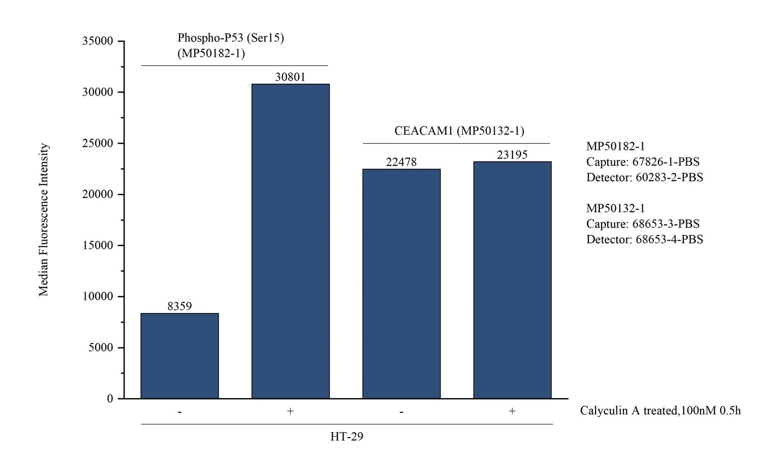
Cytometric bead array in cell lysate using MP50182-1
Cytometric bead array in cell lysate using MP50182-1, Phospho-P53 (Ser15) Monoclonal Matched Antibody Pair, PBS Only. Capture antibody: 67826-1-PBS. Detection antibody: 60283-2-PBS. Cell lysate: Non-treated HT-29 and Calyculin A treated HT-29 (30μg/well). Non-related target CEACAM1 Monoclonal Matched Antibody Pair (MP50132-1) was served as control.
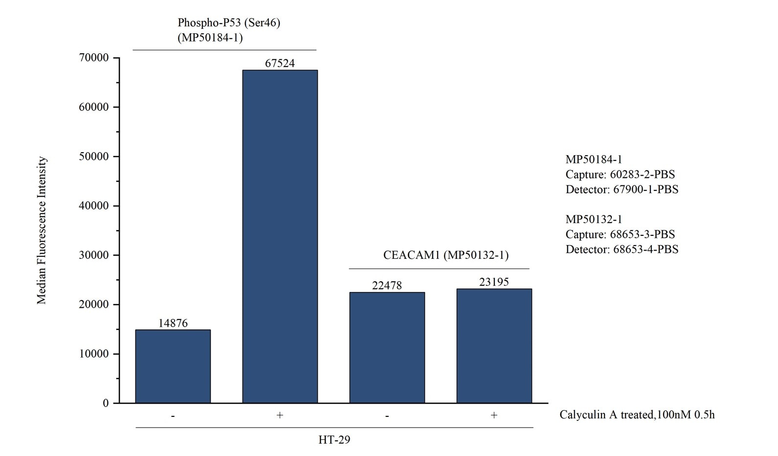
Cytometric bead array in cell lysate using MP50184-1
Cytometric bead array in cell lysate using MP50184-1, Phospho-P53 (Ser46) Monoclonal Matched Antibody Pair, PBS Only. Capture antibody: 60283-2-PBS. Detection antibody: 67900-1-PBS. Cell lysate: Non-treated HT-29 and Calyculin A treated HT-29 (30μg/well). Non-related target CEACAM1 Monoclonal Matched Antibody Pair (MP50132-1) was served as control.

