HeLa cells were subjected to SDS PAGE followed by western blot with 66698-1-Ig (OCIAD1 antibody) at dilution of 1:1000 incubated at room temperature for 1.5 hours.
HeLa cells were subjected to SDS PAGE followed by western blot with 66698-1-Ig (OCIAD1 antibody) at dilution of 1:1000 incubated at room temperature for 1.5 hours.
WB analysis of rat testis using 66698-1-Ig
rat testis tissue were subjected to SDS PAGE followed by western blot with 66698-1-Ig (OCIAD1 antibody) at dilution of 1:10000 incubated at room temperature for 1.5 hours.
rat testis tissue were subjected to SDS PAGE followed by western blot with 66698-1-Ig (OCIAD1 antibody) at dilution of 1:10000 incubated at room temperature for 1.5 hours.
WB analysis of mouse testis using 66698-1-Ig
mouse testis tissue were subjected to SDS PAGE followed by western blot with 66698-1-Ig (OCIAD1 antibody) at dilution of 1:10000 incubated at room temperature for 1.5 hours.
mouse testis tissue were subjected to SDS PAGE followed by western blot with 66698-1-Ig (OCIAD1 antibody) at dilution of 1:10000 incubated at room temperature for 1.5 hours.
IHC staining of human liver cancer using 66698-1-Ig
Immunohistochemical analysis of paraffin-embedded human liver cancer tissue slide using 66698-1-Ig (OCIAD1 antibody) at dilution of 1:200 (under 10x lens). Heat mediated antigen retrieval with Tris-EDTA buffer (pH 9.0).
Immunohistochemical analysis of paraffin-embedded human liver cancer tissue slide using 66698-1-Ig (OCIAD1 antibody) at dilution of 1:200 (under 10x lens). Heat mediated antigen retrieval with Tris-EDTA buffer (pH 9.0).
IHC staining of human liver cancer using 66698-1-Ig
Immunohistochemical analysis of paraffin-embedded human liver cancer tissue slide using 66698-1-Ig (OCIAD1 antibody) at dilution of 1:200 (under 40x lens). Heat mediated antigen retrieval with Tris-EDTA buffer (pH 9.0).
Immunohistochemical analysis of paraffin-embedded human liver cancer tissue slide using 66698-1-Ig (OCIAD1 antibody) at dilution of 1:200 (under 40x lens). Heat mediated antigen retrieval with Tris-EDTA buffer (pH 9.0).
IHC staining of human thyroid cancer using 66698-1-Ig
Immunohistochemical analysis of paraffin-embedded human thyroid cancer tissue slide using 66698-1-Ig (OCIAD1 antibody) at dilution of 1:200 (under 10x lens). Heat mediated antigen retrieval with Tris-EDTA buffer (pH 9.0).
Immunohistochemical analysis of paraffin-embedded human thyroid cancer tissue slide using 66698-1-Ig (OCIAD1 antibody) at dilution of 1:200 (under 10x lens). Heat mediated antigen retrieval with Tris-EDTA buffer (pH 9.0).
IHC staining of human thyroid cancer using 66698-1-Ig
Immunohistochemical analysis of paraffin-embedded human thyroid cancer tissue slide using 66698-1-Ig (OCIAD1 antibody) at dilution of 1:200 (under 40x lens). Heat mediated antigen retrieval with Tris-EDTA buffer (pH 9.0).
Immunohistochemical analysis of paraffin-embedded human thyroid cancer tissue slide using 66698-1-Ig (OCIAD1 antibody) at dilution of 1:200 (under 40x lens). Heat mediated antigen retrieval with Tris-EDTA buffer (pH 9.0).
IHC staining of human kidney using 66698-1-Ig
Immunohistochemical analysis of paraffin-embedded human kidney tissue slide using 66698-1-Ig (OCIAD1 antibody) at dilution of 1:200 (under 10x lens). Heat mediated antigen retrieval with Tris-EDTA buffer (pH 9.0).
Immunohistochemical analysis of paraffin-embedded human kidney tissue slide using 66698-1-Ig (OCIAD1 antibody) at dilution of 1:200 (under 10x lens). Heat mediated antigen retrieval with Tris-EDTA buffer (pH 9.0).
IHC staining of human kidney using 66698-1-Ig
Immunohistochemical analysis of paraffin-embedded human kidney tissue slide using 66698-1-Ig (OCIAD1 antibody) at dilution of 1:200 (under 40x lens). Heat mediated antigen retrieval with Tris-EDTA buffer (pH 9.0).
Immunohistochemical analysis of paraffin-embedded human kidney tissue slide using 66698-1-Ig (OCIAD1 antibody) at dilution of 1:200 (under 40x lens). Heat mediated antigen retrieval with Tris-EDTA buffer (pH 9.0).
IHC staining of human pancreas cancer using 66698-1-Ig
Immunohistochemical analysis of paraffin-embedded human pancreas cancer tissue slide using 66698-1-Ig (OCIAD1 antibody) at dilution of 1:200 (under 10x lens). Heat mediated antigen retrieval with Tris-EDTA buffer (pH 9.0).
Immunohistochemical analysis of paraffin-embedded human pancreas cancer tissue slide using 66698-1-Ig (OCIAD1 antibody) at dilution of 1:200 (under 10x lens). Heat mediated antigen retrieval with Tris-EDTA buffer (pH 9.0).
IHC staining of human pancreas cancer using 66698-1-Ig
Immunohistochemical analysis of paraffin-embedded human pancreas cancer tissue slide using 66698-1-Ig (OCIAD1 antibody) at dilution of 1:200 (under 40x lens). Heat mediated antigen retrieval with Tris-EDTA buffer (pH 9.0).
Immunohistochemical analysis of paraffin-embedded human pancreas cancer tissue slide using 66698-1-Ig (OCIAD1 antibody) at dilution of 1:200 (under 40x lens). Heat mediated antigen retrieval with Tris-EDTA buffer (pH 9.0).
IHC staining of mouse testis using 66698-1-Ig
Immunohistochemical analysis of paraffin-embedded mouse testis tissue slide using 66698-1-Ig (OCIAD1 antibody) at dilution of 1:200 (under 10x lens). Heat mediated antigen retrieval with Tris-EDTA buffer (pH 9.0).
Immunohistochemical analysis of paraffin-embedded mouse testis tissue slide using 66698-1-Ig (OCIAD1 antibody) at dilution of 1:200 (under 40x lens). Heat mediated antigen retrieval with Tris-EDTA buffer (pH 9.0).
IF Staining of human liver cancer using 66698-1-Ig
Immunofluorescent analysis of (4% PFA) fixed human liver cancer tissue using OCIAD1 antibody (66698-1-Ig, Clone: 1C10C3 ) at dilution of 1:400 and CoraLite®488-Conjugated Goat Anti-Mouse IgG(H+L).
Immunofluorescent analysis of (4% PFA) fixed human liver cancer tissue using OCIAD1 antibody (66698-1-Ig, Clone: 1C10C3 ) at dilution of 1:400 and CoraLite®488-Conjugated Goat Anti-Mouse IgG(H+L).
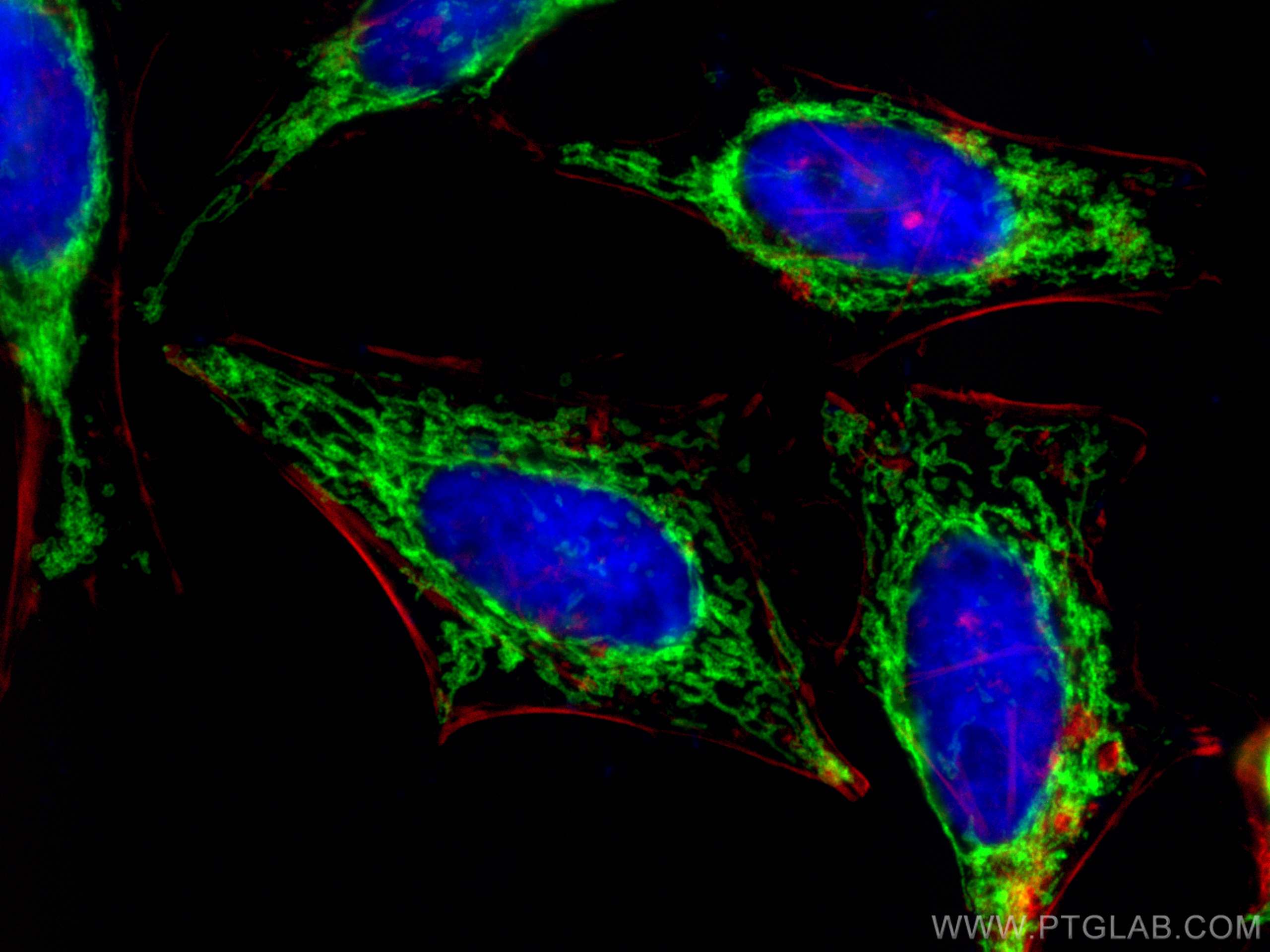
IF Staining of HeLa using 66698-1-Ig
Immunofluorescent analysis of (4% PFA) fixed HeLa cells using OCIAD1 antibody (66698-1-Ig, Clone: 1C10C3 ) at dilution of 1:400 and CoraLite®488-Conjugated Goat Anti-Mouse IgG(H+L), CL594-phalloidin (red).
Immunofluorescent analysis of (4% PFA) fixed HeLa cells using OCIAD1 antibody (66698-1-Ig, Clone: 1C10C3 ) at dilution of 1:400 and CoraLite®488-Conjugated Goat Anti-Mouse IgG(H+L), CL594-phalloidin (red).
IF Staining of HeLa using 66698-1-Ig
Immunofluorescent analysis of (-20°C Methanol) fixed HeLa cells using OCIAD1 antibody (66698-1-Ig, Clone: 1C10C3 ) at dilution of 1:800 and CoraLite®488-Conjugated Goat Anti-Mouse IgG(H+L).
Immunofluorescent analysis of (-20°C Methanol) fixed HeLa cells using OCIAD1 antibody (66698-1-Ig, Clone: 1C10C3 ) at dilution of 1:800 and CoraLite®488-Conjugated Goat Anti-Mouse IgG(H+L).
The Proteintech guarantee covers Proteintech antibodies in any species and any application, including those not listed on the datasheet. If the antibody doesn’t perform, you can receive a hassle-free refund or credit note.
HeLa cells, rat testis tissue, mouse testis tissue
Positive IHC detected in
human liver cancer tissue, human kidney tissue, human pancreas cancer tissue, human thyroid cancer tissue, mouse testis tissue Note: suggested antigen retrieval with TE buffer pH 9.0; (*) Alternatively, antigen retrieval may be performed with citrate buffer pH 6.0
Positive IF-P detected in
human liver cancer tissue, HeLa cells
Positive IF/ICC detected in
HeLa cells
Recommended dilution
Application
Dilution
Western Blot (WB)
WB : 1:1000-1:5000
Immunohistochemistry (IHC)
IHC : 1:50-1:500
Immunofluorescence (IF)-P
IF-P : 1:200-1:800
Immunofluorescence (IF)/ICC
IF/ICC : 1:200-1:800
It is recommended that this reagent should be titrated in each testing system to obtain optimal results.
Sample-dependent, Check data in validation data gallery.
PBS with 0.02% sodium azide and 50% glycerol , pH 7.3
Storage Conditions
Store at -20°C. Stable for one year after shipment. Aliquoting is unnecessary for -20oC storage. 20ul sizes contain 0.1% BSA.
Background Information
OCIAD1 was first identified by immunoscreening of an ovarian carcinoma cDNA expression library with ascites fluid from ovarian cancer patients (PMID: 11162530). OCIAD1 has been reported as a key player in ovarian cancer cell adhesion, as well as a key player in generating ovarian cancer recurrence (PMID: 18328549; 20515946). In addition to its roles in cancer, OCIAD1 participates in maintaining stem cell potency by regulating the Jak/STAT pathway (PMID: 23972987). Several alternatively spliced forms of OCIAD1 gene have been identified. The longest form (1.4 kb) is predicted to encode for a 27.6 kDa protein of 245 amino acids. This antibody detects OCIAD1 with an apparent molecular weight of ~35 kDa as has been demonstrated by several researches (PMID: 27345969; 27345976).
Genome-wide CRISPRi screening identifies OCIAD1 as a prohibitin client and regulatory determinant of mitochondrial Complex III assembly in human cells.
HeLa cells were subjected to SDS PAGE followed by western blot with 66698-1-Ig (OCIAD1 antibody) at dilution of 1:1000 incubated at room temperature for 1.5 hours.
WB analysis of rat testis using 66698-1-Ig
rat testis tissue were subjected to SDS PAGE followed by western blot with 66698-1-Ig (OCIAD1 antibody) at dilution of 1:10000 incubated at room temperature for 1.5 hours.
WB analysis of mouse testis using 66698-1-Ig
mouse testis tissue were subjected to SDS PAGE followed by western blot with 66698-1-Ig (OCIAD1 antibody) at dilution of 1:10000 incubated at room temperature for 1.5 hours.
IHC Figures
IHC staining of human liver cancer using 66698-1-Ig
Immunohistochemical analysis of paraffin-embedded human liver cancer tissue slide using 66698-1-Ig (OCIAD1 antibody) at dilution of 1:200 (under 10x lens). Heat mediated antigen retrieval with Tris-EDTA buffer (pH 9.0).
IHC staining of human liver cancer using 66698-1-Ig
Immunohistochemical analysis of paraffin-embedded human liver cancer tissue slide using 66698-1-Ig (OCIAD1 antibody) at dilution of 1:200 (under 40x lens). Heat mediated antigen retrieval with Tris-EDTA buffer (pH 9.0).
IHC staining of human thyroid cancer using 66698-1-Ig
Immunohistochemical analysis of paraffin-embedded human thyroid cancer tissue slide using 66698-1-Ig (OCIAD1 antibody) at dilution of 1:200 (under 10x lens). Heat mediated antigen retrieval with Tris-EDTA buffer (pH 9.0).
IHC staining of human thyroid cancer using 66698-1-Ig
Immunohistochemical analysis of paraffin-embedded human thyroid cancer tissue slide using 66698-1-Ig (OCIAD1 antibody) at dilution of 1:200 (under 40x lens). Heat mediated antigen retrieval with Tris-EDTA buffer (pH 9.0).
IHC staining of human kidney using 66698-1-Ig
Immunohistochemical analysis of paraffin-embedded human kidney tissue slide using 66698-1-Ig (OCIAD1 antibody) at dilution of 1:200 (under 10x lens). Heat mediated antigen retrieval with Tris-EDTA buffer (pH 9.0).
IHC staining of human kidney using 66698-1-Ig
Immunohistochemical analysis of paraffin-embedded human kidney tissue slide using 66698-1-Ig (OCIAD1 antibody) at dilution of 1:200 (under 40x lens). Heat mediated antigen retrieval with Tris-EDTA buffer (pH 9.0).
IHC staining of human pancreas cancer using 66698-1-Ig
Immunohistochemical analysis of paraffin-embedded human pancreas cancer tissue slide using 66698-1-Ig (OCIAD1 antibody) at dilution of 1:200 (under 10x lens). Heat mediated antigen retrieval with Tris-EDTA buffer (pH 9.0).
IHC staining of human pancreas cancer using 66698-1-Ig
Immunohistochemical analysis of paraffin-embedded human pancreas cancer tissue slide using 66698-1-Ig (OCIAD1 antibody) at dilution of 1:200 (under 40x lens). Heat mediated antigen retrieval with Tris-EDTA buffer (pH 9.0).
IHC staining of mouse testis using 66698-1-Ig
Immunohistochemical analysis of paraffin-embedded mouse testis tissue slide using 66698-1-Ig (OCIAD1 antibody) at dilution of 1:200 (under 10x lens). Heat mediated antigen retrieval with Tris-EDTA buffer (pH 9.0).
IHC staining of mouse testis using 66698-1-Ig
Immunohistochemical analysis of paraffin-embedded mouse testis tissue slide using 66698-1-Ig (OCIAD1 antibody) at dilution of 1:200 (under 40x lens). Heat mediated antigen retrieval with Tris-EDTA buffer (pH 9.0).
IF-P Figures
IF Staining of human liver cancer using 66698-1-Ig
Immunofluorescent analysis of (4% PFA) fixed human liver cancer tissue using OCIAD1 antibody (66698-1-Ig, Clone: 1C10C3 ) at dilution of 1:400 and CoraLite®488-Conjugated Goat Anti-Mouse IgG(H+L).
IF Staining of human liver cancer using 66698-1-Ig
Immunofluorescent analysis of (4% PFA) fixed human liver cancer tissue using OCIAD1 antibody (66698-1-Ig, Clone: 1C10C3 ) at dilution of 1:400 and CoraLite®488-Conjugated Goat Anti-Mouse IgG(H+L).
IF/ICC Figures
IF Staining of HeLa using 66698-1-Ig
Immunofluorescent analysis of (4% PFA) fixed HeLa cells using OCIAD1 antibody (66698-1-Ig, Clone: 1C10C3 ) at dilution of 1:400 and CoraLite®488-Conjugated Goat Anti-Mouse IgG(H+L), CL594-phalloidin (red).
IF Staining of HeLa using 66698-1-Ig
Immunofluorescent analysis of (-20°C Methanol) fixed HeLa cells using OCIAD1 antibody (66698-1-Ig, Clone: 1C10C3 ) at dilution of 1:800 and CoraLite®488-Conjugated Goat Anti-Mouse IgG(H+L).
The species listed in Tested Reactivity are in-house verified and applicable species. For unlisted species, please refer to the homology analysis of the immunogen sequence and related species. For rabbit polyclonal antibodies, homology >70% is recommended. For mouse monoclonal antibodies and rabbit recombinant antibodies, homology >90% is recommended. Generally, the higher the homology, the greater the applicability. However, there will be certain differences in protein expression in different species, tissues or cells. Therefore, the homology analysis results are for reference only and do not serve as a guarantee.
At Proteintech, we pride ourselves on our antibody quality, customer service and transparency. As such, we are comparing our antibodies with other vendors, enabling easy identification and comparisons of key data to help you choose the suitable antibody for your needs.
We have selected the top cited antibodies from these vendors for you to compare.
 at dilution of 1:1000 incubated at room temperature for 1.5 hours.")
 at dilution of 1:10000 incubated at room temperature for 1.5 hours.")
 at dilution of 1:10000 incubated at room temperature for 1.5 hours.")
 at dilution of 1:200 (under 10x lens). Heat mediated antigen retrieval with Tris-EDTA buffer (pH 9.0).")
 at dilution of 1:200 (under 40x lens). Heat mediated antigen retrieval with Tris-EDTA buffer (pH 9.0).")
 at dilution of 1:200 (under 10x lens). Heat mediated antigen retrieval with Tris-EDTA buffer (pH 9.0).")
 at dilution of 1:200 (under 40x lens). Heat mediated antigen retrieval with Tris-EDTA buffer (pH 9.0).")
 at dilution of 1:200 (under 10x lens). Heat mediated antigen retrieval with Tris-EDTA buffer (pH 9.0).")
 at dilution of 1:200 (under 40x lens). Heat mediated antigen retrieval with Tris-EDTA buffer (pH 9.0).")
 at dilution of 1:200 (under 10x lens). Heat mediated antigen retrieval with Tris-EDTA buffer (pH 9.0).")
 at dilution of 1:200 (under 40x lens). Heat mediated antigen retrieval with Tris-EDTA buffer (pH 9.0).")
 at dilution of 1:200 (under 10x lens). Heat mediated antigen retrieval with Tris-EDTA buffer (pH 9.0).")
 at dilution of 1:200 (under 40x lens). Heat mediated antigen retrieval with Tris-EDTA buffer (pH 9.0).")
 fixed human liver cancer tissue using OCIAD1 antibody (66698-1-Ig, Clone: 1C10C3 ) at dilution of 1:400 and CoraLite®488-Conjugated Goat Anti-Mouse IgG(H+L).")
 fixed human liver cancer tissue using OCIAD1 antibody (66698-1-Ig, Clone: 1C10C3 ) at dilution of 1:400 and CoraLite®488-Conjugated Goat Anti-Mouse IgG(H+L).")
 fixed HeLa cells using OCIAD1 antibody (66698-1-Ig, Clone: 1C10C3 ) at dilution of 1:400 and CoraLite®488-Conjugated Goat Anti-Mouse IgG(H+L), CL594-phalloidin (red).")
 fixed HeLa cells using OCIAD1 antibody (66698-1-Ig, Clone: 1C10C3 ) at dilution of 1:800 and CoraLite®488-Conjugated Goat Anti-Mouse IgG(H+L).")