at dilution of 1:4000 incubated at room temperature for 1.5 hours.")
 at dilution of 1:1000 incubated at room temperature for 1.5 hours.")
 at dilution of 1:1000 incubated at room temperature for 1.5 hours.")
 at dilution of 1:1000 incubated at room temperature for 1.5 hours.")
 at dilution of 1:1000 incubated at room temperature for 1.5 hours.")
 with mouse heart tissue lysate 3520ug.")
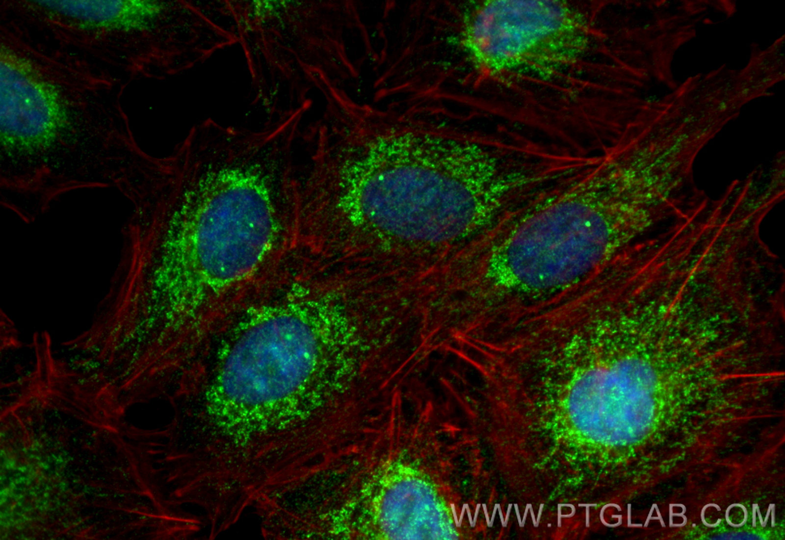
 fixed A549 cells using NLRX1 antibody (17215-1-AP) at dilution of 1:200 and CoraLite®488-Conjugated Goat Anti-Rabbit IgG(H+L) (SA00013-2), CL594-Phalloidin (red).")
 and CoraLite®488-Conjugated AffiniPure Goat Anti-Rabbit IgG(H+L) at dilution 1:1000 (red), or 0.4 ug Isotype Control. Cells were fixed with 4% PFA and permeabilized with Flow Cytometry Perm Buffer.")
Tested Applications
| Positive WB detected in | HEK-293 cells, HepG2 cells, mouse skeletal muscle tissue, MCF-7 cells, HeLa cells, mouse colon tissue, mouse heart tissue, THP-1 cells, rat colon tissue, rat heart tissue |
| Positive IP detected in | mouse heart tissue |
| Positive IF/ICC detected in | A549 cells |
| Positive FC (Intra) detected in | HepG2 cells |
Recommended dilution
| Application | Dilution |
|---|---|
| Western Blot (WB) | WB : 1:1000-1:8000 |
| Immunoprecipitation (IP) | IP : 0.5-4.0 ug for 1.0-3.0 mg of total protein lysate |
| Immunofluorescence (IF)/ICC | IF/ICC : 1:50-1:500 |
| Flow Cytometry (FC) (INTRA) | FC (INTRA) : 0.40 ug per 10^6 cells in a 100 µl suspension |
| It is recommended that this reagent should be titrated in each testing system to obtain optimal results. | |
| Sample-dependent, Check data in validation data gallery. | |
Published Applications
| KD/KO | See 8 publications below |
| WB | See 23 publications below |
| IHC | See 3 publications below |
| IF | See 8 publications below |
| IP | See 1 publications below |
| CoIP | See 2 publications below |
Product Information
17215-1-AP targets NLRX1 in WB, IHC, IF/ICC, FC (Intra), IP, CoIP, ELISA applications and shows reactivity with human, mouse, rat samples.
| Tested Reactivity | human, mouse, rat |
| Cited Reactivity | human, mouse, rat |
| Host / Isotype | Rabbit / IgG |
| Class | Polyclonal |
| Type | Antibody |
| Immunogen |
CatNo: Ag11000 Product name: Recombinant human NLRX1 protein Source: e coli.-derived, PGEX-4T Tag: GST Domain: 359-708 aa of BC013199 Sequence: EAVAQAMVLEMFREEDYYNDDVLDQMGASILGVEGPRRHPDEPPEDEVFELFPMFMGGLLSAHNRAVLAQLGCPIKNLDALENAQAIKKKLGKLGRQVLPPSELLDHLFFHYEFQNQRFSAEVLSSLRQLNLAGVRMTPVKCTVVAAVLGSGRHALDEVNLASCQLDPAGLRTLLPVFLRARKLGLQLNSLGPEACKDLRDLLLHDQCQITTLRLSNNPLTEAGVAVLMEGLAGNTSVTHLSLLHTGLGDEGLELLAAQLDRNRQLQELNVAYNGAGDTAALALARAAREHPSLELLQGVAIQMCWKLPLLPYAHLWTPRMPSHWCFLLILMPPLPQWYDGLVAPRGRCT Predict reactive species |
| Full Name | NLR family member X1 |
| Calculated Molecular Weight | 975 aa, 108 kDa |
| Observed Molecular Weight | 100-110 kDa |
| GenBank Accession Number | BC013199 |
| Gene Symbol | NLRX1 |
| Gene ID (NCBI) | 79671 |
| RRID | AB_2236031 |
| Conjugate | Unconjugated |
| Form | Liquid |
| Purification Method | Antigen affinity purification |
| UNIPROT ID | Q86UT6 |
| Storage Buffer | PBS with 0.02% sodium azide and 50% glycerol, pH 7.3. |
| Storage Conditions | Store at -20°C. Stable for one year after shipment. Aliquoting is unnecessary for -20oC storage. 20ul sizes contain 0.1% BSA. |
Background Information
NLRX1 (Nucleotide-binding oligomerization domain, leucine-rich repeat containing X1) is a member of the NOD-like receptor (NLR) family and is unique among NLRs due to its localization to the mitochondrial matrix. NLRX1 is a negative regulator of innate immunity, particularly in viral infections. It interacts with the mitochondrial antiviral signaling protein (MAVS) to attenuate antiviral responses. NLRX1 has been implicated in various diseases, including multiple sclerosis, colorectal cancer, and ischemia-reperfusion injury. Its role in controlling inflammation and mitochondrial function makes it a potential therapeutic target.
Protocols
| Product Specific Protocols | |
|---|---|
| FC protocol for NLRX1 antibody 17215-1-AP | Download protocol |
| IF protocol for NLRX1 antibody 17215-1-AP | Download protocol |
| IP protocol for NLRX1 antibody 17215-1-AP | Download protocol |
| WB protocol for NLRX1 antibody 17215-1-AP | Download protocol |
| Standard Protocols | |
|---|---|
| Click here to view our Standard Protocols |
Publications
| Species | Application | Title |
|---|---|---|
Mol Cell Mitochondrial protein import stress regulates the LC3 lipidation step of mitophagy through NLRX1 and RRBP1.
| ||
Cell Death Differ NLRX1 mediated impaired microglial phagocytosis of NETs in cerebral ischemia and reperfusion injury | ||
J Exp Med NLRX1 dampens oxidative stress and apoptosis in tissue injury via control of mitochondrial activity.
| ||