Various lysates were subjected to SDS PAGE followed by western blot with 68321-1-Ig (MTHFD1L antibody) at dilution of 1:10000 incubated at room temperature for 1.5 hours. The membrane was stripped and reblotted with HRP-conjugated Lamin B1 Monoclonal antibody (HRP-66095) as loading control.
Various lysates were subjected to SDS PAGE followed by western blot with 68321-1-Ig (MTHFD1L antibody) at dilution of 1:10000 incubated at room temperature for 1.5 hours. The membrane was stripped and reblotted with HRP-conjugated Lamin B1 Monoclonal antibody (HRP-66095) as loading control.
WB analysis using 68321-1-Ig
Various lysates were subjected to SDS PAGE followed by western blot with 68321-1-Ig (MTHFD1L antibody) at dilution of 1:10000 incubated at room temperature for 1.5 hours. The membrane was stripped and reblotted with HRP-conjugated Lamin B1 Monoclonal antibody (HRP-66095) as loading control.
Various lysates were subjected to SDS PAGE followed by western blot with 68321-1-Ig (MTHFD1L antibody) at dilution of 1:10000 incubated at room temperature for 1.5 hours. The membrane was stripped and reblotted with HRP-conjugated Lamin B1 Monoclonal antibody (HRP-66095) as loading control.
IHC staining of mouse liver using 68321-1-Ig
Immunohistochemical analysis of paraffin-embedded mouse liver tissue slide using 68321-1-Ig (MTHFD1L antibody) at dilution of 1:500 (under 10x lens). Heat mediated antigen retrieval with Tris-EDTA buffer (pH 9.0).
Immunohistochemical analysis of paraffin-embedded mouse liver tissue slide using 68321-1-Ig (MTHFD1L antibody) at dilution of 1:500 (under 40x lens). Heat mediated antigen retrieval with Tris-EDTA buffer (pH 9.0).
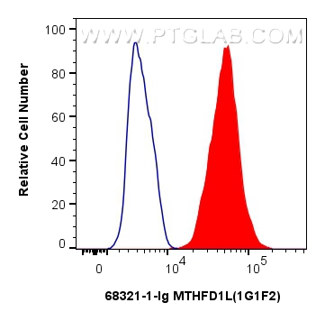
FC experiment of HepG2 using 68321-1-Ig
1x10^6 HepG2 cells were intracellularly stained with 0.4 ug MTHFD1L Monoclonal antibody (68321-1-Ig, Clone:1G1F2) and CoraLite®488-Conjugated Goat Anti-Mouse IgG(H+L) (SA00013-1)(red), or 0.4 ug Mouse IgG2b isotype control Mouse McAb (66360-3-Ig, Clone: 11B8C4) (blue). Cells were fixed with 4% PFA and permeabilized with Flow Cytometry Perm Buffer (PF00011-C).
1x10^6 HepG2 cells were intracellularly stained with 0.4 ug MTHFD1L Monoclonal antibody (68321-1-Ig, Clone:1G1F2) and CoraLite®488-Conjugated Goat Anti-Mouse IgG(H+L) (SA00013-1)(red), or 0.4 ug Mouse IgG2b isotype control Mouse McAb (66360-3-Ig, Clone: 11B8C4) (blue). Cells were fixed with 4% PFA and permeabilized with Flow Cytometry Perm Buffer (PF00011-C).
The Proteintech guarantee covers Proteintech antibodies in any species and any application, including those not listed on the datasheet. If the antibody doesn’t perform, you can receive a hassle-free refund or credit note.
U2OS cells, rabbit testis tissue, LNCaP cells, HeLa cells, HEK-293 cells, HepG2 cells, rat testis tissue
Positive IHC detected in
mouse liver tissue Note: suggested antigen retrieval with TE buffer pH 9.0; (*) Alternatively, antigen retrieval may be performed with citrate buffer pH 6.0
Positive FC (Intra) detected in
HepG2 cells
Recommended dilution
Application
Dilution
Western Blot (WB)
WB : 1:5000-1:50000
Immunohistochemistry (IHC)
IHC : 1:250-1:1000
Flow Cytometry (FC) (INTRA)
FC (INTRA) : 0.40 ug per 10^6 cells in a 100 µl suspension
It is recommended that this reagent should be titrated in each testing system to obtain optimal results.
Sample-dependent, Check data in validation data gallery.
Product Information
68321-1-Ig targets MTHFD1L in WB, IHC, FC (Intra), ELISA applications and shows reactivity with human, mouse, rat, rabbit samples.
PBS with 0.02% sodium azide and 50% glycerol, pH 7.3.
Storage Conditions
Store at -20°C. Stable for one year after shipment. Aliquoting is unnecessary for -20oC storage. 20ul sizes contain 0.1% BSA.
Background Information
MTHFD1L(Monofunctional C1-tetrahydrofolate synthase, mitochondrial) is also named as FTHFSDC1(Formyltetrahydrofolate synthetase). MTHFD1L enzyme is present in mitochondria from normal embryonic tissues and embryonic fibroblast cell lines, and embryonic mitochondria possess the ability to synthesize formate from glycine. It catalyzes the final step in the mitochondrial conversion of 1-C units to formate in embryos. Moreover, MTHFD1L levels were substantially higher in embryonic mitochondria than in adult liver mitochondria and embryonic mitochondria exhibited greater formate production(PMID:19948730). It has 2 isoforms produced by alternative splicing.
Various lysates were subjected to SDS PAGE followed by western blot with 68321-1-Ig (MTHFD1L antibody) at dilution of 1:10000 incubated at room temperature for 1.5 hours. The membrane was stripped and reblotted with HRP-conjugated Lamin B1 Monoclonal antibody (HRP-66095) as loading control.
WB analysis using 68321-1-Ig
Various lysates were subjected to SDS PAGE followed by western blot with 68321-1-Ig (MTHFD1L antibody) at dilution of 1:10000 incubated at room temperature for 1.5 hours. The membrane was stripped and reblotted with HRP-conjugated Lamin B1 Monoclonal antibody (HRP-66095) as loading control.
IHC Figures
IHC staining of mouse liver using 68321-1-Ig
Immunohistochemical analysis of paraffin-embedded mouse liver tissue slide using 68321-1-Ig (MTHFD1L antibody) at dilution of 1:500 (under 10x lens). Heat mediated antigen retrieval with Tris-EDTA buffer (pH 9.0).
IHC staining of mouse liver using 68321-1-Ig
Immunohistochemical analysis of paraffin-embedded mouse liver tissue slide using 68321-1-Ig (MTHFD1L antibody) at dilution of 1:500 (under 40x lens). Heat mediated antigen retrieval with Tris-EDTA buffer (pH 9.0).
FC (INTRA) Figures
FC experiment of HepG2 using 68321-1-Ig
1x10^6 HepG2 cells were intracellularly stained with 0.4 ug MTHFD1L Monoclonal antibody (68321-1-Ig, Clone:1G1F2) and CoraLite®488-Conjugated Goat Anti-Mouse IgG(H+L) (SA00013-1)(red), or 0.4 ug Mouse IgG2b isotype control Mouse McAb (66360-3-Ig, Clone: 11B8C4) (blue). Cells were fixed with 4% PFA and permeabilized with Flow Cytometry Perm Buffer (PF00011-C).
The species listed in Tested Reactivity are in-house verified and applicable species. For unlisted species, please refer to the homology analysis of the immunogen sequence and related species. For rabbit polyclonal antibodies, homology >70% is recommended. For mouse monoclonal antibodies and rabbit recombinant antibodies, homology >90% is recommended. Generally, the higher the homology, the greater the applicability. However, there will be certain differences in protein expression in different species, tissues or cells. Therefore, the homology analysis results are for reference only and do not serve as a guarantee.
At Proteintech, we pride ourselves on our antibody quality, customer service and transparency. As such, we are comparing our antibodies with other vendors, enabling easy identification and comparisons of key data to help you choose the suitable antibody for your needs.
We have selected the top cited antibodies from these vendors for you to compare.
Proteintech
MTHFD1L Monoclonal antibody
Catalog Number
68321-1-Ig
Citations
-
Dilutions
WB : 1:5000-1:50000 IHC : 1:250-1:1000 FC (INTRA) : 0.40 ug per 10^6 cells in a 100 µl suspension
Applications
WB, IHC, FC (Intra), ELISA
Reactivity
human, mouse, rat, rabbit
Product Guarantee
Covers any species including not listed on datasheet
Covers any applications including not listed on datasheet
 at dilution of 1:10000 incubated at room temperature for 1.5 hours. The membrane was stripped and reblotted with HRP-conjugated Lamin B1 Monoclonal antibody (HRP-66095) as loading control.")
 at dilution of 1:10000 incubated at room temperature for 1.5 hours. The membrane was stripped and reblotted with HRP-conjugated Lamin B1 Monoclonal antibody (HRP-66095) as loading control.")
 at dilution of 1:500 (under 10x lens). Heat mediated antigen retrieval with Tris-EDTA buffer (pH 9.0).")
 at dilution of 1:500 (under 40x lens). Heat mediated antigen retrieval with Tris-EDTA buffer (pH 9.0).")
 and CoraLite®488-Conjugated Goat Anti-Mouse IgG(H+L) (SA00013-1)(red), or 0.4 ug Mouse IgG2b isotype control Mouse McAb (66360-3-Ig, Clone: 11B8C4) (blue). Cells were fixed with 4% PFA and permeabilized with Flow Cytometry Perm Buffer (PF00011-C).")