Various lysates were subjected to SDS PAGE followed by western blot with 14795-1-AP (MRPL12 antibody) at dilution of 1:2000 incubated at room temperature for 1.5 hours.
Various lysates were subjected to SDS PAGE followed by western blot with 14795-1-AP (MRPL12 antibody) at dilution of 1:2000 incubated at room temperature for 1.5 hours.
WB analysis of HeLa using 14795-1-AP
WB result of MRPL12 antibody (14795-1-AP; 1:2000; incubated at room temperature for 1.5 hours) with sh-Control and sh-MRPL12 transfected HeLa cells.
IP result of anti-MRPL12 (IP:14795-1-AP, 4ug; Detection:14795-1-AP 1:500) with HeLa cells lysate 2000ug.
IHC staining of human colon cancer using 14795-1-AP
Immunohistochemical analysis of paraffin-embedded human colon cancer tissue slide using 14795-1-AP (MRPL12 antibody) at dilution of 1:200 (under 10x lens). Heat mediated antigen retrieval with Tris-EDTA buffer (pH 9.0).
Immunohistochemical analysis of paraffin-embedded human colon cancer tissue slide using 14795-1-AP (MRPL12 antibody) at dilution of 1:200 (under 10x lens). Heat mediated antigen retrieval with Tris-EDTA buffer (pH 9.0).
IHC staining of human colon cancer using 14795-1-AP
Immunohistochemical analysis of paraffin-embedded human colon cancer tissue slide using 14795-1-AP (MRPL12 antibody) at dilution of 1:200 (under 40x lens). Heat mediated antigen retrieval with Tris-EDTA buffer (pH 9.0).
Immunohistochemical analysis of paraffin-embedded human colon cancer tissue slide using 14795-1-AP (MRPL12 antibody) at dilution of 1:200 (under 40x lens). Heat mediated antigen retrieval with Tris-EDTA buffer (pH 9.0).
IF Staining of HepG2 using 14795-1-AP
Immunofluorescent analysis of (4% PFA) fixed HepG2 cells using MRPL12 antibody (14795-1-AP) at dilution of 1:200 and CoraLite®488-Conjugated AffiniPure Goat Anti-Rabbit IgG(H+L), CL594-Phalloidin (red).
Immunofluorescent analysis of (4% PFA) fixed HepG2 cells using MRPL12 antibody (14795-1-AP) at dilution of 1:200 and CoraLite®488-Conjugated AffiniPure Goat Anti-Rabbit IgG(H+L), CL594-Phalloidin (red).
IF Staining of HepG2 using 14795-1-AP
Immunofluorescent analysis of (4% PFA) fixed HepG2 cells using MRPL12 antibody (14795-1-AP) at dilution of 1:500 and CoraLite®488-Conjugated AffiniPure Goat Anti-Rabbit IgG(H+L), CL594-Phalloidin (red).
Immunofluorescent analysis of (4% PFA) fixed HepG2 cells using MRPL12 antibody (14795-1-AP) at dilution of 1:500 and CoraLite®488-Conjugated AffiniPure Goat Anti-Rabbit IgG(H+L), CL594-Phalloidin (red).
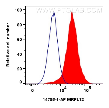
FC experiment of HeLa using 14795-1-AP
1x10^6 HeLa cells were intracellularly stained with 0.4 ug MRPL12 Polyclonal antibody (14795-1-AP) and CoraLite®488-Conjugated Goat Anti-Rabbit IgG(H+L) (SA00013-2)(red), or 0.4 ug Rabbit IgG control Rabbit PolyAb (30000-0-AP) (blue). Cells were fixed with 4% PFA and permeabilized with Flow Cytometry Perm Buffer (PF00011-C).
1x10^6 HeLa cells were intracellularly stained with 0.4 ug MRPL12 Polyclonal antibody (14795-1-AP) and CoraLite®488-Conjugated Goat Anti-Rabbit IgG(H+L) (SA00013-2)(red), or 0.4 ug Rabbit IgG control Rabbit PolyAb (30000-0-AP) (blue). Cells were fixed with 4% PFA and permeabilized with Flow Cytometry Perm Buffer (PF00011-C).
The Proteintech guarantee covers Proteintech antibodies in any species and any application, including those not listed on the datasheet. If the antibody doesn’t perform, you can receive a hassle-free refund or credit note.
human colon cancer tissue Note: suggested antigen retrieval with TE buffer pH 9.0; (*) Alternatively, antigen retrieval may be performed with citrate buffer pH 6.0
Positive IF/ICC detected in
HepG2 cells
Positive FC (Intra) detected in
HeLa cells
Recommended dilution
Application
Dilution
Western Blot (WB)
WB : 1:1000-1:4000
Immunoprecipitation (IP)
IP : 0.5-4.0 ug for 1.0-3.0 mg of total protein lysate
Immunohistochemistry (IHC)
IHC : 1:50-1:500
Immunofluorescence (IF)/ICC
IF/ICC : 1:250-1:1000
Flow Cytometry (FC) (INTRA)
FC (INTRA) : 0.40 ug per 10^6 cells in a 100 µl suspension
It is recommended that this reagent should be titrated in each testing system to obtain optimal results.
Sample-dependent, Check data in validation data gallery.
PBS with 0.02% sodium azide and 50% glycerol , pH 7.3
Storage Conditions
Store at -20°C. Stable for one year after shipment. Aliquoting is unnecessary for -20oC storage. 20ul sizes contain 0.1% BSA.
Background Information
Mammalian MRPL12 was the first mitochondrial ribosomal protein to be characterized, and is encoded by a nuclear gene. The protein forms a homodimer, localizes predominantly to mitochondria and binds to the large mitochondrial ribosomal subunit [PMID:862670]. It is required for CycD/Cdk4-induced cell growth. In addition, forming a ribosomal stalk with MRPL7, MRPL12 is required for the recruitment of translation elongation factors Tu and G to the large ribosomal subunit [PMID:12370014]. It also a part of a novel regulatory mechanism that coordinates mitochondrial transcription with translation and/or ribosome biogenesis during human mitochondrial gene expression. The antibody reacts specifically with 21kDa MRPL12 protein.
Various lysates were subjected to SDS PAGE followed by western blot with 14795-1-AP (MRPL12 antibody) at dilution of 1:2000 incubated at room temperature for 1.5 hours.
WB analysis of HeLa using 14795-1-AP
WB result of MRPL12 antibody (14795-1-AP; 1:2000; incubated at room temperature for 1.5 hours) with sh-Control and sh-MRPL12 transfected HeLa cells.
IHC Figures
IHC staining of human colon cancer using 14795-1-AP
Immunohistochemical analysis of paraffin-embedded human colon cancer tissue slide using 14795-1-AP (MRPL12 antibody) at dilution of 1:200 (under 10x lens). Heat mediated antigen retrieval with Tris-EDTA buffer (pH 9.0).
IHC staining of human colon cancer using 14795-1-AP
Immunohistochemical analysis of paraffin-embedded human colon cancer tissue slide using 14795-1-AP (MRPL12 antibody) at dilution of 1:200 (under 40x lens). Heat mediated antigen retrieval with Tris-EDTA buffer (pH 9.0).
IP Figures
IP experiment of HeLa using 14795-1-AP
IP result of anti-MRPL12 (IP:14795-1-AP, 4ug; Detection:14795-1-AP 1:500) with HeLa cells lysate 2000ug.
IF/ICC Figures
IF Staining of HepG2 using 14795-1-AP
Immunofluorescent analysis of (4% PFA) fixed HepG2 cells using MRPL12 antibody (14795-1-AP) at dilution of 1:200 and CoraLite®488-Conjugated AffiniPure Goat Anti-Rabbit IgG(H+L), CL594-Phalloidin (red).
IF Staining of HepG2 using 14795-1-AP
Immunofluorescent analysis of (4% PFA) fixed HepG2 cells using MRPL12 antibody (14795-1-AP) at dilution of 1:500 and CoraLite®488-Conjugated AffiniPure Goat Anti-Rabbit IgG(H+L), CL594-Phalloidin (red).
IF Staining of HepG2 using 14795-1-AP
Immunofluorescent analysis of (4% PFA) fixed HepG2 cells using MRPL12 antibody (14795-1-AP) at dilution of 1:500 and CoraLite®488-Conjugated AffiniPure Goat Anti-Rabbit IgG(H+L), CL594-Phalloidin (red).
FC (INTRA) Figures
FC experiment of HeLa using 14795-1-AP
1x10^6 HeLa cells were intracellularly stained with 0.4 ug MRPL12 Polyclonal antibody (14795-1-AP) and CoraLite®488-Conjugated Goat Anti-Rabbit IgG(H+L) (SA00013-2)(red), or 0.4 ug Rabbit IgG control Rabbit PolyAb (30000-0-AP) (blue). Cells were fixed with 4% PFA and permeabilized with Flow Cytometry Perm Buffer (PF00011-C).
The species listed in Tested Reactivity are in-house verified and applicable species. For unlisted species, please refer to the homology analysis of the immunogen sequence and related species. For rabbit polyclonal antibodies, homology >70% is recommended. For mouse monoclonal antibodies and rabbit recombinant antibodies, homology >90% is recommended. Generally, the higher the homology, the greater the applicability. However, there will be certain differences in protein expression in different species, tissues or cells. Therefore, the homology analysis results are for reference only and do not serve as a guarantee.
At Proteintech, we pride ourselves on our antibody quality, customer service and transparency. As such, we are comparing our antibodies with other vendors, enabling easy identification and comparisons of key data to help you choose the suitable antibody for your needs.
We have selected the top cited antibodies from these vendors for you to compare.
Proteintech
KD/KO VALIDATED
MRPL12 Polyclonal antibody
Catalog Number
14795-1-AP
Citations
34
Dilutions
WB : 1:1000-1:4000 IP : 0.5-4.0 ug for IP and 0.5-4.0 ug for 1.0-3.0 mg of total protein lysate for WB IHC : 1:50-1:500 IF/ICC : 1:250-1:1000 FC (INTRA) : 0.40 ug per 10^6 cells in a 100 µl suspension
Applications
WB, IHC, IF/ICC, FC (Intra), IP, ELISA
Reactivity
human, mouse, rat, zebrafish
Product Guarantee
Covers any species including not listed on datasheet
Covers any applications including not listed on datasheet
 at dilution of 1:2000 incubated at room temperature for 1.5 hours.")
 with sh-Control and sh-MRPL12 transfected HeLa cells.")
 with HeLa cells lysate 2000ug.")
 at dilution of 1:200 (under 10x lens). Heat mediated antigen retrieval with Tris-EDTA buffer (pH 9.0).")
 at dilution of 1:200 (under 40x lens). Heat mediated antigen retrieval with Tris-EDTA buffer (pH 9.0).")
 fixed HepG2 cells using MRPL12 antibody (14795-1-AP) at dilution of 1:200 and CoraLite®488-Conjugated AffiniPure Goat Anti-Rabbit IgG(H+L), CL594-Phalloidin (red).")
 fixed HepG2 cells using MRPL12 antibody (14795-1-AP) at dilution of 1:500 and CoraLite®488-Conjugated AffiniPure Goat Anti-Rabbit IgG(H+L), CL594-Phalloidin (red).")
 fixed HepG2 cells using MRPL12 antibody (14795-1-AP) at dilution of 1:500 and CoraLite®488-Conjugated AffiniPure Goat Anti-Rabbit IgG(H+L), CL594-Phalloidin (red).")
 and CoraLite®488-Conjugated Goat Anti-Rabbit IgG(H+L) (SA00013-2)(red), or 0.4 ug Rabbit IgG control Rabbit PolyAb (30000-0-AP) (blue). Cells were fixed with 4% PFA and permeabilized with Flow Cytometry Perm Buffer (PF00011-C).")