Various lysates were subjected to SDS PAGE followed by western blot with 67974-1-Ig (LIMK1 antibody) at dilution of 1:5000 incubated at room temperature for 1.5 hours. The membrane was stripped and reblotted with HRP-conjugated Beta Actin Monoclonal antibody (HRP-66009) as loading control.
Various lysates were subjected to SDS PAGE followed by western blot with 67974-1-Ig (LIMK1 antibody) at dilution of 1:5000 incubated at room temperature for 1.5 hours. The membrane was stripped and reblotted with HRP-conjugated Beta Actin Monoclonal antibody (HRP-66009) as loading control.
WB analysis using 67974-1-Ig
Various lysates were subjected to SDS PAGE followed by western blot with 67974-1-Ig (LIMK1 antibody) at dilution of 1:5000 incubated at room temperature for 1.5 hours. The membrane was stripped and reblotted with HRP-conjugated Beta Actin Monoclonal antibody (HRP-66009) as loading control.
Various lysates were subjected to SDS PAGE followed by western blot with 67974-1-Ig (LIMK1 antibody) at dilution of 1:5000 incubated at room temperature for 1.5 hours. The membrane was stripped and reblotted with HRP-conjugated Beta Actin Monoclonal antibody (HRP-66009) as loading control.
WB analysis of HeLa using 67974-1-Ig
WB result of LIMK1 antibody (67974-1-Ig; 1:2000; incubated at room temperature for 1.5 hours) with sh-Control and sh-LIMK1 transfected HeLa cells.
WB result of LIMK1 antibody (67974-1-Ig; 1:2000; incubated at room temperature for 1.5 hours) with sh-Control and sh-LIMK1 transfected HeLa cells.
WB analysis of MCF-7 using 67974-1-Ig
MCF-7 cells were subjected to SDS PAGE followed by western blot with 67974-1-Ig (LIMK1 antibody) at dilution of 1:5000 incubated at room temperature for 1.5 hours.
MCF-7 cells were subjected to SDS PAGE followed by western blot with 67974-1-Ig (LIMK1 antibody) at dilution of 1:5000 incubated at room temperature for 1.5 hours.
WB analysis of SH-SY5Y using 67974-1-Ig
SH-SY5Y cells were subjected to SDS PAGE followed by western blot with 67974-1-Ig (LIMK1 antibody) at dilution of 1:5000 incubated at room temperature for 1.5 hours.
SH-SY5Y cells were subjected to SDS PAGE followed by western blot with 67974-1-Ig (LIMK1 antibody) at dilution of 1:5000 incubated at room temperature for 1.5 hours.
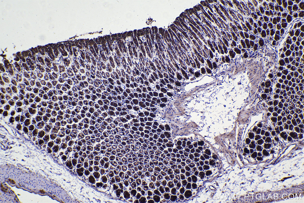
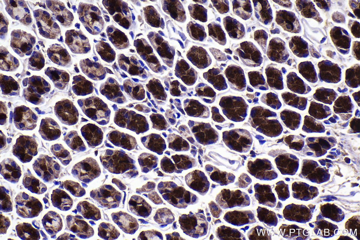
IHC staining of mouse stomach using 67974-1-Ig
Immunohistochemical analysis of paraffin-embedded mouse stomach tissue slide using 67974-1-Ig (LIMK1 antibody) at dilution of 1:2000 (under 10x lens). Heat mediated antigen retrieval with Tris-EDTA buffer (pH 9.0).
Immunohistochemical analysis of paraffin-embedded mouse stomach tissue slide using 67974-1-Ig (LIMK1 antibody) at dilution of 1:2000 (under 40x lens). Heat mediated antigen retrieval with Tris-EDTA buffer (pH 9.0).
IF Staining of HeLa using 67974-1-Ig
Immunofluorescent analysis of (-20°C Methanol) fixed HeLa cells using LIMK1 antibody (67974-1-Ig, Clone: 2B11E9 ) at dilution of 1:1000 and CoraLite®488-Conjugated AffiniPure Goat Anti-Mouse IgG(H+L).
Immunofluorescent analysis of (-20°C Methanol) fixed HeLa cells using LIMK1 antibody (67974-1-Ig, Clone: 2B11E9 ) at dilution of 1:1000 and CoraLite®488-Conjugated AffiniPure Goat Anti-Mouse IgG(H+L).
The Proteintech guarantee covers Proteintech antibodies in any species and any application, including those not listed on the datasheet. If the antibody doesn’t perform, you can receive a hassle-free refund or credit note.
mouse stomach tissue Note: suggested antigen retrieval with TE buffer pH 9.0; (*) Alternatively, antigen retrieval may be performed with citrate buffer pH 6.0
Positive IF/ICC detected in
HeLa cells
Recommended dilution
Application
Dilution
Western Blot (WB)
WB : 1:2000-1:10000
Immunohistochemistry (IHC)
IHC : 1:1000-1:4000
Immunofluorescence (IF)/ICC
IF/ICC : 1:500-1:2000
It is recommended that this reagent should be titrated in each testing system to obtain optimal results.
Sample-dependent, Check data in validation data gallery.
PBS with 0.02% sodium azide and 50% glycerol , pH 7.3
Storage Conditions
Store at -20°C. Stable for one year after shipment. Aliquoting is unnecessary for -20oC storage. 20ul sizes contain 0.1% BSA.
Background Information
LIMK1,also named LIMK, belongs to the protein kinase superfamily and TKL Ser/Thr protein kinase family. LIMK1 is a protein kinase which regulates actin filament dynamics. Phosphorylates and inactivates the actin binding/depolymerizing factor cofilin, thereby stabilizing the actin cytoskeleton. Isoform 3 has a dominant negative effect on actin cytoskeletal changes. LIMK1 may be involved in brain development. The antibody has no cross-reaction with LIMK2.
Various lysates were subjected to SDS PAGE followed by western blot with 67974-1-Ig (LIMK1 antibody) at dilution of 1:5000 incubated at room temperature for 1.5 hours. The membrane was stripped and reblotted with HRP-conjugated Beta Actin Monoclonal antibody (HRP-66009) as loading control.
WB analysis using 67974-1-Ig
Various lysates were subjected to SDS PAGE followed by western blot with 67974-1-Ig (LIMK1 antibody) at dilution of 1:5000 incubated at room temperature for 1.5 hours. The membrane was stripped and reblotted with HRP-conjugated Beta Actin Monoclonal antibody (HRP-66009) as loading control.
WB analysis of HeLa using 67974-1-Ig
WB result of LIMK1 antibody (67974-1-Ig; 1:2000; incubated at room temperature for 1.5 hours) with sh-Control and sh-LIMK1 transfected HeLa cells.
WB analysis of MCF-7 using 67974-1-Ig
MCF-7 cells were subjected to SDS PAGE followed by western blot with 67974-1-Ig (LIMK1 antibody) at dilution of 1:5000 incubated at room temperature for 1.5 hours.
WB analysis of SH-SY5Y using 67974-1-Ig
SH-SY5Y cells were subjected to SDS PAGE followed by western blot with 67974-1-Ig (LIMK1 antibody) at dilution of 1:5000 incubated at room temperature for 1.5 hours.
IHC Figures
IHC staining of mouse stomach using 67974-1-Ig
Immunohistochemical analysis of paraffin-embedded mouse stomach tissue slide using 67974-1-Ig (LIMK1 antibody) at dilution of 1:2000 (under 10x lens). Heat mediated antigen retrieval with Tris-EDTA buffer (pH 9.0).
IHC staining of mouse stomach using 67974-1-Ig
Immunohistochemical analysis of paraffin-embedded mouse stomach tissue slide using 67974-1-Ig (LIMK1 antibody) at dilution of 1:2000 (under 40x lens). Heat mediated antigen retrieval with Tris-EDTA buffer (pH 9.0).
IF/ICC Figures
IF Staining of HeLa using 67974-1-Ig
Immunofluorescent analysis of (-20°C Methanol) fixed HeLa cells using LIMK1 antibody (67974-1-Ig, Clone: 2B11E9 ) at dilution of 1:1000 and CoraLite®488-Conjugated AffiniPure Goat Anti-Mouse IgG(H+L).
The species listed in Tested Reactivity are in-house verified and applicable species. For unlisted species, please refer to the homology analysis of the immunogen sequence and related species. For rabbit polyclonal antibodies, homology >70% is recommended. For mouse monoclonal antibodies and rabbit recombinant antibodies, homology >90% is recommended. Generally, the higher the homology, the greater the applicability. However, there will be certain differences in protein expression in different species, tissues or cells. Therefore, the homology analysis results are for reference only and do not serve as a guarantee.
At Proteintech, we pride ourselves on our antibody quality, customer service and transparency. As such, we are comparing our antibodies with other vendors, enabling easy identification and comparisons of key data to help you choose the suitable antibody for your needs.
We have selected the top cited antibodies from these vendors for you to compare.
 at dilution of 1:5000 incubated at room temperature for 1.5 hours. The membrane was stripped and reblotted with HRP-conjugated Beta Actin Monoclonal antibody (HRP-66009) as loading control.")
 at dilution of 1:5000 incubated at room temperature for 1.5 hours. The membrane was stripped and reblotted with HRP-conjugated Beta Actin Monoclonal antibody (HRP-66009) as loading control.")
 with sh-Control and sh-LIMK1 transfected HeLa cells.")
 at dilution of 1:5000 incubated at room temperature for 1.5 hours.")
 at dilution of 1:5000 incubated at room temperature for 1.5 hours.")
 at dilution of 1:2000 (under 10x lens). Heat mediated antigen retrieval with Tris-EDTA buffer (pH 9.0).")
 at dilution of 1:2000 (under 40x lens). Heat mediated antigen retrieval with Tris-EDTA buffer (pH 9.0).")
 fixed HeLa cells using LIMK1 antibody (67974-1-Ig, Clone: 2B11E9 ) at dilution of 1:1000 and CoraLite®488-Conjugated AffiniPure Goat Anti-Mouse IgG(H+L).")