at dilution of 1:3000 incubated at room temperature for 1.5 hours.")
 at dilution of 1:2000 incubated at room temperature for 1.5 hours.")
 at dilution of 1:2000 incubated at room temperature for 1.5 hours.")
 at dilution of 1:2000 incubated at room temperature for 1.5 hours.")
 at dilution of 1:2000 incubated at room temperature for 1.5 hours.")
 at dilution of 1:2000 incubated at room temperature for 1.5 hours.")
 at dilution of 1:200 (under 10x lens. Heat mediated antigen retrieval with Tris-EDTA buffer (pH 9.0).")
 at dilution of 1:200 (under 40x lens. Heat mediated antigen retrieval with Tris-EDTA buffer (pH 9.0).")
 at dilution of 1:800 (under 10x lens. Heat mediated antigen retrieval with Tris-EDTA buffer (pH 9.0).")
 at dilution of 1:800 (under 40x lens. Heat mediated antigen retrieval with Tris-EDTA buffer (pH 9.0).")
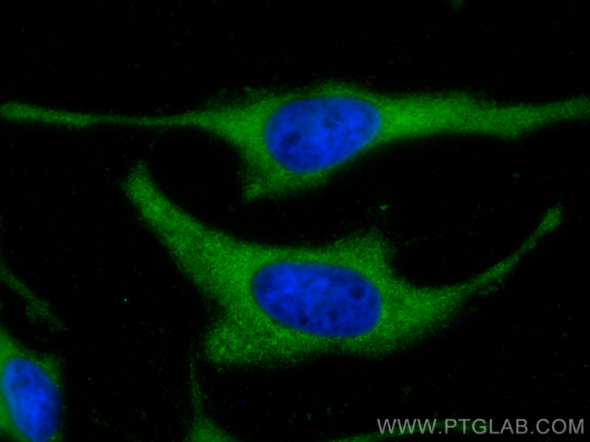
 fixed HeLa cells using ATG13 antibody (66708-1-Ig, Clone: 1C3A7 ) at dilution of 1:800 and CoraLite®488-Conjugated AffiniPure Goat Anti-Mouse IgG(H+L).")
 and CoraLite®488-Conjugated AffiniPure Goat Anti-Mouse IgG(H+L) at dilution 1:1000 (red), or 0.4 ug Control Antibody. Cells were fixed with 4% PFA and permeabilized with Flow Cytometry Perm Buffer.")
Tested Applications
| Positive WB detected in | HEK-293 cells, BGC-823 cells, NIH/3T3 cells, pig brain tissue, rat brain tissue, mouse brain tissue, HeLa cells, Jurkat cells |
| Positive IHC detected in | mouse testis tissue, mouse brain tissue Note: suggested antigen retrieval with TE buffer pH 9.0; (*) Alternatively, antigen retrieval may be performed with citrate buffer pH 6.0 |
| Positive IF/ICC detected in | HeLa cells |
| Positive FC (Intra) detected in | HeLa cells |
Recommended dilution
| Application | Dilution |
|---|---|
| Western Blot (WB) | WB : 1:1000-1:6000 |
| Immunohistochemistry (IHC) | IHC : 1:50-1:500 |
| Immunofluorescence (IF)/ICC | IF/ICC : 1:400-1:1600 |
| Flow Cytometry (FC) (INTRA) | FC (INTRA) : 0.40 ug per 10^6 cells in a 100 µl suspension |
| It is recommended that this reagent should be titrated in each testing system to obtain optimal results. | |
| Sample-dependent, Check data in validation data gallery. | |
Published Applications
| WB | See 2 publications below |
Product Information
66708-1-Ig targets ATG13 in WB, IHC, IF/ICC, FC (Intra), ELISA applications and shows reactivity with human, mouse, rat, pig samples.
| Tested Reactivity | human, mouse, rat, pig |
| Cited Reactivity | human, mouse |
| Host / Isotype | Mouse / IgG1 |
| Class | Monoclonal |
| Type | Antibody |
| Immunogen |
CatNo: Ag12968 Product name: Recombinant human KIAA0652 protein Source: e coli.-derived, PET28a Tag: 6*His Domain: 131-481 aa of BC001331 Sequence: ITRVTPAYRLSRKQGHEYVILYRIYFGEVQLSGLGEGFQTVRVGTVGTPVGTITLSCAYRINLAFMSTRQFERTPPIMGIIIDHFVDRPYPSSSPMHPCNYRTAGEDTGVIYPSVEDSQEVCTTSFSTSPPSQLMVPGKEGGVPLAPNQPVHGTQADQERLATCTPSDRTHCAATPSSSEDTETVSNSSEGRASPHDVLETIFVRKVGAFVNKPINQVTLTSLDIPFAMFAPKNLELEDTDPMVNPPDSPETESPLQGSLHSDGSSGGSSGNTHDDFVMIDFKPAFSKDDILPMDLGTFYREFQNPPQLSSLSIDIGAQSMAEDLDSLPEKLAVHEKNVREFDAFVETLQ* Predict reactive species |
| Full Name | KIAA0652 |
| Calculated Molecular Weight | 57 kDa |
| Observed Molecular Weight | 57 kDa |
| GenBank Accession Number | BC001331 |
| Gene Symbol | ATG13 |
| Gene ID (NCBI) | 9776 |
| RRID | AB_2882060 |
| Conjugate | Unconjugated |
| Form | Liquid |
| Purification Method | Protein G purification |
| UNIPROT ID | O75143 |
| Storage Buffer | PBS with 0.02% sodium azide and 50% glycerol, pH 7.3. |
| Storage Conditions | Store at -20°C. Stable for one year after shipment. Aliquoting is unnecessary for -20oC storage. 20ul sizes contain 0.1% BSA. |
Background Information
ATG13 is one component protein of the ULK1 complex which is required for autophagosome formation and mitophagy. ATG13 has two nutrient regulatory phosphorylation sites and the phosphorylation status of ATG13 affect regulation of autophagy by modulating enzyme activity and cellular localization of ULK1. Besides, it has been reported the nonautophagic function of ATG13 on cardiac development for ATG13-deficient embryos show myocardial growth defects.(PMID:27387056, 26801615, 26644405)
Protocols
| Product Specific Protocols | |
|---|---|
| FC protocol for ATG13 antibody 66708-1-Ig | Download protocol |
| IF protocol for ATG13 antibody 66708-1-Ig | Download protocol |
| IHC protocol for ATG13 antibody 66708-1-Ig | Download protocol |
| WB protocol for ATG13 antibody 66708-1-Ig | Download protocol |
| Standard Protocols | |
|---|---|
| Click here to view our Standard Protocols |
Publications
| Species | Application | Title |
|---|---|---|
Cell Rep Destabilization of TP53 by USP10 is essential for neonatal autophagy and survival | ||
Cancer Chemother Pharmacol Avermectin B1 mediates antitumor activity and induces autophagy in osteosarcoma through the AMPK/ULK1 signaling pathway |