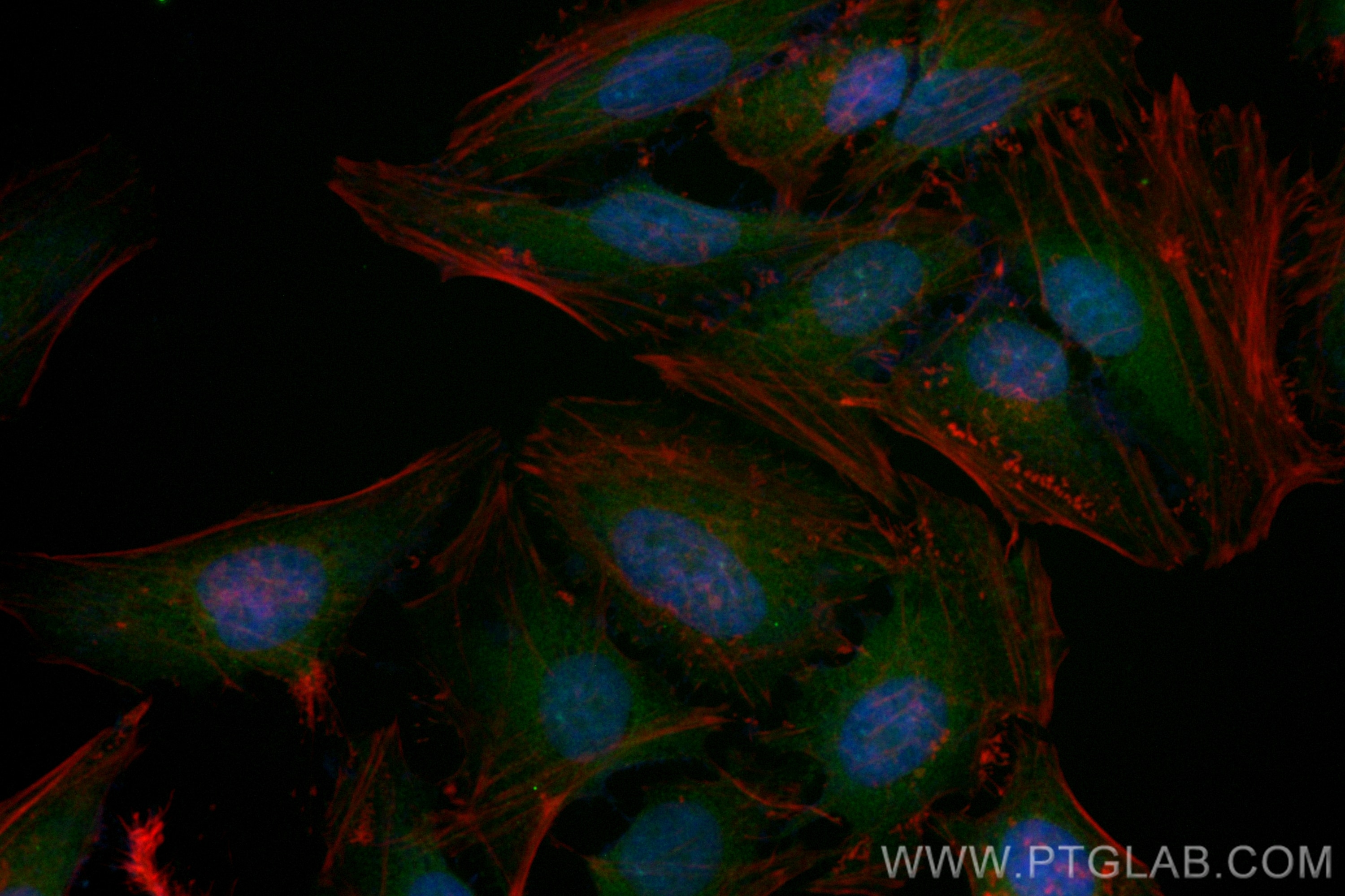
Tested Applications
| Positive IF/ICC detected in | HeLa cells |
Recommended dilution
| Application | Dilution |
|---|---|
| Immunofluorescence (IF)/ICC | IF/ICC : 1:50-1:500 |
| It is recommended that this reagent should be titrated in each testing system to obtain optimal results. | |
| Sample-dependent, Check data in validation data gallery. | |
Product Information
CL488-80744 targets KEAP1 in IF/ICC applications and shows reactivity with human, mouse, rat samples.
| Tested Reactivity | human, mouse, rat |
| Host / Isotype | Rabbit / IgG |
| Class | Recombinant |
| Type | Antibody |
| Immunogen |
CatNo: Ag0779 Product name: Recombinant human KEAP1 protein Source: e coli.-derived, PGEX-4T Tag: GST Domain: 325-624 aa of BC002930 Sequence: GRLIYTAGGYFRQSLSYLEAYNPSDGTWLRLADLQVPRSGLAGCVVGGLLYAVGGRNNSPDGNTDSSALDCYNPMTNQWSPCAPMSVPRNRIGVGVIDGHIYAVGGSHGCIHHNSVERYEPERDEWHLVAPMLTRRIGVGVAVLNRLLYAVGGFDGTNRLNSAECYYPERNEWRMITAMNTIRSGAGVCVLHNCIYAAGGYDGQDQLNSVERYDVETETWTFVAPMKHRRSALGITVHQGRIYVLGGYDGHTFLDSVECYDPDTDTWSEVTRMTSGRSGVGVAVTMEPCRKQIDQQNCTC Predict reactive species |
| Full Name | kelch-like ECH-associated protein 1 |
| Calculated Molecular Weight | 624 aa, 70 kDa |
| Observed Molecular Weight | 55~70 kDa |
| GenBank Accession Number | BC002930 |
| Gene Symbol | KEAP1 |
| Gene ID (NCBI) | 9817 |
| RRID | AB_3673038 |
| Conjugate | CoraLite® Plus 488 Fluorescent Dye |
| Excitation/Emission Maxima Wavelengths | 493 nm / 522 nm |
| Form | Liquid |
| Purification Method | Protein A purification |
| UNIPROT ID | Q14145 |
| Storage Buffer | PBS with 50% glycerol, 0.05% Proclin300, 0.5% BSA, pH 7.3. |
| Storage Conditions | Store at -20°C. Avoid exposure to light. Stable for one year after shipment. Aliquoting is unnecessary for -20oC storage. |
Background Information
Kelch-like ECH-associated protein 1 (KEAP1) is a negative regulator of nuclear factor erythroid 2-related factor 2 (Nrf2), a transcription factor governing the antioxidant response.
What is the molecular weight of KEAP1 protein? Are there any isoforms of KEAP1?
The molecular weight of KEAP1 protein is 70 kDa. The KEAP1 gene gives rise only to protein isoforms, but mutations of KEAP1 protein have been found in various cancer types.
What is the subcellular localization of KEAP1?
KEAP1 resides in the cytoplasm, where it binds to Nrf2, targeting it for degradation and preventing translocation of Nrf2 to the nucleus.
How does KEAP1 control Nrf2 levels? Is KEAP1 post-translationally modified?
KEAP1 is rich in reactive cysteine residues, whose thiol groups play a role in binding to CUL3 and the polyubiquitination of Nrf2, which leads to degradation of Nrf2 via the proteasome system. During oxidative stress, electrophiles and reactive oxygen species (ROS) modify the KEAP1 thiol groups, reducing the affinity of KEAP1 to CUL3 and the stabilization of Nrf2. Nrf2 then translocates to the nucleus, where it binds to the antioxidant responsive elements (AREs) and induces the expression of antioxidant proteins (PMID: 16354693).
How to measure oxidative stress using KEAP1 and Nrf2 proteins as a readout
Under basal conditions (unstressed cells), a detectable KEAP1 protein level is observed. Oxidative stress modifies KEAP1 protein activity by increasing the Nrf2 protein levels. This can be measured, for example, using western blotting (PMID: 27697860). KEAP1 protein levels are not altered by oxidative stress.
What is the role of the KEAP1-Nrf2 pathway in health and disease?
The KEAP-Nrf2 pathway plays a vital role in redox homeostasis and cryoprotection. Inhibition of KEAP1 activity leads to the activation of Nrf2 and increase the response to oxidative stress and anti-inflammatory effects (PMID: 29717933). The activation of Nrf2 can be beneficial in the case of metabolic diseases, such as diabetes, as well as neurodegenerative diseases such as Parkinson's and Alzheimer's diseases. However, the increased activation of Nrf2 is also known to promote tumor growth and metastasis. Mutations in both KEAP1 and Nrf2 were found in various solid tumor types.
Protocols
| Product Specific Protocols | |
|---|---|
| IF protocol for CL Plus 488 KEAP1 antibody CL488-80744 | Download protocol |
| Standard Protocols | |
|---|---|
| Click here to view our Standard Protocols |


