and IHC Detect Kit for Rabbit Primary Antibody (PK10009).")
 and IHC Detect Kit for Rabbit Primary Antibody (PK10009).")
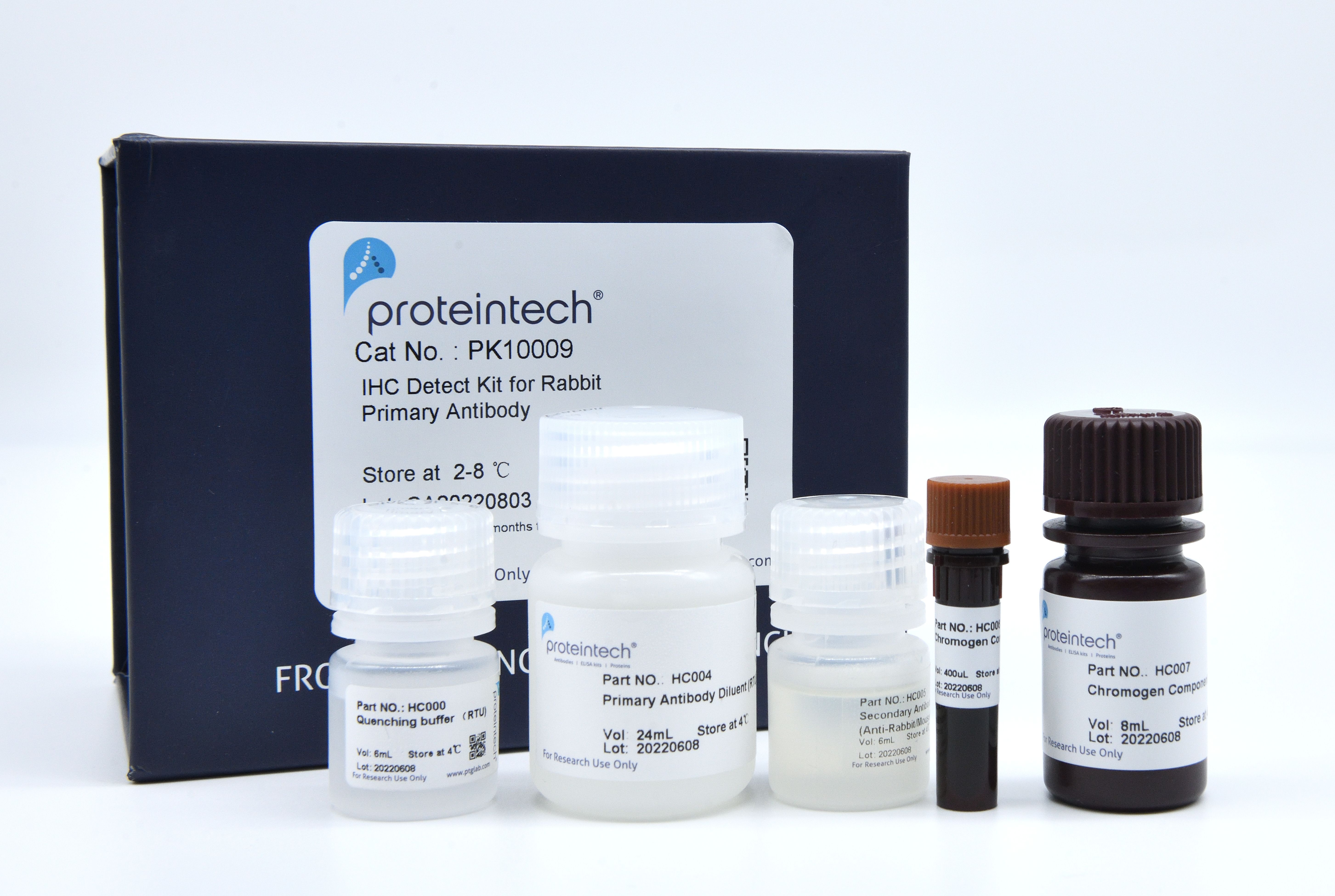
Product Information
The PK10009 kit is designed to facilitate signal detection during IHC analysis of FFPE tissue samples using primary antibodies raised in rabbits. The kit consists of Polymer HRP-Goat anti-Rabbit Secondary Antibody and an ultra-sensitive Chromogen for highly sensitive detection of rabbit primary antibodies. The kit also contains an optimized primary antibody dilution buffer that preserves primary antibody activity and helps lower non-specific background staining.
Note:
*If antigen retrieval buffer, counter staining reagent and mounting media are needed, please see our IHC Prep & Detect Kit for Rabbit Primary Antibody (Cat.NO. PK10017).
Components
| Part NO. | Component | Size |
| HC000 | Quenching Buffer (RTU) | 6 mL |
| HC004 | Primary Antibody Diluent (RTU) | 24 mL |
| HC005R | Polymer HRP-Goat anti-Rabbit Secondary Antibody | 6 mL |
| HC006 | Chromogen Component A | 0.4 mL |
| HC007 | Chromogen Component B | 8 mL |
Size: 60 tests (may vary depending on tissue size).
Storage
All the reagents are stored at 2-8°C. The kit is stable for 6 months from the date of receipt.
Usage
1. Prepare slides from tissues sections following routine methods. Deparaffinize tissues with xylene and re-hydrate using a decreasing ethanol gradient.
2. Perform antigen retrieval following the manufacturer’s recommendations of the primary antibody.
3. Quenching (optional): Based on our experience, endogenous peroxidase quenching is not necessary in most cases and can sometimes even have adverse effects on staining. However, we have included a quenching reagent in this kit since the quenching step is widely recommended in publications.
3.1 Wash the slides with 1x wash buffer. Drain off the liquid on the slides and absorb any residual buffer around the tissue section (making the slides as dry as possible). Use a hydrophobic IHC pen to draw a circle on the glass slides around tissue sections (optional).
3.2 Apply 30-80 uL (depending on the size of the tissue) of the ready-to-use Quenching Buffer (Part NO. HC000) to the slide, covering the entire tissue. Leave the slides at room temperature for 10 minutes. Briefly wash the slides with 1x wash buffer after quenching.
4. Block the tissues with blocking buffer (e.g., 3% BSA in PBS) for 30 minutes at room temperature. Drain the liquid off the slides and absorb the blocking buffer around the tissue section.
* The Primary Antibody Diluent (Part NO. HC004) could also be used as alternative of blocking buffer.
5. Use the Primary Antibody Diluent (Part NO. HC004) in the kit to dilute your primary antibody (ONLY primary antibodies raised in rabbits should be used with this kit). Apply 80-200 uL (depending on the size of the tissue) of the diluted primary antibody to the slide, covering the entire tissue. Incubate at room temperature in a wet box with a lid for 1 hour.
6. Wash the slides three times with 1x wash buffer following primary antibody incubation. Drain the liquid off the slides and absorb any residual buffer around the tissue section. Apply 30-80 uL (depending on the size of the tissue) of the ready-to-use Polymer HRP-Goat anti-Rabbit Secondary Antibody (Part NO. HC005R) to the slide, covering the entire tissue. Incubate at room temperature in a wet box with a lid for 30 minutes.
7. Wash the slides three times with 1x wash buffer following secondary antibody incubation. Drain the liquid off the slides and absorb any residual buffer around the tissue section. Use pipette to prepare an appropriate volume of Chromogen in a microcentrifuge tube. Ratio of solutions Chromogen Component A (Part NO. HC006) to Chromogen Component B (Part NO. HC007) is 1:20.
* Depending on the size of the tissue sample, approximately 30-80 ul/slide is needed.
Use pipette to add enough Chromogen to cover the tissue. Lay slides flat on the benchtop at room temperature for 5 minutes, or until a brown color develops. If color is visible, stop reaction early by washing with 1x wash buffer.
8. Perform counter staining with hematoxylin.
9. Wash the slides with wash buffer or pure water. Dehydrate tissues using an increasing ethanol gradient followed by xylene. Mount the slides with cover slides and analyze with a light microscope once they are fully dried.
Application Note
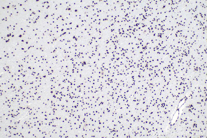
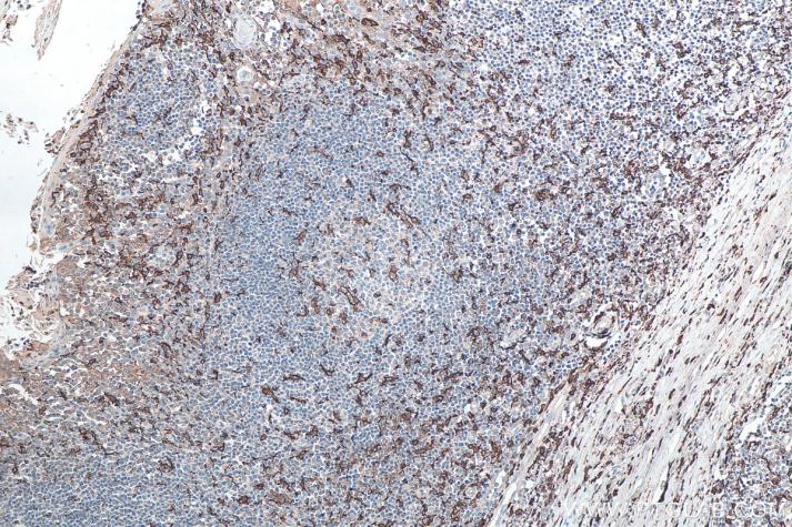
This kit is recommended for FFPE tissues and not for frozen sections.
Cited in Article as
PK10009, IHC Detect Kit for Rabbit Primary Antibody, Proteintech, IL, USA
Documentation
| SDS |
|---|
| IHC Detect Kit for Rabbit Primary Antibody |
| Datasheet |
|---|
| IHC Detect Kit for Rabbit Primary Antibody Datasheet |
Publications
| Application | Title |
|---|---|
Chem Biol Interact Targeting PKM2 improves the gemcitabine sensitivity of intrahepatic cholangiocarcinoma cells via inhibiting β-catenin signaling pathway | |
Front Immunol Direct Inhibition of GSDMD by PEITC Reduces Hepatocyte Pyroptosis and Alleviates Acute Liver Injury in Mice. | |
Bioengineered Rapamycin ameliorates chronic intermittent hypoxia and sleep deprivation-induced renal damage via the mammalian target of rapamycin (mTOR)/NOD-like receptor protein 3 (NLRP3) signaling pathway. | |
Biomater Adv Injectable tissue-engineered human cartilage matrix composite fibrin glue for regeneration of articular cartilage defects | |
Mol Cell Biochem RBM15-mediated the m6A modification of MAT2A promotes osteosarcoma cell proliferation, metastasis and suppresses ferroptosis |