Various lysates were subjected to SDS PAGE followed by western blot with 82973-1-RR (HMGB1 antibody) at dilution of 1:10000 incubated at room temperature for 1.5 hours.
Various lysates were subjected to SDS PAGE followed by western blot with 82973-1-RR (HMGB1 antibody) at dilution of 1:10000 incubated at room temperature for 1.5 hours.
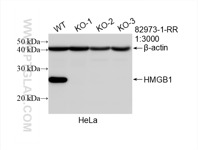
WB analysis of HeLa using 82973-1-RR
WB result of HMGB1 antibody (82973-1-RR; 1:3000; incubated at room temperature for 1.5 hours) with negative control and HMGB1 knockout HeLa cells.
WB result of HMGB1 antibody (82973-1-RR; 1:3000; incubated at room temperature for 1.5 hours) with negative control and HMGB1 knockout HeLa cells.
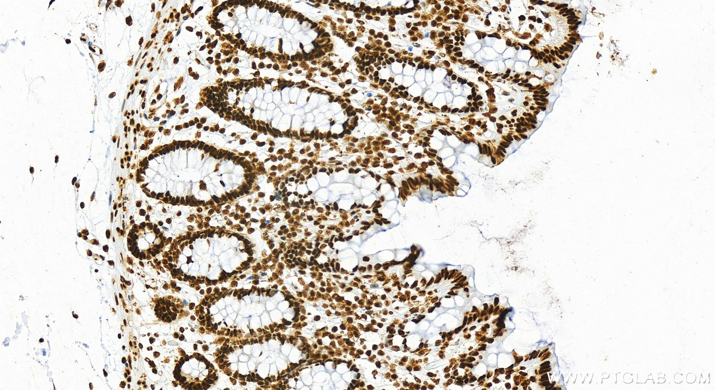
IHC staining of human normal colon using 82973-1-RR
Immunohistochemical analysis of paraffin-embedded human colon tissue slide using 82973-1-RR (HMGB1 antibody) at dilution of 1:2000 (under 20x lens). Heat mediated antigen retrieval with Tris-EDTA buffer (pH 9.0).
Immunohistochemical analysis of paraffin-embedded human colon tissue slide using 82973-1-RR (HMGB1 antibody) at dilution of 1:2000 (under 20x lens). Heat mediated antigen retrieval with Tris-EDTA buffer (pH 9.0).
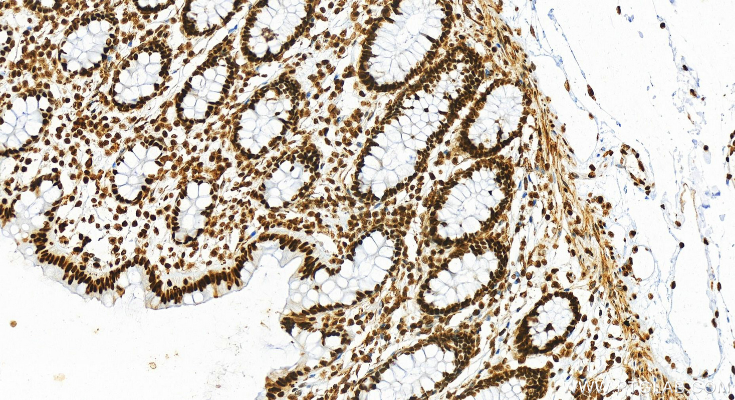
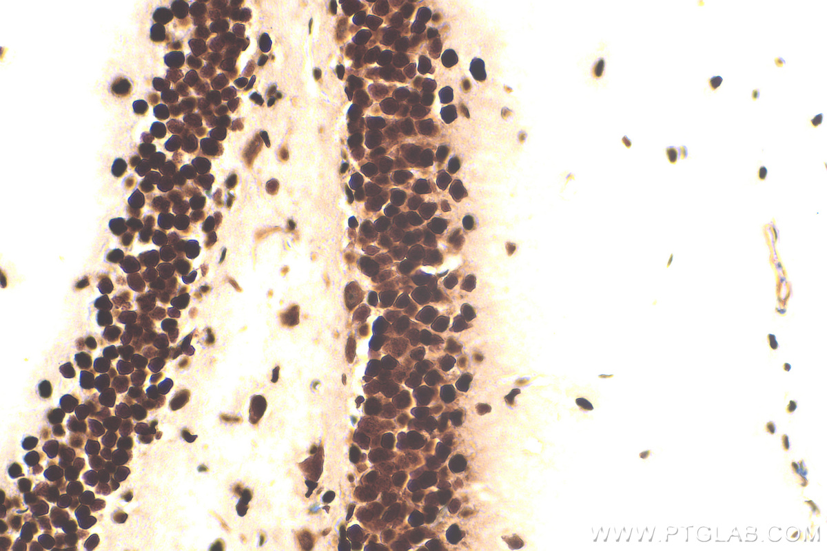
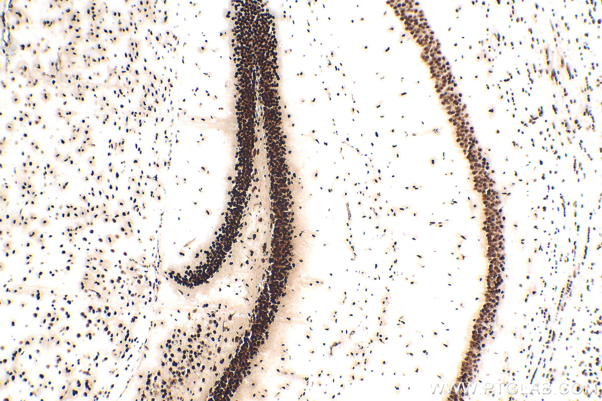
IHC staining of mouse brain using 82973-1-RR
Immunohistochemical analysis of paraffin-embedded mouse brain tissue slide using 82973-1-RR (HMGB1 antibody) at dilution of 1:1000 (under 40x lens). Heat mediated antigen retrieval with Tris-EDTA buffer (pH 9.0).
Immunohistochemical analysis of paraffin-embedded mouse brain tissue slide using 82973-1-RR (HMGB1 antibody) at dilution of 1:1000 (under 10x lens). Heat mediated antigen retrieval with Tris-EDTA buffer (pH 9.0).
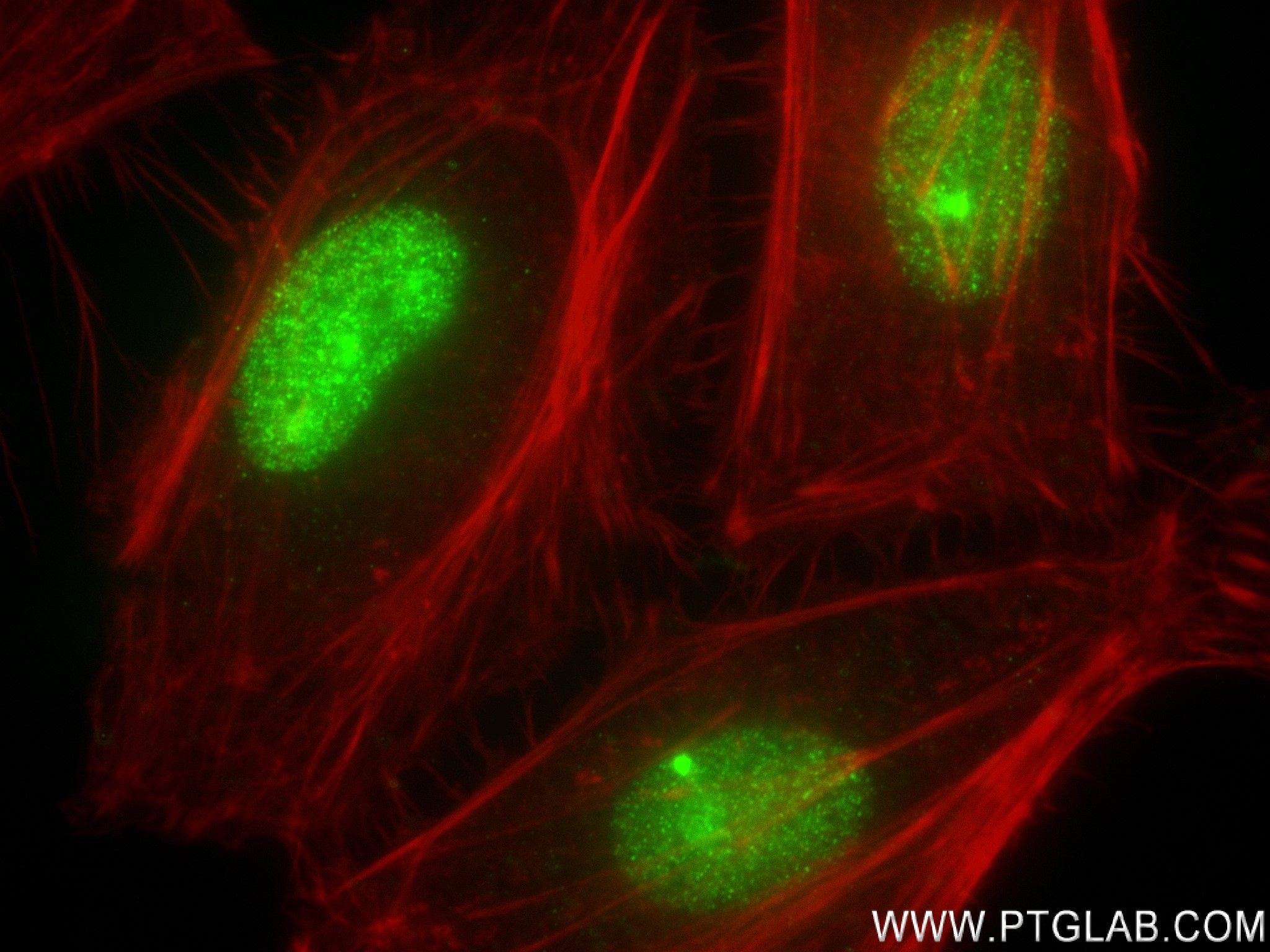
IF Staining of HeLa using 82973-1-RR
Immunofluorescent analysis of (4% PFA) fixed HeLa cells using HMGB1 antibody (82973-1-RR, Clone: 230182G6 ) at dilution of 1:250 and CoraLite®488-Conjugated AffiniPure Goat Anti-Rabbit IgG(H+L) (SA00013-2), CL594-Phalloidin (red).
Immunofluorescent analysis of (4% PFA) fixed HeLa cells using HMGB1 antibody (82973-1-RR, Clone: 230182G6 ) at dilution of 1:250 and CoraLite®488-Conjugated AffiniPure Goat Anti-Rabbit IgG(H+L) (SA00013-2), CL594-Phalloidin (red).
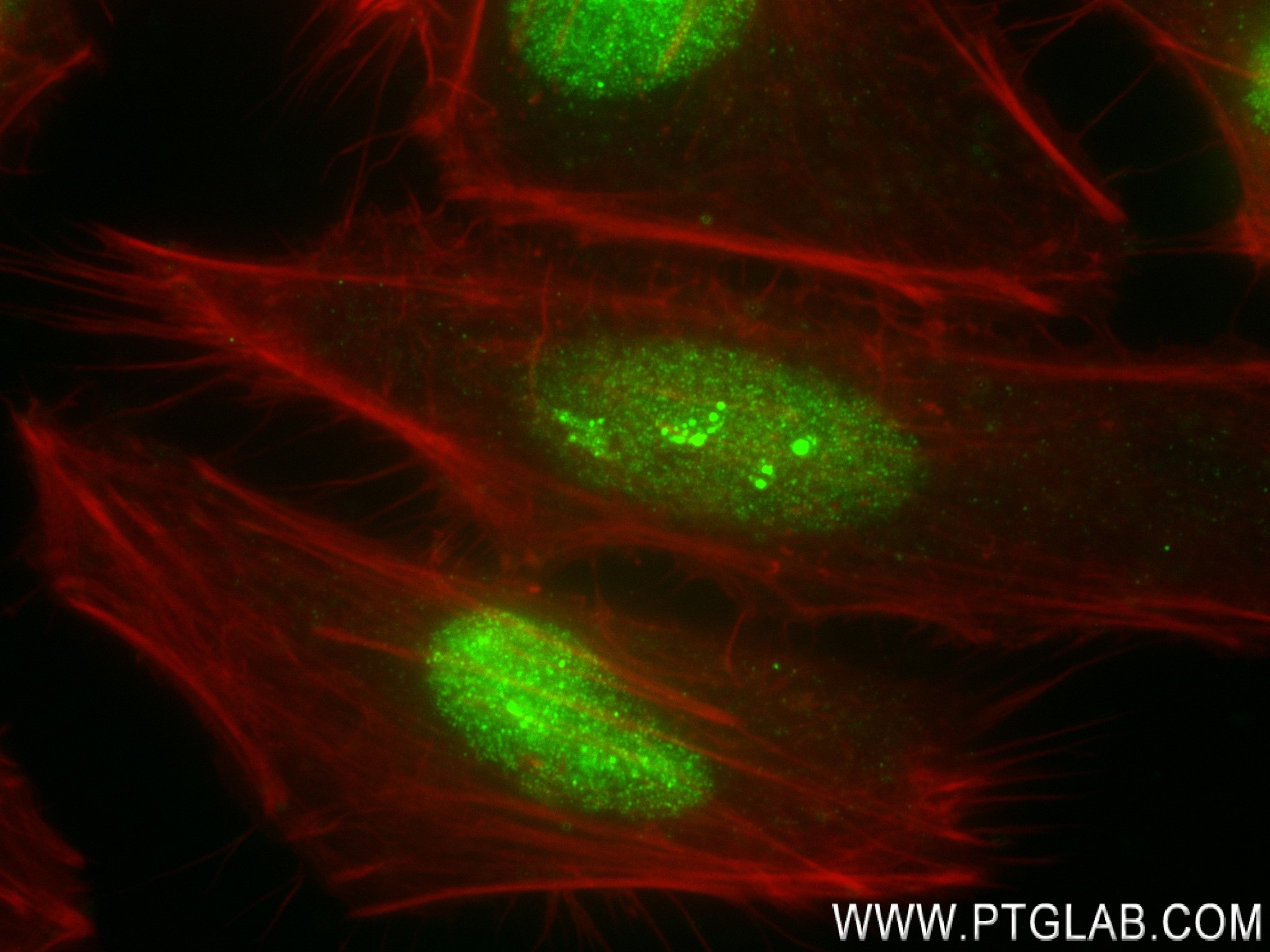
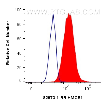
FC experiment of HeLa using 82973-1-RR
1x10^6 HeLa cells were intracellularly stained with 0.4 ug HMGB1 Recombinant antibody (82973-1-RR, Clone:230182G6) and CoraLite®488-Conjugated AffiniPure Goat Anti-Rabbit IgG(H+L) (SA00013-2)(red), or 0.4 ug Isotype Control (blue). Cells were fixed and permeabilized with Transcription Factor Staining Buffer Kit (PF00011).
1x10^6 HeLa cells were intracellularly stained with 0.4 ug HMGB1 Recombinant antibody (82973-1-RR, Clone:230182G6) and CoraLite®488-Conjugated AffiniPure Goat Anti-Rabbit IgG(H+L) (SA00013-2)(red), or 0.4 ug Isotype Control (blue). Cells were fixed and permeabilized with Transcription Factor Staining Buffer Kit (PF00011).
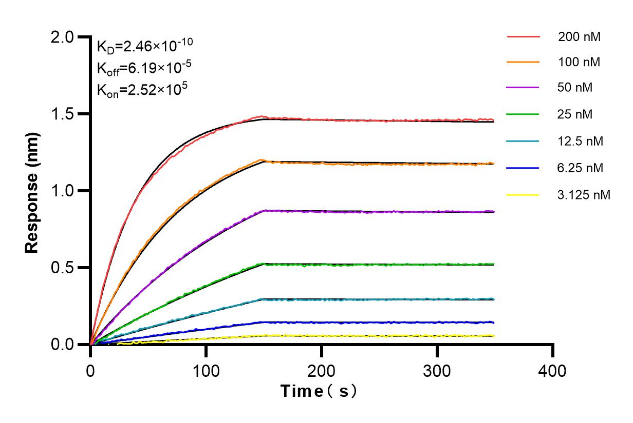
Affinity and Kinetic Characterization of 82973-1-RR
Biolayer interferometry (BLl) kinetic assays of 82973-1-RR against Human HMGB1 were performed. The affinity constant is 0.246 nM.
The Proteintech guarantee covers Proteintech antibodies in any species and any application, including those not listed on the datasheet. If the antibody doesn’t perform, you can receive a hassle-free refund or credit note.
L02 cells, HeLa cells, Jurkat cells, HEK-293 cells, K-562 cells, HCT 116 cells, mouse brain tissue, rat brain tissue
Positive IHC detected in
human colon tissue, mouse brain tissue Note: suggested antigen retrieval with TE buffer pH 9.0; (*) Alternatively, antigen retrieval may be performed with citrate buffer pH 6.0
Positive IF/ICC detected in
HeLa cells
Positive FC (Intra) detected in
HeLa cells
Recommended dilution
Application
Dilution
Western Blot (WB)
WB : 1:5000-1:50000
Immunohistochemistry (IHC)
IHC : 1:1000-1:4000
Immunofluorescence (IF)/ICC
IF/ICC : 1:125-1:500
Flow Cytometry (FC) (INTRA)
FC (INTRA) : 0.40 ug per 10^6 cells in a 100 µl suspension
It is recommended that this reagent should be titrated in each testing system to obtain optimal results.
Sample-dependent, Check data in validation data gallery.
PBS with 0.02% sodium azide and 50% glycerol pH 7.3.
Storage Conditions
Store at -20°C. Stable for one year after shipment. Aliquoting is unnecessary for -20oC storage. 20ul sizes contain 0.1% BSA.
Background Information
The HMG (high mobility group) proteins are nonhistone chromosomal proteins that is present in almost all eukaryotic cells, and it functions to stabilize NUCLEOSOME formation and acts as a transcription-factor-like protein that regulates the expression of several genes[PMID: 18160415]. Once injury, infection or other inflammatory stimuli, activated macrophages, mature dendritic cells and natural killer cells can secret HMGB1, which act as a crucial cytokine[PMID: 20163887]. HMGB1 also involved in V(D)J recombination by acting as a cofactor of the RAG complex, stimulating cleavage and RAG protein binding at the 23 bp spacer of conserved recombination signal sequences (RSS)[PMID: 19360789 ]. Act as a Heparin-binding protein that has a role in the extension of neurite-type cytoplasmic processes in developing cells. HMGB1 (high mobility group box 1) modulates gene expression in the nucleus, but certain immune cells secrete HMGB1 as an extracellular Alarmin to signal tissue damage.The nuclear HMGB1 relocalizes to the extracellular milieu in senescent human and mouse cells in culture and in vivo, which stimulated cytokine secretion through TLR-4 signaling (23649808).
Various lysates were subjected to SDS PAGE followed by western blot with 82973-1-RR (HMGB1 antibody) at dilution of 1:10000 incubated at room temperature for 1.5 hours.
WB analysis of HeLa using 82973-1-RR
WB result of HMGB1 antibody (82973-1-RR; 1:3000; incubated at room temperature for 1.5 hours) with negative control and HMGB1 knockout HeLa cells.
IHC Figures
IHC staining of human normal colon using 82973-1-RR
Immunohistochemical analysis of paraffin-embedded human colon tissue slide using 82973-1-RR (HMGB1 antibody) at dilution of 1:2000 (under 20x lens). Heat mediated antigen retrieval with Tris-EDTA buffer (pH 9.0).
IHC staining of human normal colon using 82973-1-RR
Immunohistochemical analysis of paraffin-embedded human colon tissue slide using 82973-1-RR (HMGB1 antibody) at dilution of 1:2000 (under 20x lens). Heat mediated antigen retrieval with Tris-EDTA buffer (pH 9.0).
IHC staining of mouse brain using 82973-1-RR
Immunohistochemical analysis of paraffin-embedded mouse brain tissue slide using 82973-1-RR (HMGB1 antibody) at dilution of 1:1000 (under 40x lens). Heat mediated antigen retrieval with Tris-EDTA buffer (pH 9.0).
IHC staining of mouse brain using 82973-1-RR
Immunohistochemical analysis of paraffin-embedded mouse brain tissue slide using 82973-1-RR (HMGB1 antibody) at dilution of 1:1000 (under 10x lens). Heat mediated antigen retrieval with Tris-EDTA buffer (pH 9.0).
IF/ICC Figures
IF Staining of HeLa using 82973-1-RR
Immunofluorescent analysis of (4% PFA) fixed HeLa cells using HMGB1 antibody (82973-1-RR, Clone: 230182G6 ) at dilution of 1:250 and CoraLite®488-Conjugated AffiniPure Goat Anti-Rabbit IgG(H+L) (SA00013-2), CL594-Phalloidin (red).
IF Staining of HeLa using 82973-1-RR
Immunofluorescent analysis of (4% PFA) fixed HeLa cells using HMGB1 antibody (82973-1-RR, Clone: 230182G6 ) at dilution of 1:250 and CoraLite®488-Conjugated AffiniPure Goat Anti-Rabbit IgG(H+L) (SA00013-2), CL594-Phalloidin (red).
FC (INTRA) Figures
FC experiment of HeLa using 82973-1-RR
1x10^6 HeLa cells were intracellularly stained with 0.4 ug HMGB1 Recombinant antibody (82973-1-RR, Clone:230182G6) and CoraLite®488-Conjugated AffiniPure Goat Anti-Rabbit IgG(H+L) (SA00013-2)(red), or 0.4 ug Isotype Control (blue). Cells were fixed and permeabilized with Transcription Factor Staining Buffer Kit (PF00011).
AFFINITY Figures
Affinity and Kinetic Characterization of 82973-1-RR
Biolayer interferometry (BLl) kinetic assays of 82973-1-RR against Human HMGB1 were performed. The affinity constant is 0.246 nM.
The species listed in Tested Reactivity are in-house verified and applicable species. For unlisted species, please refer to the homology analysis of the immunogen sequence and related species. For rabbit polyclonal antibodies, homology >70% is recommended. For mouse monoclonal antibodies and rabbit recombinant antibodies, homology >90% is recommended. Generally, the higher the homology, the greater the applicability. However, there will be certain differences in protein expression in different species, tissues or cells. Therefore, the homology analysis results are for reference only and do not serve as a guarantee.
At Proteintech, we pride ourselves on our antibody quality, customer service and transparency. As such, we are comparing our antibodies with other vendors, enabling easy identification and comparisons of key data to help you choose the suitable antibody for your needs.
We have selected the top cited antibodies from these vendors for you to compare.
Proteintech
KD/KO VALIDATED
HMGB1 Recombinant antibody
Catalog Number
82973-1-RR
Citations
1
Dilutions
WB : 1:5000-1:50000 IHC : 1:1000-1:4000 IF/ICC : 1:125-1:500 FC (INTRA) : 0.40 ug per 10^6 cells in a 100 µl suspension
Applications
WB, IHC, IF/ICC, FC (Intra), ELISA
Reactivity
human, mouse, rat
Product Guarantee
Covers any species including not listed on datasheet
Covers any applications including not listed on datasheet
 at dilution of 1:10000 incubated at room temperature for 1.5 hours.")
 with negative control and HMGB1 knockout HeLa cells.")
 at dilution of 1:2000 (under 20x lens). Heat mediated antigen retrieval with Tris-EDTA buffer (pH 9.0).")
 at dilution of 1:2000 (under 20x lens). Heat mediated antigen retrieval with Tris-EDTA buffer (pH 9.0).")
 at dilution of 1:1000 (under 40x lens). Heat mediated antigen retrieval with Tris-EDTA buffer (pH 9.0).")
 at dilution of 1:1000 (under 10x lens). Heat mediated antigen retrieval with Tris-EDTA buffer (pH 9.0).")
 fixed HeLa cells using HMGB1 antibody (82973-1-RR, Clone: 230182G6 ) at dilution of 1:250 and CoraLite®488-Conjugated AffiniPure Goat Anti-Rabbit IgG(H+L) (SA00013-2), CL594-Phalloidin (red).")
 fixed HeLa cells using HMGB1 antibody (82973-1-RR, Clone: 230182G6 ) at dilution of 1:250 and CoraLite®488-Conjugated AffiniPure Goat Anti-Rabbit IgG(H+L) (SA00013-2), CL594-Phalloidin (red).")
 and CoraLite®488-Conjugated AffiniPure Goat Anti-Rabbit IgG(H+L) (SA00013-2)(red), or 0.4 ug Isotype Control (blue). Cells were fixed and permeabilized with Transcription Factor Staining Buffer Kit (PF00011).")
 kinetic assays of 82973-1-RR against Human HMGB1 were performed. The affinity constant is 0.246 nM.")