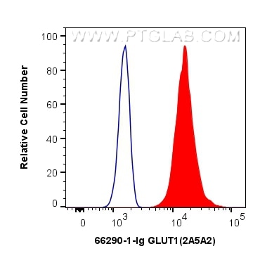

Tested Applications
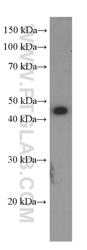
| Positive WB detected in | HEK-293 cells |

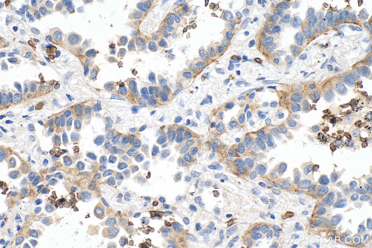
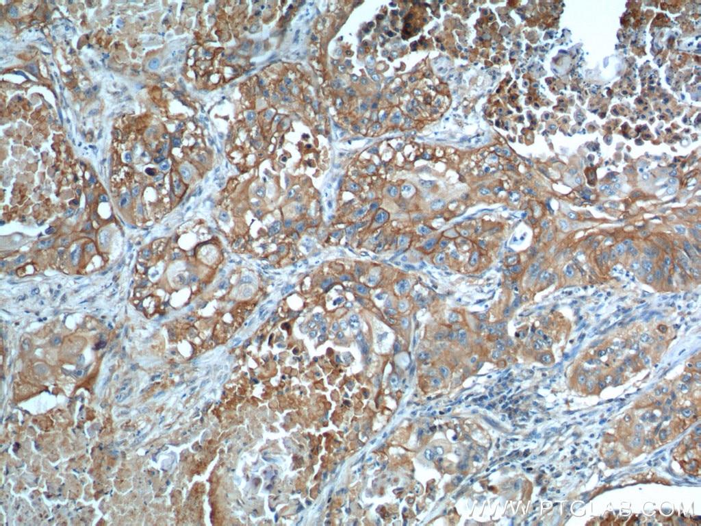
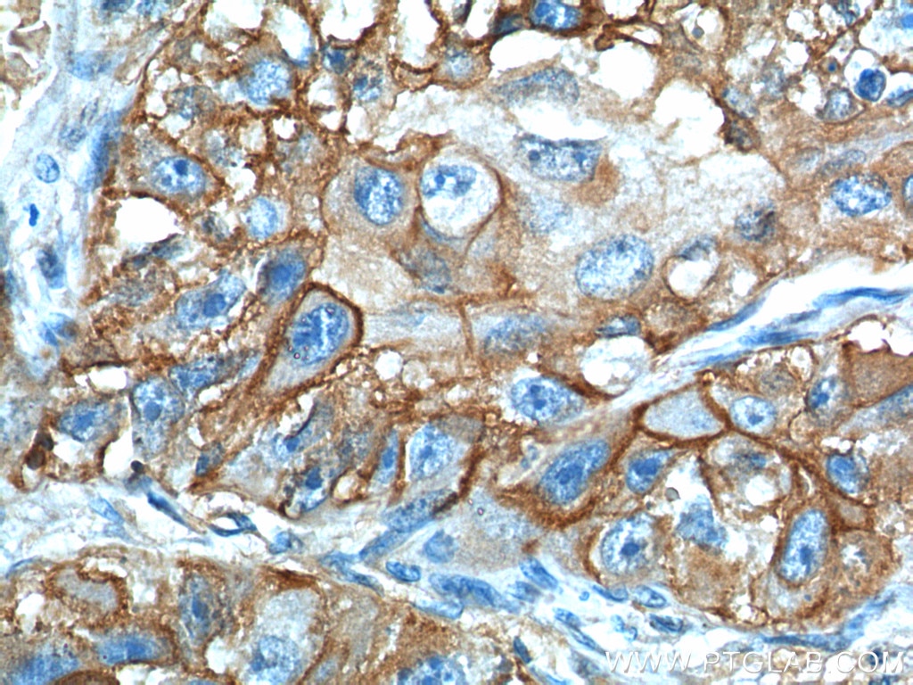
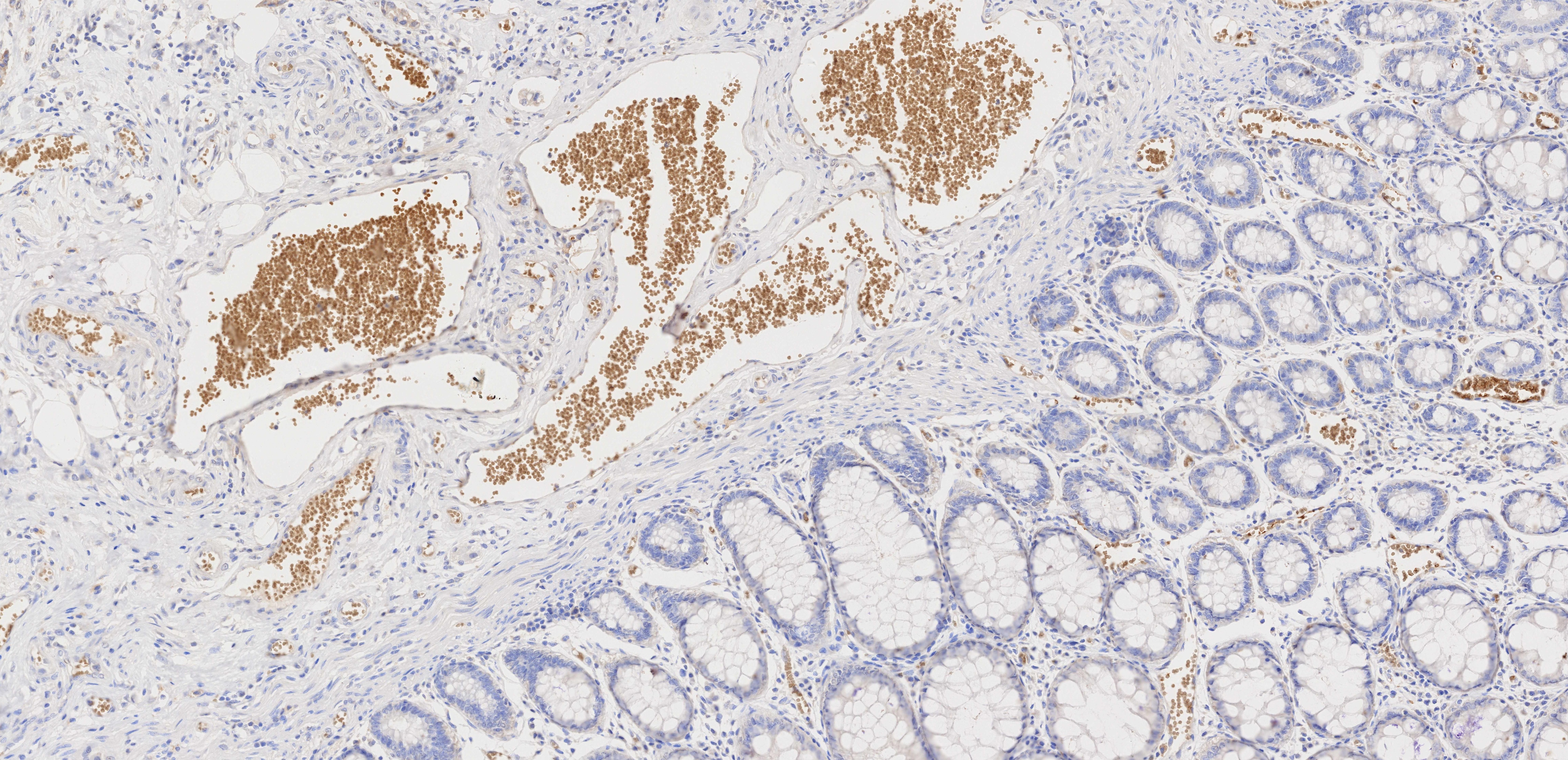
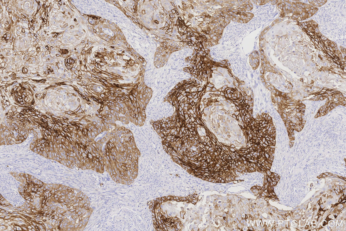
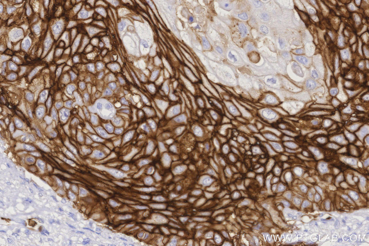
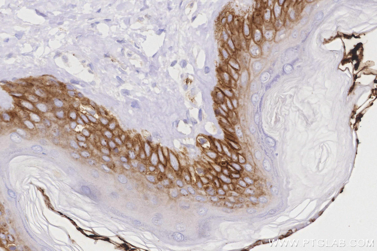
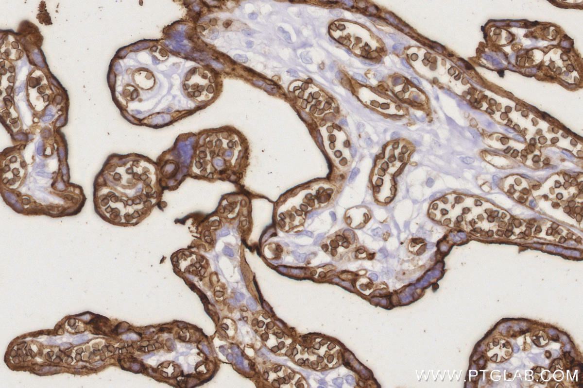
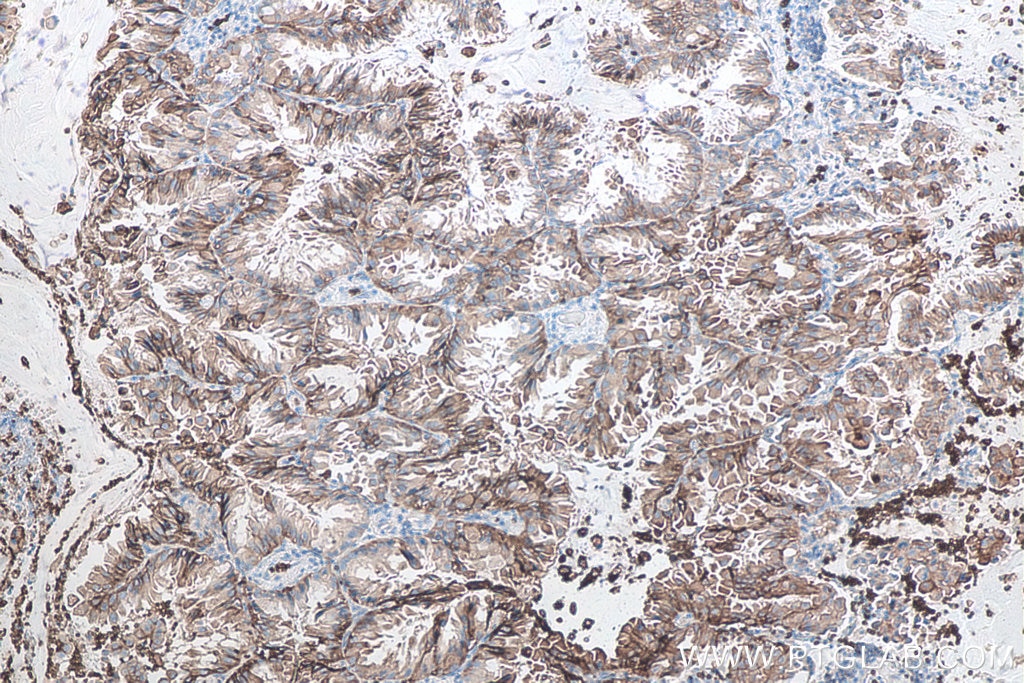
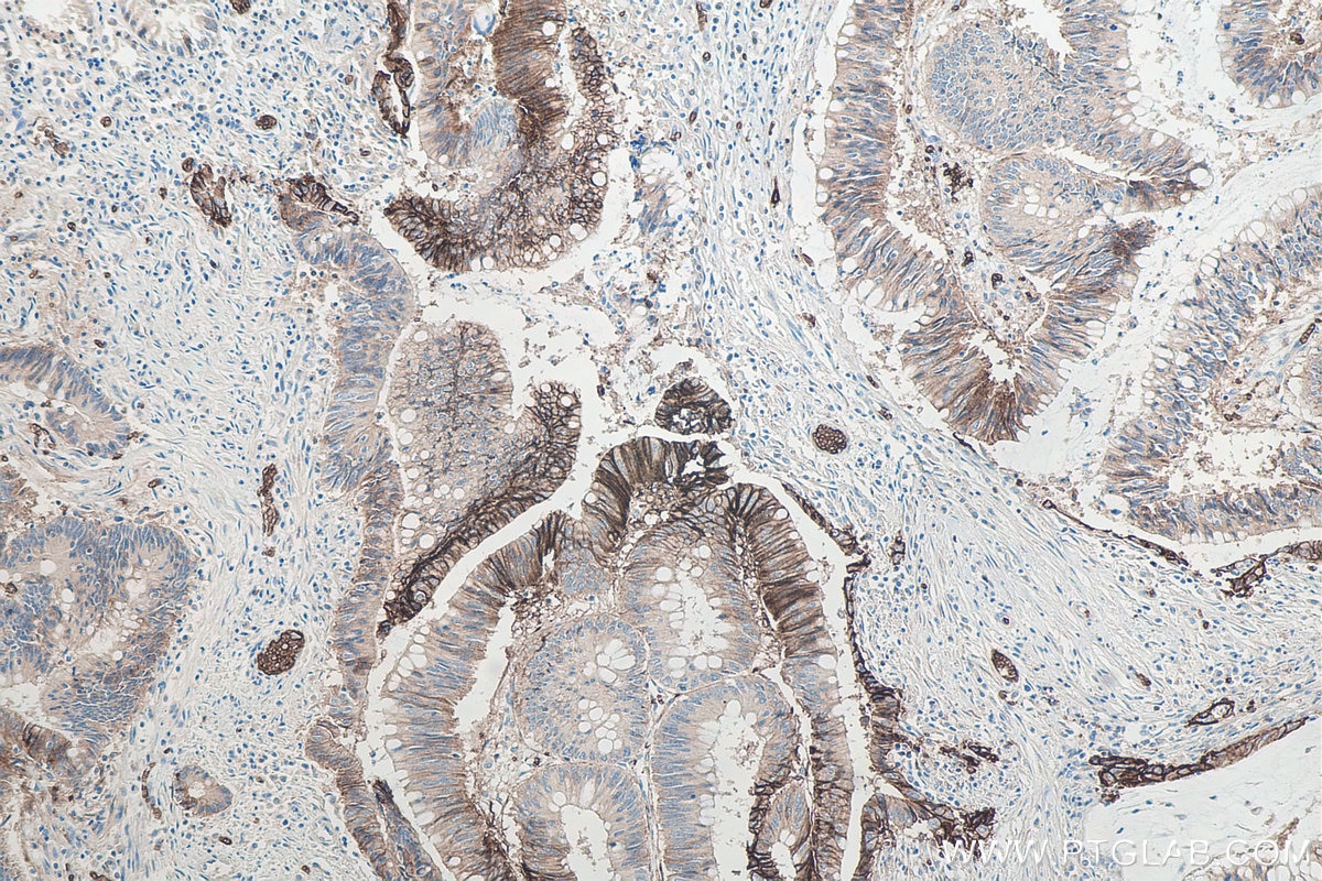
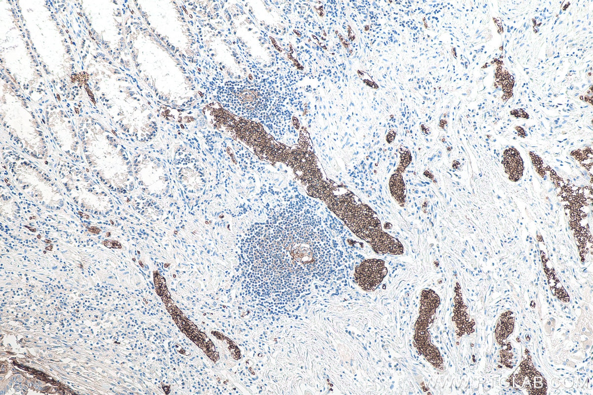
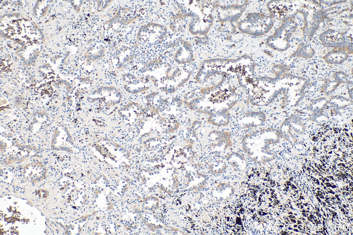
| Positive IHC detected in | human lung cancer tissue, human skin tissue, human rectal cancer tissue, human cervical cancer tissue, human lung cancer tissue, human colon cancer tissue, human placenta tissue Note: suggested antigen retrieval with TE buffer pH 9.0; (*) Alternatively, antigen retrieval may be performed with citrate buffer pH 6.0 |
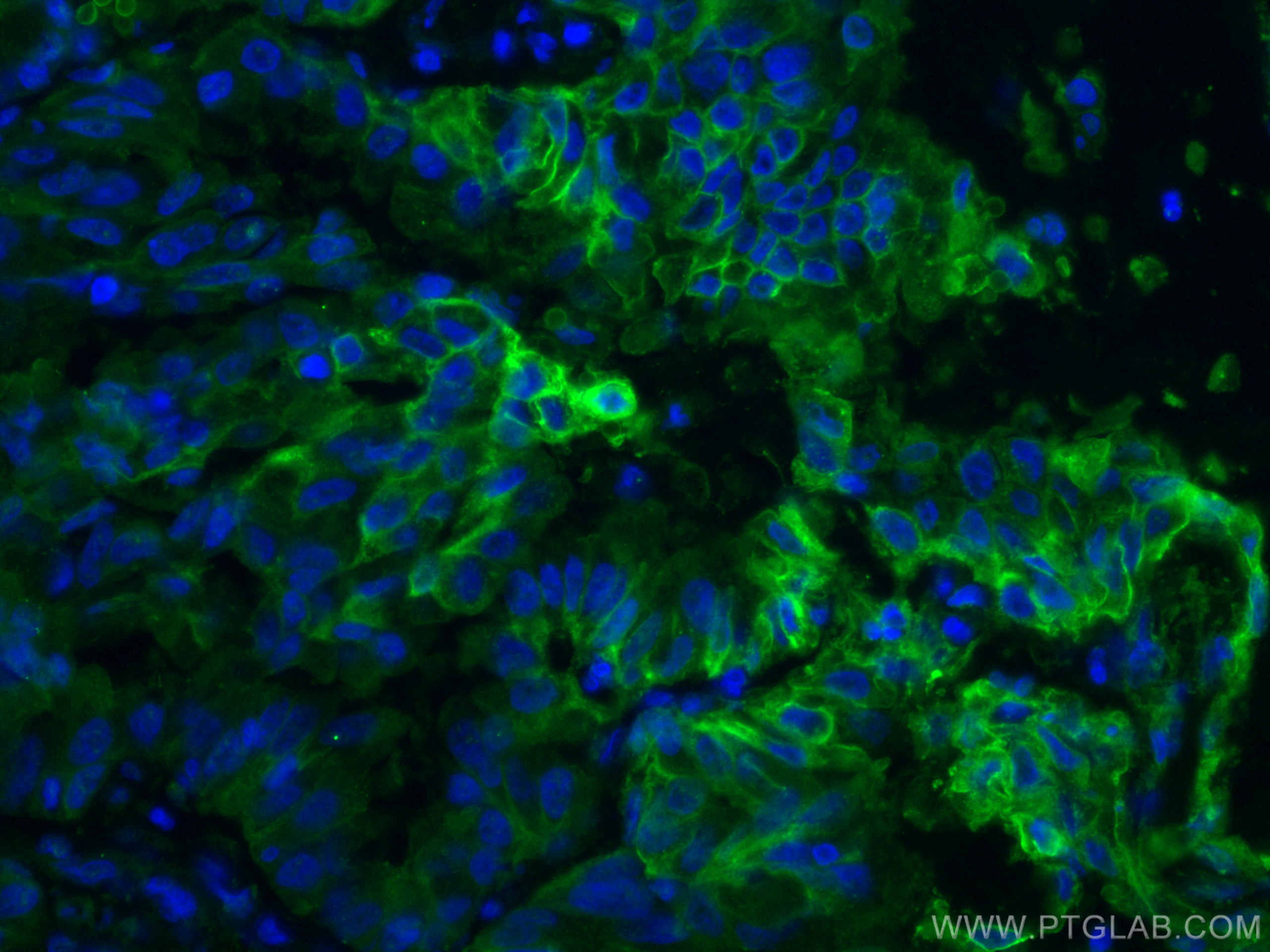
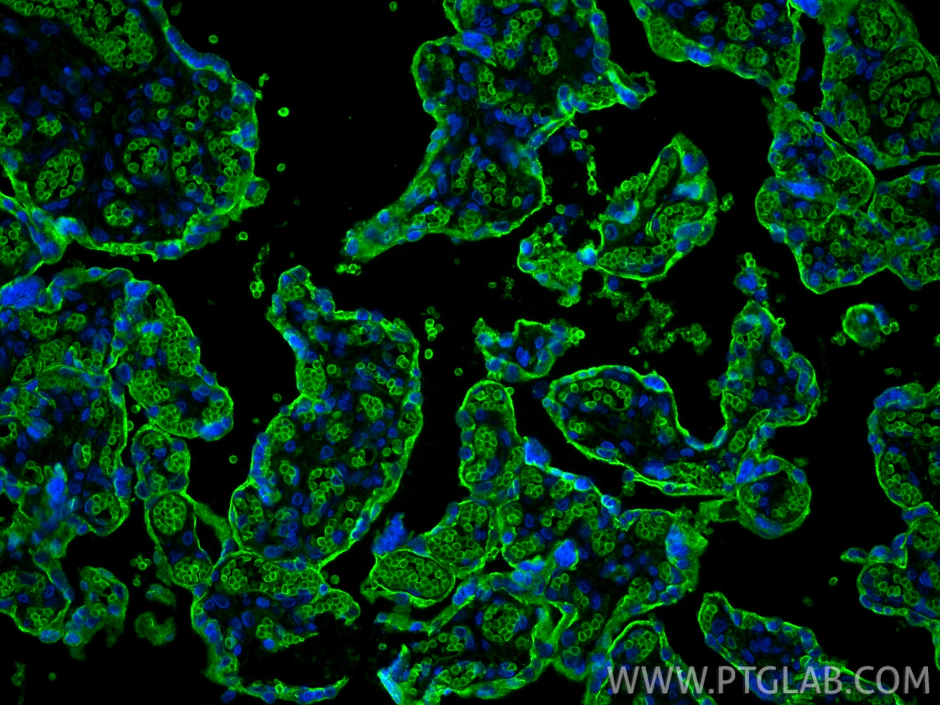
| Positive IF-P detected in | human lung cancer tissue, human placenta tissue |
| Positive FC (Intra) detected in | Jurkat cells |
Recommended dilution
| Application | Dilution |
|---|---|
| Western Blot (WB) | WB : 1:500-1:3000 |
| Immunohistochemistry (IHC) | IHC : 1:1000-1:4000 |
| Immunofluorescence (IF)-P | IF-P : 1:200-1:800 |
| Flow Cytometry (FC) (INTRA) | FC (INTRA) : 0.40 ug per 10^6 cells in a 100 µl suspension |
| It is recommended that this reagent should be titrated in each testing system to obtain optimal results. | |
| Sample-dependent, Check data in validation data gallery. | |
Product Information
66290-1-Ig targets GLUT1 in WB, IHC, IF-P, FC (Intra), ELISA applications and shows reactivity with human samples.
| Tested Reactivity | human |
| Cited Reactivity | human, rat |
| Host / Isotype | Mouse / IgG1 |
| Class | Monoclonal |
| Type | Antibody |
| Immunogen |
CatNo: Ag17108 Product name: Recombinant human SLC2A1,GLUT1 protein Source: e coli.-derived, PET28a Tag: 6*His Domain: 216-280 aa of BC121804 Sequence: INRNEENRAKSVLKKLRGTADVTHDLQEMKEESRQMMREKKVTILELFRSPAYRQPILIAVVLQL Predict reactive species |
| Full Name | solute carrier family 2 (facilitated glucose transporter), member 1 |
| Calculated Molecular Weight | 492 aa, 54 kDa |
| Observed Molecular Weight | 45-55 kDa |
| GenBank Accession Number | BC121804 |
| Gene Symbol | GLUT1 |
| Gene ID (NCBI) | 6513 |
| RRID | AB_2881673 |
| Conjugate | Unconjugated |
| Form | Liquid |
| Purification Method | Protein G purification |
| UNIPROT ID | P11166 |
| Storage Buffer | PBS with 0.02% sodium azide and 50% glycerol, pH 7.3. |
| Storage Conditions | Store at -20°C. Stable for one year after shipment. Aliquoting is unnecessary for -20oC storage. 20ul sizes contain 0.1% BSA. |
Background Information
Glucose transporter 1 (GLUT1), also known as solute carrier family 2, facilitated glucose transporter member 1 (SLC2A1), is a uniporter protein responsible for the transport of glucose in many cell types and across the blood-brain barrier.
What is the molecular weight of GLUT1? Is GLUT1 post-translationally modified?
There are two forms of GLUT1 transporter that differ in their molecular weight. The 45-kDa form is found in glial cells, while the 55-kDa form is present in the endothelial cells regulating glucose transport over the blood-brain and blood-tissue barriers (PMID: 9630522). N-glycosylation of asparagine at position 42 is the only known post-translation modification of GLUT1 (PMID: 3839598).
What is the subcellular localization of GLUT1?
Glucose transporters, including GLUT1, are multiple-pass integral membrane proteins. GLUT1 is present at the plasma membrane but is also a subject of recycling between plasma membrane and endosomes.
What molecules can be transported by GLUT1?
The main substrate of GLUT1 transport is glucose, but it can also transport galactose, mannose, glucosamine, and reduced ascorbate.
What is the tissue expression pattern of GLUT1?
GLUT1 is expressed by many cell types but the highest levels are observed in erythrocytes and in the central nervous system (astrocytes). GLUT1 is responsible for glucose transfer across the blood-brain and blood-tissue barriers, including placental transport.
Protocols
| Product Specific Protocols | |
|---|---|
| FC protocol for GLUT1 antibody 66290-1-Ig | Download protocol |
| IF protocol for GLUT1 antibody 66290-1-Ig | Download protocol |
| IHC protocol for GLUT1 antibody 66290-1-Ig | Download protocol |
| WB protocol for GLUT1 antibody 66290-1-Ig | Download protocol |
| Standard Protocols | |
|---|---|
| Click here to view our Standard Protocols |
Publications
What published studies show
The anti-GLUT1 (SLC2A1) antibody (Cat# 66290-1-Ig) has 106 citations published in journals including Nature Nanotechnology, Nature Communications, Cell Metabolism, Molecular Cell, ACS Nano, Brain, Molecular Cancer, and Advanced Science. Research fields encompass cancer glycolysis/Warburg effect, neurodegeneration (ALS, Alzheimer's, Parkinson's), stroke, immune metabolic reprogramming, diabetes, reproductive biology, inflammation, ferroptosis, and nanomedicine. Applications are predominantly Western blot, supplemented by IHC and IF.
| Species | Application | Title |
|---|---|---|
Nat Nanotechnol Nanoparticles for inducing Gaucher disease-like damage in cancer cells
| ||
Cell Metab Acetate enables metabolic fitness and cognitive performance during sleep disruption | ||
Cell Metab Di-methylation of CD147-K234 Promotes the Progression of NSCLC by Enhancing Lactate Export. | ||
Adv Mater Manganese Galvanic Cells Intervene in Tumor Metabolism to Reinforce cGAS-STING Activation for Bidirectional Synergistic Hydrogen-Immunotherapy | ||
Clin Mol Hepatol UBE2S promotes glycolysis in hepatocellular carcinoma by enhancing E3 enzyme-independent polyubiquitination of VHL |
Reviews
What customers say
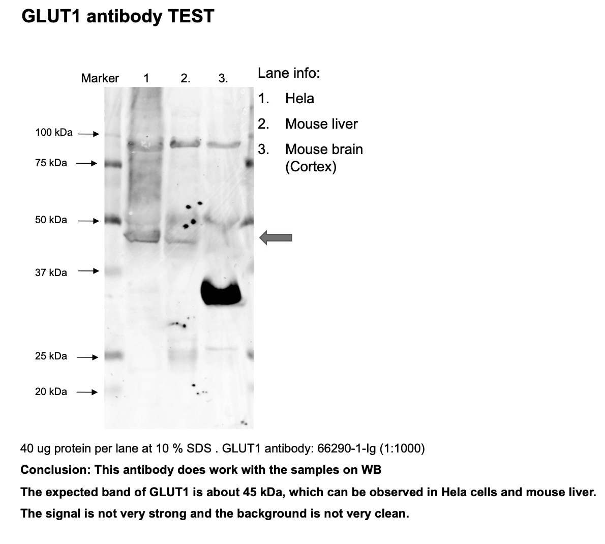
Both reviewers used this antibody for Western Blot with mixed results. One user (rated 2/5) reported weak signal and unclear background at 1:1000 dilution in HeLa cells and mouse tissues. Another user (rated 3/5) found it only worked at 1:500, well above the recommended 1:1500–1:3000 range, to visualize bands in fallopian tube epithelial cells. Overall, users suggest the antibody requires optimization and may not perform as expected under standard protocol conditions.
The reviews below have been submitted by verified Proteintech customers who received an incentive for providing their feedback.
Hua (Verified Customer) (09-06-2023) | Works ok in WB. Signal is weak and background is not very clean.
 |
Iru (Verified Customer) (04-23-2019) | This antibody works ok-ish, even though the protocol says use 1:1500 to 1:3000, I have to use 1:500 to see the bands.
|