at dilution of 1:10000 incubated at room temperature for 1.5 hours.")
 at dilution of 1:10000 incubated at room temperature for 1.5 hours.")
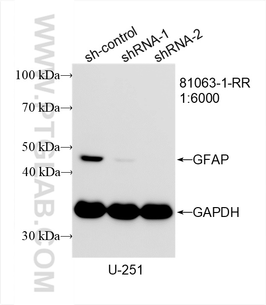
 with sh-Control and sh-GFAP transfected U-251 cells.")
 at dilution of 1:9900 (under 10x lens). Heat mediated antigen retrieval with Tris-EDTA buffer (pH 9.0).")
 at dilution of 1:9900 (under 40x lens). Heat mediated antigen retrieval with Tris-EDTA buffer (pH 9.0).")
 fixed rat brain tissue using GFAP antibody (81063-1-RR, Clone: 4C6 ) at dilution of 1:200 and CoraLite®488-Conjugated AffiniPure Goat Anti-Rabbit IgG(H+L).")
 fixed mouse brain tissue using GFAP antibody (81063-1-RR, Clone: 4C6 ) at dilution of 1:200 and CoraLite®488-Conjugated AffiniPure Goat Anti-Rabbit IgG(H+L).")
 fixed mouse brain tissue using GFAP antibody (81063-1-RR, Clone: 4C6 ) at dilution of 1:200 and CoraLite®488-Conjugated AffiniPure Goat Anti-Rabbit IgG(H+L).")
 fixed frozen OCT-embedded mouse brain tissue using GFAP antibody (81063-1-RR, Clone: 4C6 ) at dilution of 1:200 and CoraLite®488-Conjugated Goat Anti-Rabbit IgG(H+L) (SA00013-2).")
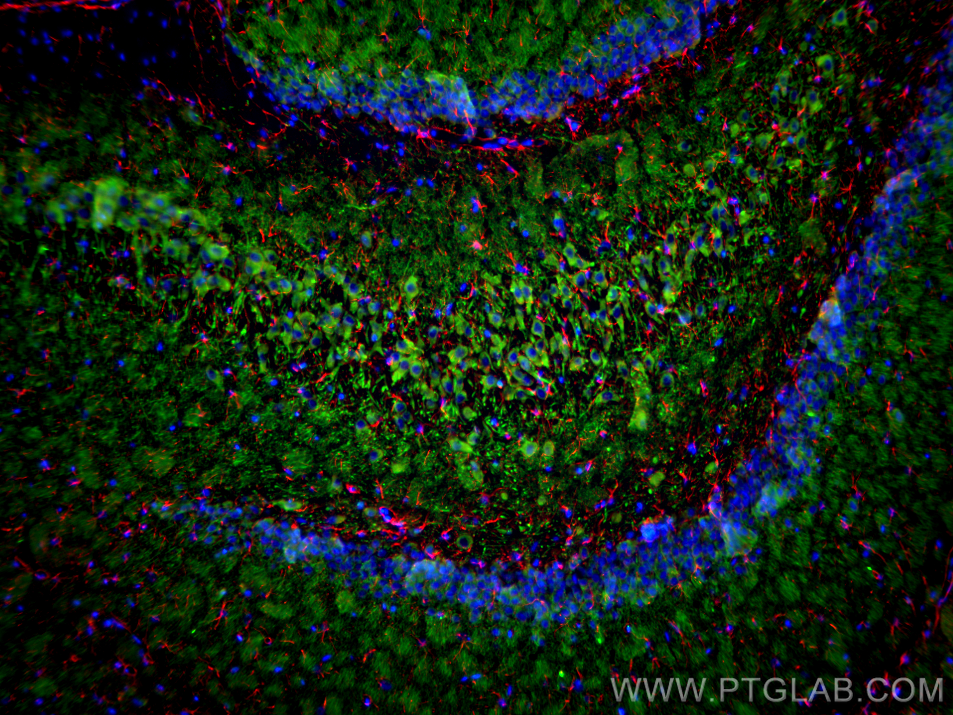
 fixed frozen OCT-embedded rat brain tissue using GFAP antibody (81063-1-RR, Clone: 4C6 ) at dilution of 1:200 and CoraLite®594-Conjugated Goat Anti-Rabbit IgG(H+L) (SA00013-4), MAP2 antibody (67015-1-Ig, Clone: 1C3E6, green).")
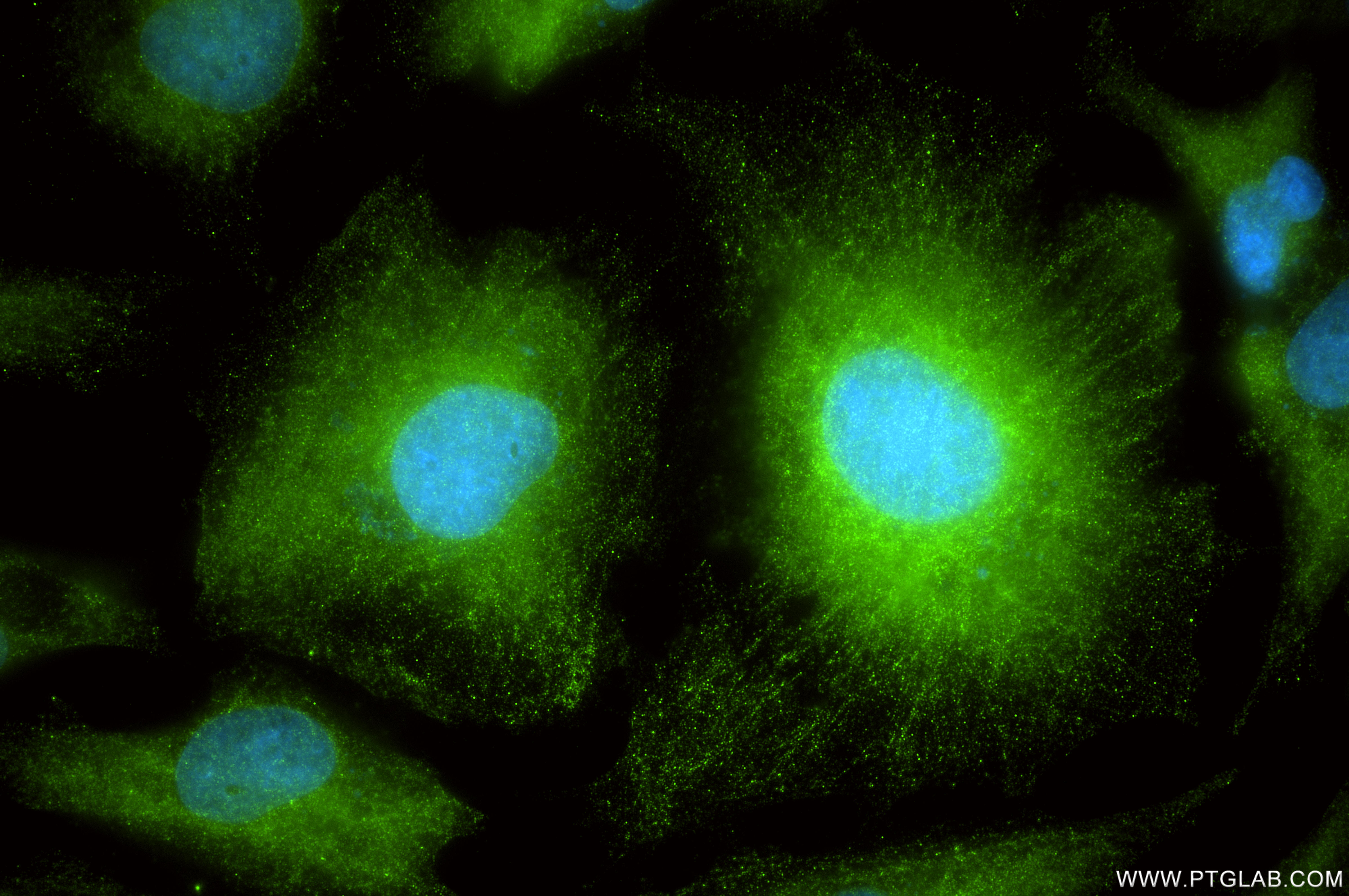
 fixed U-251 cells using GFAP antibody (81063-1-RR, Clone: 4C6 ) at dilution of 1:400 and CoraLite®488-Conjugated Goat Anti-Rabbit IgG(H+L) (SA00013-2).")
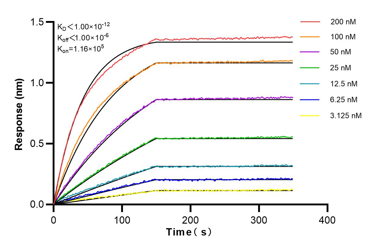
 kinetic assays of 81077-1-RR against Human GFAP were performed. The affinity constant is below 1 pM.")
Tested Applications
| Positive WB detected in | U-251 cells, rat brain tissue, mouse brain tissue, pig brain tissue |
| Positive IHC detected in | rat brain tissue Note: suggested antigen retrieval with TE buffer pH 9.0; (*) Alternatively, antigen retrieval may be performed with citrate buffer pH 6.0 |
| Positive IF-P detected in | rat brain tissue, mouse brain tissue |
| Positive IF-Fro detected in | mouse brain tissue, rat brain tissue |
| Positive IF/ICC detected in | U-251 cells |
Recommended dilution
| Application | Dilution |
|---|---|
| Western Blot (WB) | WB : 1:5000-1:50000 |
| Immunohistochemistry (IHC) | IHC : 1:4950-1:19800 |
| Immunofluorescence (IF)-P | IF-P : 1:50-1:500 |
| Immunofluorescence (IF)-FRO | IF-FRO : 1:50-1:500 |
| Immunofluorescence (IF)/ICC | IF/ICC : 1:200-1:800 |
| It is recommended that this reagent should be titrated in each testing system to obtain optimal results. | |
| Sample-dependent, Check data in validation data gallery. | |
Product Information
81063-1-RR targets GFAP in WB, IHC, IF/ICC, IF-P, IF-Fro, ELISA applications and shows reactivity with human, mouse, rat, pig samples.
| Tested Reactivity | human, mouse, rat, pig |
| Host / Isotype | Rabbit / IgG |
| Class | Recombinant |
| Type | Antibody |
| Immunogen |
CatNo: Ag10423 Product name: Recombinant human GFAP protein Source: e coli.-derived, PGEX-4T Tag: GST Domain: 83-432 aa of BC013596 Sequence: YIEKVRFLEQQNKALAAELNQLRAKEPTKLADVYQAELRELRLRLDQLTANSARLEVERDNLAQDLATVRQKLQDETNLRLEAENNLAAYRQEADEATLARLDLERKIESLEEEIRFLRKIHEEEVRELQEQLARQQVHVELDVAKPDLTAALKEIRTQYEAMASSNMHEAEEWYRSKFADLTDAAARNAELLRQAKHEANDYRRQLQSLTCDLESLRGTNESLERQMREQEERHVREAASYQEALARLEEEGQSLKDEMARHLQEYQDLLNVKLALDIEIATYRKLLEGEENRITIPVQTFSNLQIRETSLDTKSVSEGHLKRNIVVKTVEMRDGEVIKESKQEHKDVM Predict reactive species |
| Full Name | glial fibrillary acidic protein |
| Calculated Molecular Weight | 432 aa, 50 kDa |
| Observed Molecular Weight | 45-50 kDa |
| GenBank Accession Number | BC013596 |
| Gene Symbol | GFAP |
| Gene ID (NCBI) | 2670 |
| RRID | AB_2923699 |
| Conjugate | Unconjugated |
| Form | Liquid |
| Purification Method | Protein A purification |
| UNIPROT ID | P14136 |
| Storage Buffer | PBS with 0.02% sodium azide and 50% glycerol, pH 7.3. |
| Storage Conditions | Store at -20°C. Stable for one year after shipment. Aliquoting is unnecessary for -20oC storage. 20ul sizes contain 0.1% BSA. |
Background Information
GFAP (Glial fibrillary acidic protein ), an intermediate-filament (IF) protein , is specifically expressed in cells of astroglial lineage and is widely used to mark astroglia in the brain. It is also used as a marker for intracranial and intraspinal tumors arising from astrocytes.
Protocols
| Product Specific Protocols | |
|---|---|
| IF protocol for GFAP antibody 81063-1-RR | Download protocol |
| IHC protocol for GFAP antibody 81063-1-RR | Download protocol |
| WB protocol for GFAP antibody 81063-1-RR | Download protocol |
| Standard Protocols | |
|---|---|
| Click here to view our Standard Protocols |
Reviews
The reviews below have been submitted by verified Proteintech customers who received an incentive for providing their feedback.
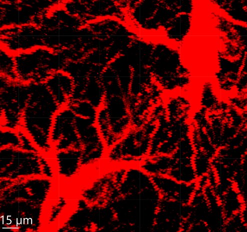
FH Kateryna (Verified Customer) (01-23-2025) | We use the GFAP antibody to visualize astrocytes via immunostaining in mouse brain tissue. The mouse brain tissue is fixed, brain sections are 40 μm thick, and blocked with 3% normal goat serum. The incubation with the primary antibody (GFAP) is performed at 4°C overnight, followed by the corresponding Alexa 633-labeled anti-rabbit fluorescent secondary antibody.
 |