WB analysis of mouse skeletal muscle using 66012-1-Ig
mouse skeletal muscle tissue were subjected to SDS PAGE followed by western blot with 66012-1-Ig (GEFT antibody) at dilution of 1:1000 incubated at room temperature for 1.5 hours.
mouse skeletal muscle tissue were subjected to SDS PAGE followed by western blot with 66012-1-Ig (GEFT antibody) at dilution of 1:1000 incubated at room temperature for 1.5 hours.
WB analysis of rat skeletal muscle using 66012-1-Ig
rat skeletal muscle tissue were subjected to SDS PAGE followed by western blot with 66012-1-Ig (GEFT antibody) at dilution of 1:1000 incubated at room temperature for 1.5 hours.
rat skeletal muscle tissue were subjected to SDS PAGE followed by western blot with 66012-1-Ig (GEFT antibody) at dilution of 1:1000 incubated at room temperature for 1.5 hours.
WB analysis of human brain using 66012-1-Ig
human brain tissue were subjected to SDS PAGE followed by western blot with 66012-1-Ig (GEFT antibody) at dilution of 1:1000 incubated at room temperature for 1.5 hours.
human brain tissue were subjected to SDS PAGE followed by western blot with 66012-1-Ig (GEFT antibody) at dilution of 1:1000 incubated at room temperature for 1.5 hours.
WB analysis of mouse brain using 66012-1-Ig
mouse brain tissue were subjected to SDS PAGE followed by western blot with 66012-1-Ig (GEFT antibody) at dilution of 1:1000 incubated at room temperature for 1.5 hours.
mouse brain tissue were subjected to SDS PAGE followed by western blot with 66012-1-Ig (GEFT antibody) at dilution of 1:1000 incubated at room temperature for 1.5 hours.
WB analysis of rat brain using 66012-1-Ig
rat brain tissue were subjected to SDS PAGE followed by western blot with 66012-1-Ig (GEFT antibody) at dilution of 1:1000 incubated at room temperature for 1.5 hours.
rat brain tissue were subjected to SDS PAGE followed by western blot with 66012-1-Ig (GEFT antibody) at dilution of 1:1000 incubated at room temperature for 1.5 hours.
WB analysis of human brain using 66012-1-Ig
human brain tissue were subjected to SDS PAGE followed by western blot with 66012-1-Ig (GEFT antibody) at dilution of 1:1000 incubated at room temperature for 1.5 hours.
human brain tissue were subjected to SDS PAGE followed by western blot with 66012-1-Ig (GEFT antibody) at dilution of 1:1000 incubated at room temperature for 1.5 hours.
IHC staining of human heart using 66012-1-Ig
Immunohistochemical analysis of paraffin-embedded human heart using 66012-1-Ig(GEFT antibody) at dilution of 1:50 (under 10x lens).
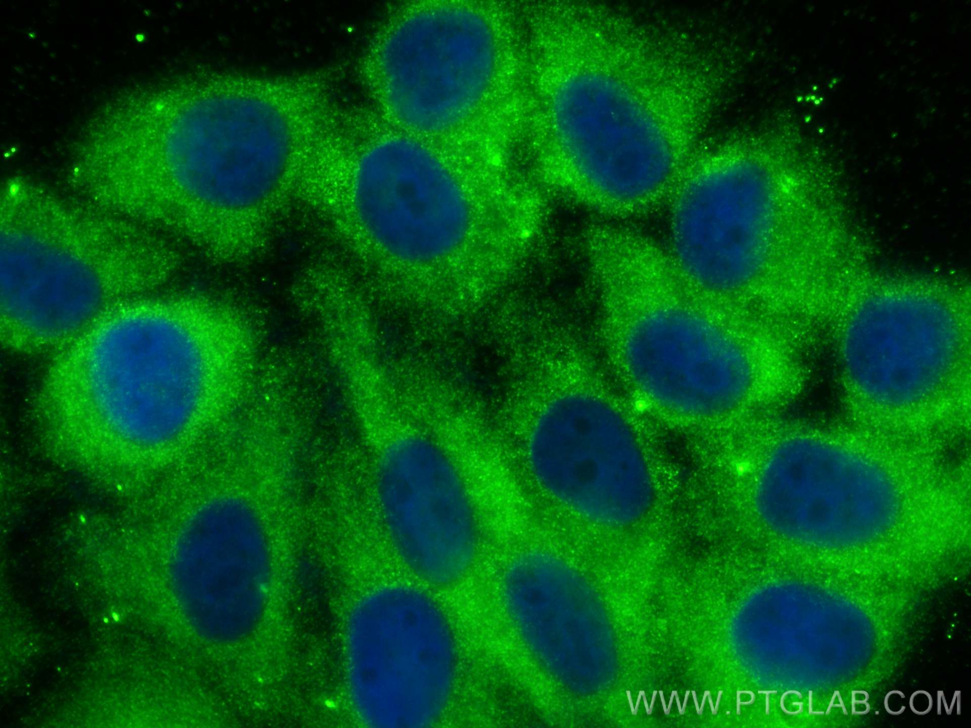
Immunofluorescent analysis of (-20°C Ethanol) fixed HepG2 cells using GEFT antibody (66012-1-Ig, Clone: 5A11F8 ) at dilution of 1:800 and CoraLite®488-Conjugated AffiniPure Goat Anti-Mouse IgG(H+L) (SA00013-1).
FC experiment of HEK-293 using 66012-1-Ig
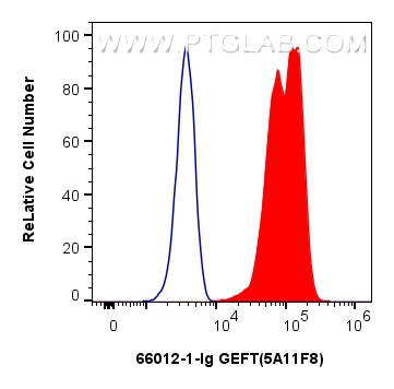
1x10^6 HEK-293 cells were intracellularly stained with 0.8 ug GEFT Monoclonal antibody (66012-1-Ig, Clone:5A11F8) and CoraLite®488-Conjugated AffiniPure Goat Anti-Mouse IgG(H+L) (SA00013-1)(red), or 0.8 ug Mouse IgG2a isotype control Mouse McAb (66360-2-Ig, Clone: 11A1B2) (blue). Cells were fixed with 4% PFA and permeabilized with Flow Cytometry Perm Buffer (PF00011-C).
1x10^6 HEK-293 cells were intracellularly stained with 0.8 ug GEFT Monoclonal antibody (66012-1-Ig, Clone:5A11F8) and CoraLite®488-Conjugated AffiniPure Goat Anti-Mouse IgG(H+L) (SA00013-1)(red), or 0.8 ug Mouse IgG2a isotype control Mouse McAb (66360-2-Ig, Clone: 11A1B2) (blue). Cells were fixed with 4% PFA and permeabilized with Flow Cytometry Perm Buffer (PF00011-C).
The Proteintech guarantee covers Proteintech antibodies in any species and any application, including those not listed on the datasheet. If the antibody doesn’t perform, you can receive a hassle-free refund or credit note.
mouse skeletal muscle tissue, human brain tissue, mouse brain tissue, rat brain tissue, rat skeletal muscle tissue
Positive IHC detected in
human heart tissue, human brain tissue Note: suggested antigen retrieval with TE buffer pH 9.0; (*) Alternatively, antigen retrieval may be performed with citrate buffer pH 6.0
Positive IF/ICC detected in
HepG2 cells
Positive FC (Intra) detected in
HEK-293 cells
Recommended dilution
Application
Dilution
Western Blot (WB)
WB : 1:500-1:2000
Immunohistochemistry (IHC)
IHC : 1:20-1:200
Immunofluorescence (IF)/ICC
IF/ICC : 1:400-1:1600
Flow Cytometry (FC) (INTRA)
FC (INTRA) : 0.80 ug per 10^6 cells in a 100 µl suspension
It is recommended that this reagent should be titrated in each testing system to obtain optimal results.
Sample-dependent, Check data in validation data gallery.
PBS with 0.02% sodium azide and 50% glycerol , pH 7.3
Storage Conditions
Store at -20°C. Stable for one year after shipment. Aliquoting is unnecessary for -20oC storage. 20ul sizes contain 0.1% BSA.
Background Information
GEFT, also named as p63RhoGEF, may play a role in actin cytoskeleton reorganization in different tissues since its activation induces formation of actin stress fibers. It works as a guanine nucleotide exchange factor for Rho family of small GTPases. GETF links specifically G alpha q/11-coupled receptors to RHOA activation. GEFT may be an important regulator of processes involved in axon and dendrite formation. In neurons seems to be an exchange factor primarily for RAC1. It is involved in skeletal myogenesis. GEFT is expressed as three isoforms (64, 53 and 68kDa).
WB analysis of mouse skeletal muscle using 66012-1-Ig
mouse skeletal muscle tissue were subjected to SDS PAGE followed by western blot with 66012-1-Ig (GEFT antibody) at dilution of 1:1000 incubated at room temperature for 1.5 hours.
WB analysis of rat skeletal muscle using 66012-1-Ig
rat skeletal muscle tissue were subjected to SDS PAGE followed by western blot with 66012-1-Ig (GEFT antibody) at dilution of 1:1000 incubated at room temperature for 1.5 hours.
WB analysis of human brain using 66012-1-Ig
human brain tissue were subjected to SDS PAGE followed by western blot with 66012-1-Ig (GEFT antibody) at dilution of 1:1000 incubated at room temperature for 1.5 hours.
WB analysis of mouse brain using 66012-1-Ig
mouse brain tissue were subjected to SDS PAGE followed by western blot with 66012-1-Ig (GEFT antibody) at dilution of 1:1000 incubated at room temperature for 1.5 hours.
WB analysis of rat brain using 66012-1-Ig
rat brain tissue were subjected to SDS PAGE followed by western blot with 66012-1-Ig (GEFT antibody) at dilution of 1:1000 incubated at room temperature for 1.5 hours.
WB analysis of human brain using 66012-1-Ig
human brain tissue were subjected to SDS PAGE followed by western blot with 66012-1-Ig (GEFT antibody) at dilution of 1:1000 incubated at room temperature for 1.5 hours.
IHC Figures
IHC staining of human heart using 66012-1-Ig
Immunohistochemical analysis of paraffin-embedded human heart using 66012-1-Ig(GEFT antibody) at dilution of 1:50 (under 10x lens).
IHC staining of human heart using 66012-1-Ig
Immunohistochemical analysis of paraffin-embedded human heart using 66012-1-Ig(GEFT antibody) at dilution of 1:50 (under 40x lens).
IHC staining of human brain using 66012-1-Ig
Immunohistochemical analysis of paraffin-embedded human brain using 66012-1-Ig(GEFT antibody) at dilution of 1:50 (under 10x lens).
IHC staining of human brain using 66012-1-Ig
Immunohistochemical analysis of paraffin-embedded human brain using 66012-1-Ig(GEFT antibody) at dilution of 1:50 (under 40x lens).
IF/ICC Figures
IF Staining of HepG2 using 66012-1-Ig
Immunofluorescent analysis of (-20°C Ethanol) fixed HepG2 cells using GEFT antibody (66012-1-Ig, Clone: 5A11F8 ) at dilution of 1:800 and CoraLite®488-Conjugated AffiniPure Goat Anti-Mouse IgG(H+L) (SA00013-1).
FC (INTRA) Figures
FC experiment of HEK-293 using 66012-1-Ig
1x10^6 HEK-293 cells were intracellularly stained with 0.8 ug GEFT Monoclonal antibody (66012-1-Ig, Clone:5A11F8) and CoraLite®488-Conjugated AffiniPure Goat Anti-Mouse IgG(H+L) (SA00013-1)(red), or 0.8 ug Mouse IgG2a isotype control Mouse McAb (66360-2-Ig, Clone: 11A1B2) (blue). Cells were fixed with 4% PFA and permeabilized with Flow Cytometry Perm Buffer (PF00011-C).
The species listed in Tested Reactivity are in-house verified and applicable species. For unlisted species, please refer to the homology analysis of the immunogen sequence and related species. For rabbit polyclonal antibodies, homology >70% is recommended. For mouse monoclonal antibodies and rabbit recombinant antibodies, homology >90% is recommended. Generally, the higher the homology, the greater the applicability. However, there will be certain differences in protein expression in different species, tissues or cells. Therefore, the homology analysis results are for reference only and do not serve as a guarantee.
At Proteintech, we pride ourselves on our antibody quality, customer service and transparency. As such, we are comparing our antibodies with other vendors, enabling easy identification and comparisons of key data to help you choose the suitable antibody for your needs.
We have selected the top cited antibodies from these vendors for you to compare.
Proteintech
GEFT Monoclonal antibody
Catalog Number
66012-1-Ig
Citations
1
Dilutions
WB : 1:500-1:2000 IHC : 1:20-1:200 IF/ICC : 1:400-1:1600 FC (INTRA) : 0.80 ug per 10^6 cells in a 100 µl suspension
Applications
WB, IHC, IF/ICC, FC (Intra), ELISA
Reactivity
human, mouse, rat, pig, rabbit
Product Guarantee
Covers any species including not listed on datasheet
Covers any applications including not listed on datasheet
 at dilution of 1:1000 incubated at room temperature for 1.5 hours.")
 at dilution of 1:1000 incubated at room temperature for 1.5 hours.")
 at dilution of 1:1000 incubated at room temperature for 1.5 hours.")
 at dilution of 1:1000 incubated at room temperature for 1.5 hours.")
 at dilution of 1:1000 incubated at room temperature for 1.5 hours.")
 at dilution of 1:1000 incubated at room temperature for 1.5 hours.")
 at dilution of 1:50 (under 10x lens).")
 at dilution of 1:50 (under 40x lens).")
 at dilution of 1:50 (under 10x lens).")
 at dilution of 1:50 (under 40x lens).")
 fixed HepG2 cells using GEFT antibody (66012-1-Ig, Clone: 5A11F8 ) at dilution of 1:800 and CoraLite®488-Conjugated AffiniPure Goat Anti-Mouse IgG(H+L) (SA00013-1).")
 and CoraLite®488-Conjugated AffiniPure Goat Anti-Mouse IgG(H+L) (SA00013-1)(red), or 0.8 ug Mouse IgG2a isotype control Mouse McAb (66360-2-Ig, Clone: 11A1B2) (blue). Cells were fixed with 4% PFA and permeabilized with Flow Cytometry Perm Buffer (PF00011-C).")