Tested Applications
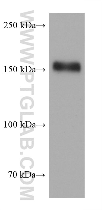
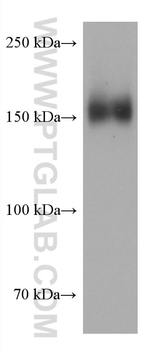
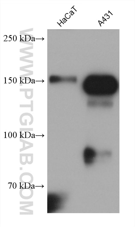
| Positive WB detected in | SCaBER cells, HaCaT cells, MDA-MB-468 cells, A431 cells |
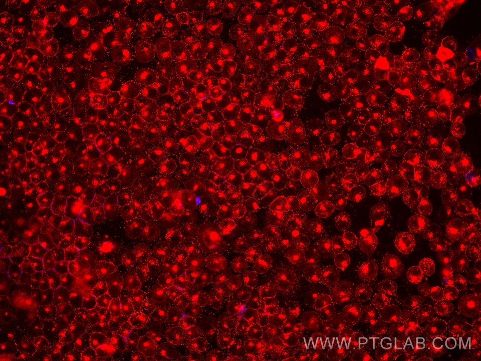
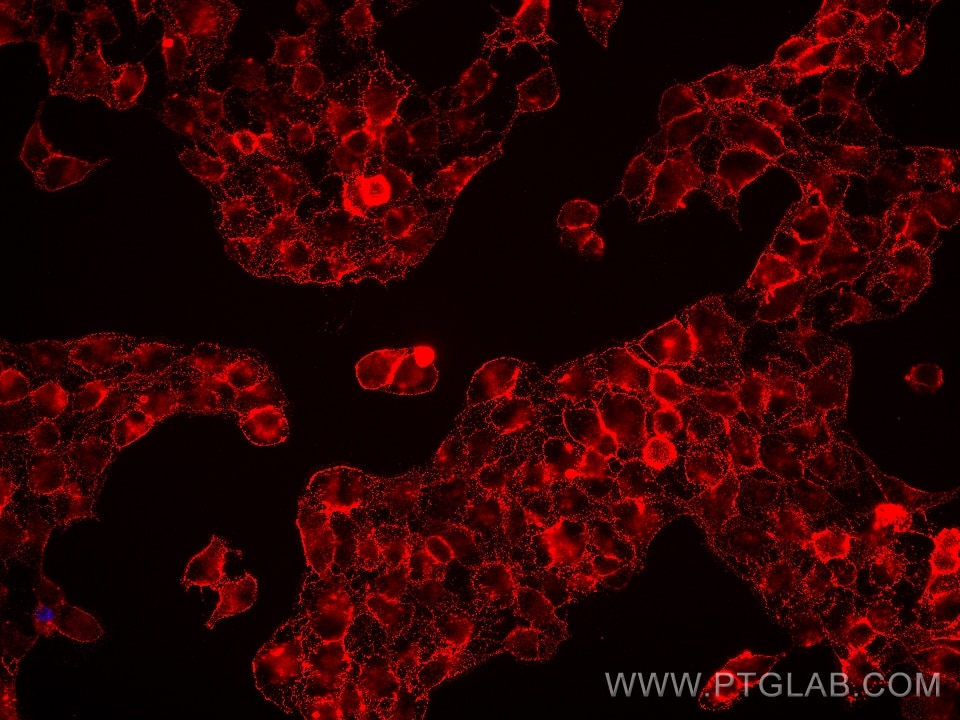
| Positive IF/ICC detected in | HaCaT cells, A431 cells |
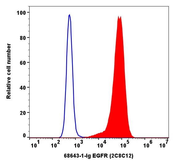
| Positive FC detected in | A431 cells |
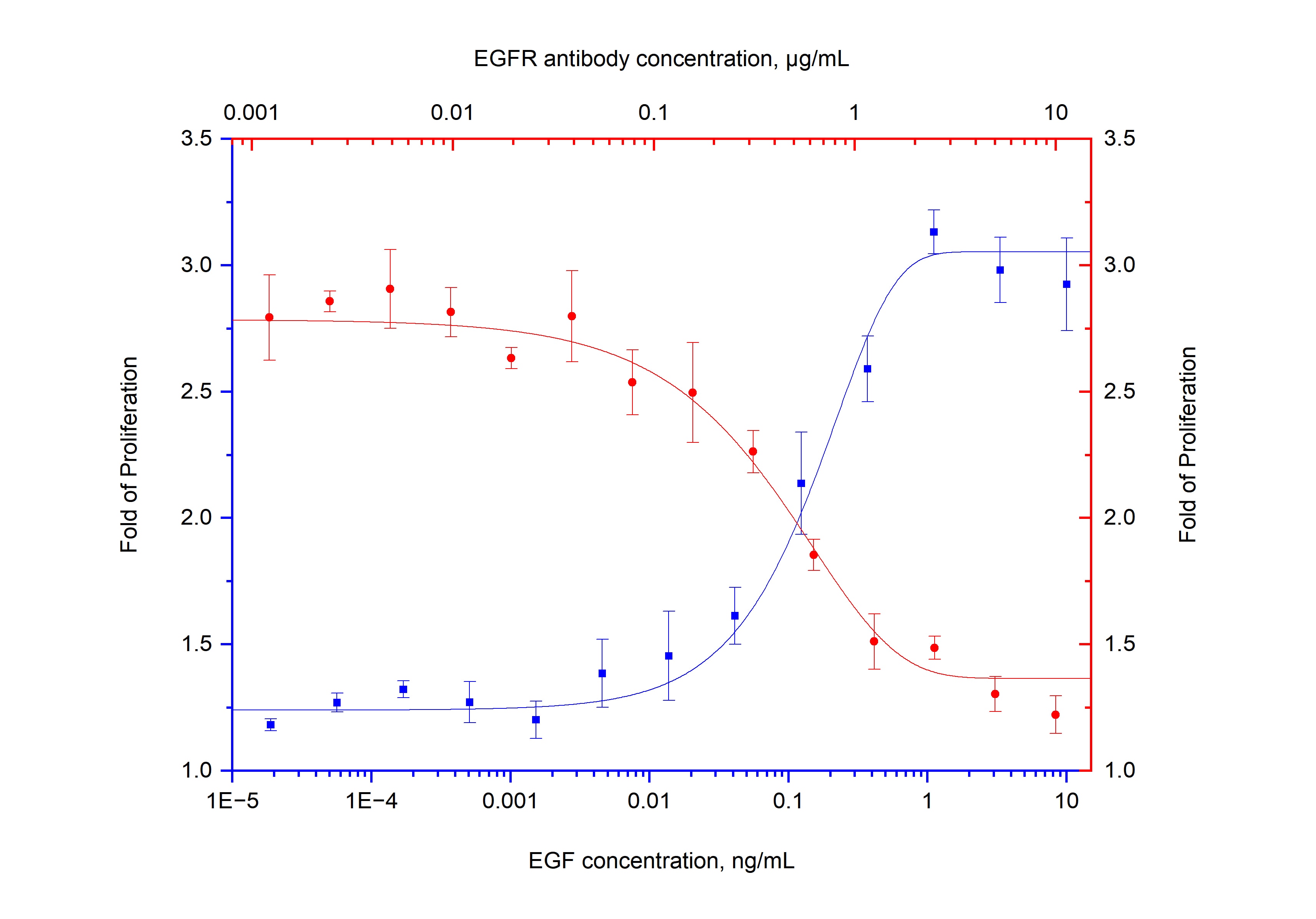
| Positive Blocking detected in | HeLa cells |
Recommended dilution
| Application | Dilution |
|---|---|
| Western Blot (WB) | WB : 1:5000-1:50000 |
| Immunofluorescence (IF)/ICC | IF/ICC : 1:500-1:2000 |
| Flow Cytometry (FC) | FC : 0.20 ug per 10^6 cells in a 100 µl suspension |
| BLOCKING | BLOCKING : 1:10-1:100 |
| It is recommended that this reagent should be titrated in each testing system to obtain optimal results. | |
| Sample-dependent, Check data in validation data gallery. | |
Product Information
68643-1-Ig targets EGFR in WB, IF/ICC, FC, ELISA, Blocking applications and shows reactivity with human samples.
| Tested Reactivity | human |
| Host / Isotype | Mouse / IgG1 |
| Class | Monoclonal |
| Type | Antibody |
| Immunogen |
CatNo: Ag24947 Product name: Recombinant human EGFR protein Source: e coli.-derived, PET30a Tag: 6*His Domain: 291-534 aa of BC094761 Sequence: VCNGIGIGEFKDSLSINATNIKHFKNCTSISGDLHILPVAFRGDSFTHTPPLDPQELDILKTVKEITGFLLIQAWPENRTDLHAFENLEIIRGRTKQHGQFSLAVVSLNITSLGLRSLKEISDGDVIISGNKNLCYANTINWKKLFGTSGQKTKIISNRGENSCKATGQVCHALCSPEGCWGPEPRDCVSCRNVSRGRECVDKCNLLEGEPREFVENSECIQCHPECLPQAMNITCTGRGPDNC Predict reactive species |
| Full Name | epidermal growth factor receptor (erythroblastic leukemia viral (v-erb-b) oncogene homolog, avian) |
| Calculated Molecular Weight | 134kd |
| Observed Molecular Weight | 160 kDa |
| GenBank Accession Number | NM_005228.5 |
| Gene Symbol | EGFR |
| Gene ID (NCBI) | 1956 |
| RRID | AB_3670387 |
| Conjugate | Unconjugated |
| Form | Liquid |
| Purification Method | Protein G purification |
| UNIPROT ID | P00533 |
| Storage Buffer | PBS with 0.02% sodium azide and 50% glycerol, pH 7.3. |
| Storage Conditions | Store at -20°C. Stable for one year after shipment. Aliquoting is unnecessary for -20oC storage. 20ul sizes contain 0.1% BSA. |
Background Information
EGFR, also named ERBB1, is a cell-surface receptor for members of the epidermal growth factor family (EGF-family) of extracellular protein ligands. Binding of the protein to a ligand induces receptor dimerization and tyrosine autophosphorylation and leads to cell proliferation. The gene resides on chromosome 7p12, encoding a 170 kDa membrane-associated glycoprotein. Recent studies have shown EGFR plays a critical role in cancer development and progression, including cell proliferation, apoptosis, angiogenesis, and metastatic spread. Mutations in this gene are associated with lung cancer. When stained with live cells, EGFR can be transported into the cell interior, which is consistent with the literature report (PMID: 18084617).
Protocols
| Product Specific Protocols | |
|---|---|
| FC protocol for EGFR antibody 68643-1-Ig | Download protocol |
| IF protocol for EGFR antibody 68643-1-Ig | Download protocol |
| WB protocol for EGFR antibody 68643-1-Ig | Download protocol |
| Standard Protocols | |
|---|---|
| Click here to view our Standard Protocols |

