
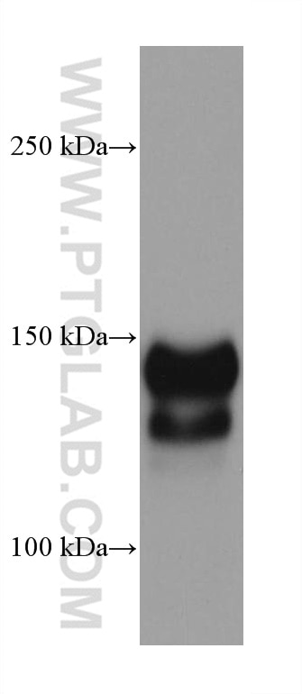
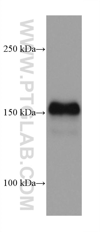
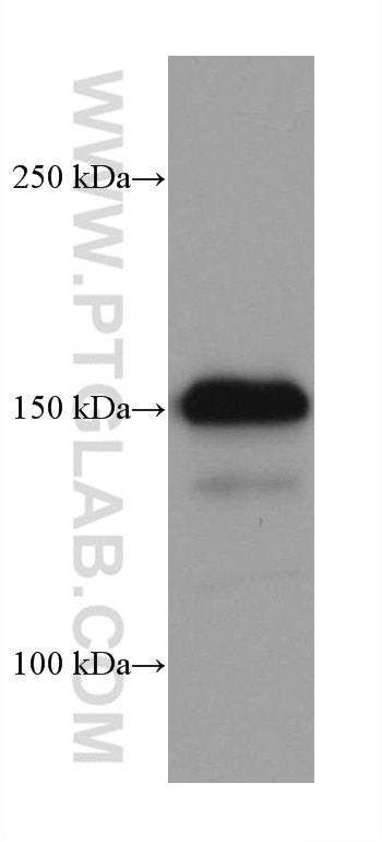
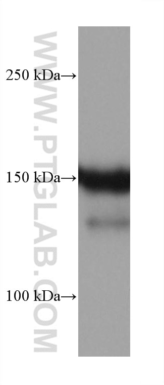

Tested Applications
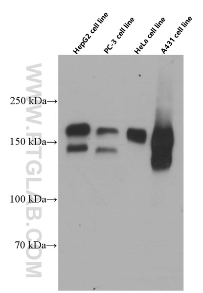
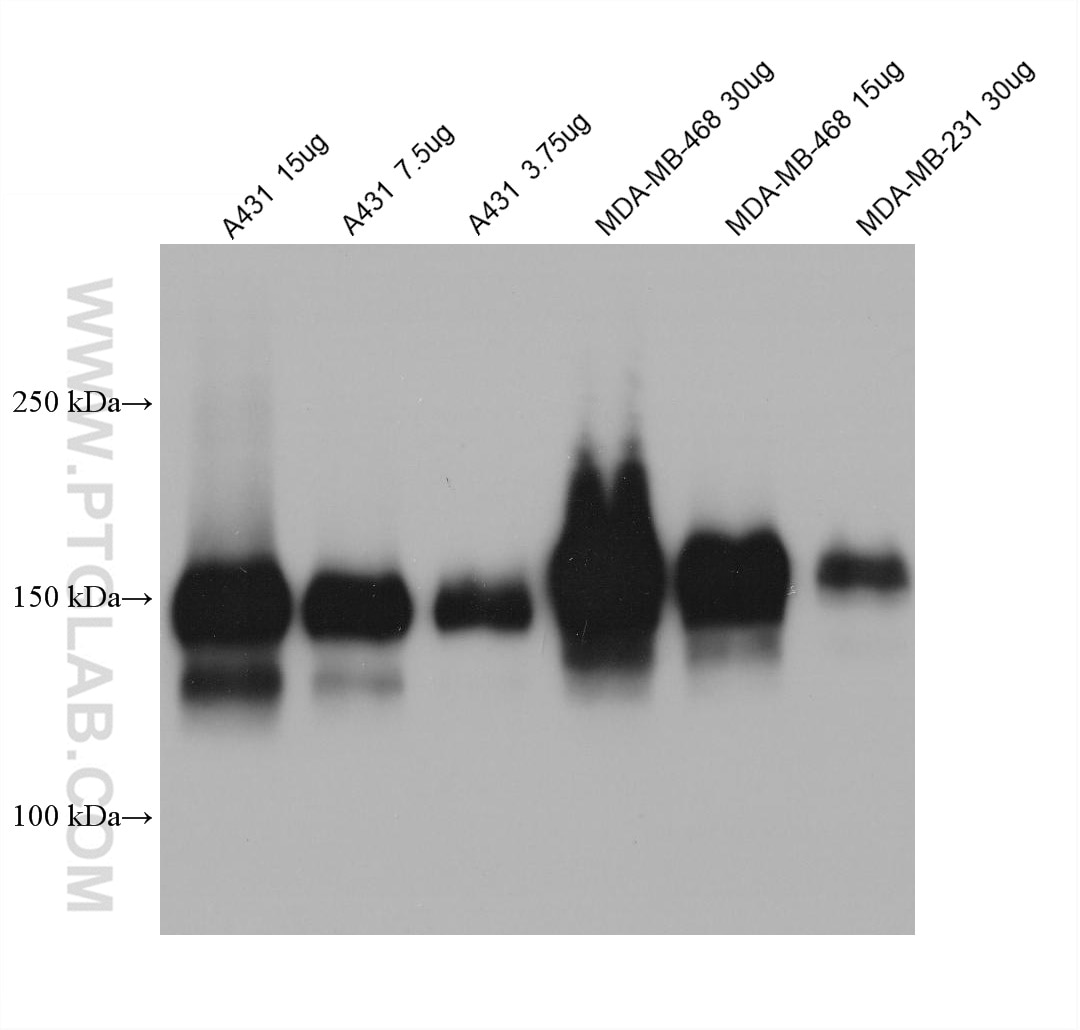
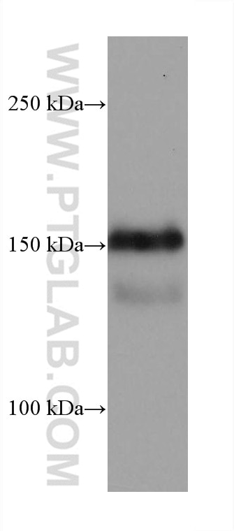
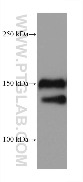
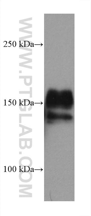
| Positive WB detected in | A431 cells, EC109 cells, A549 cells, HeLa cells, HepG2 cells, LNCaP cells, PC-3 cells, SKOV-3 cells, MDA-MB-468 cells, MDA-MB-231 cells, PC-3 clles |
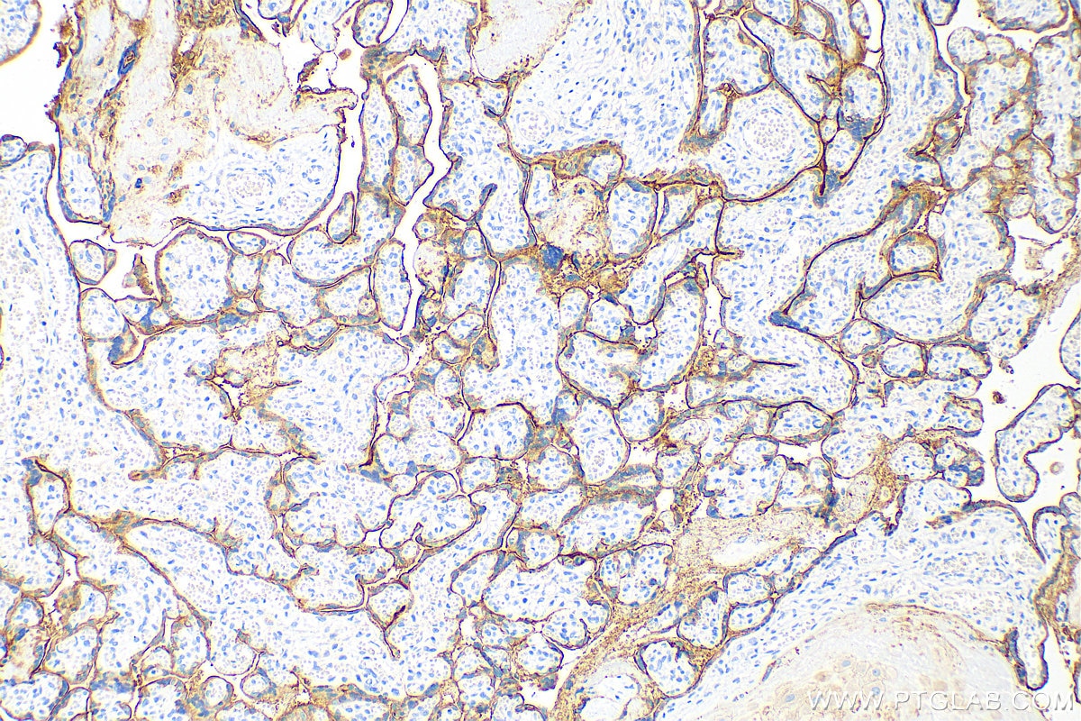
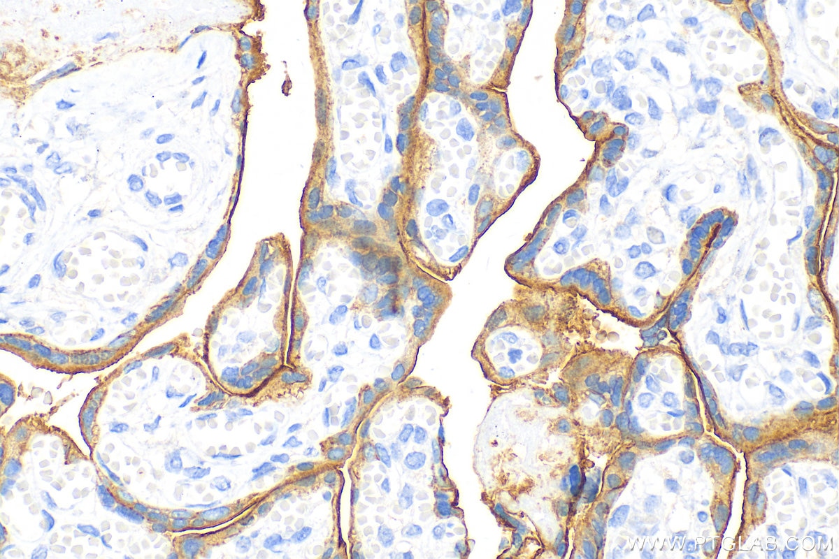
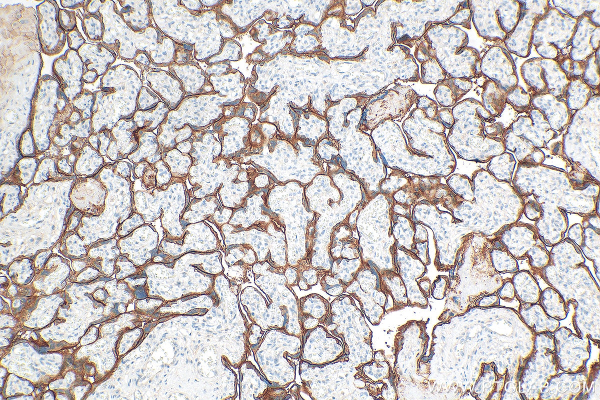
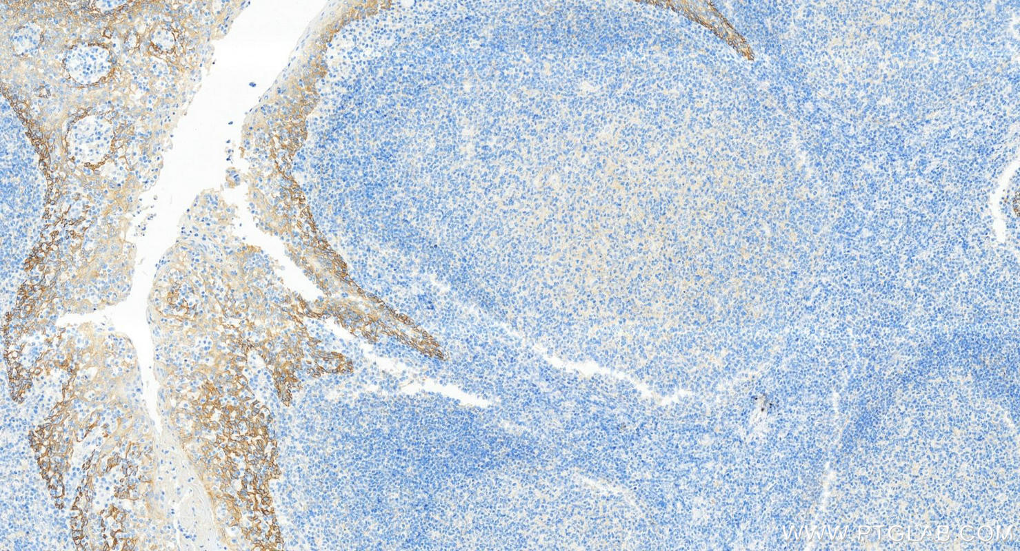
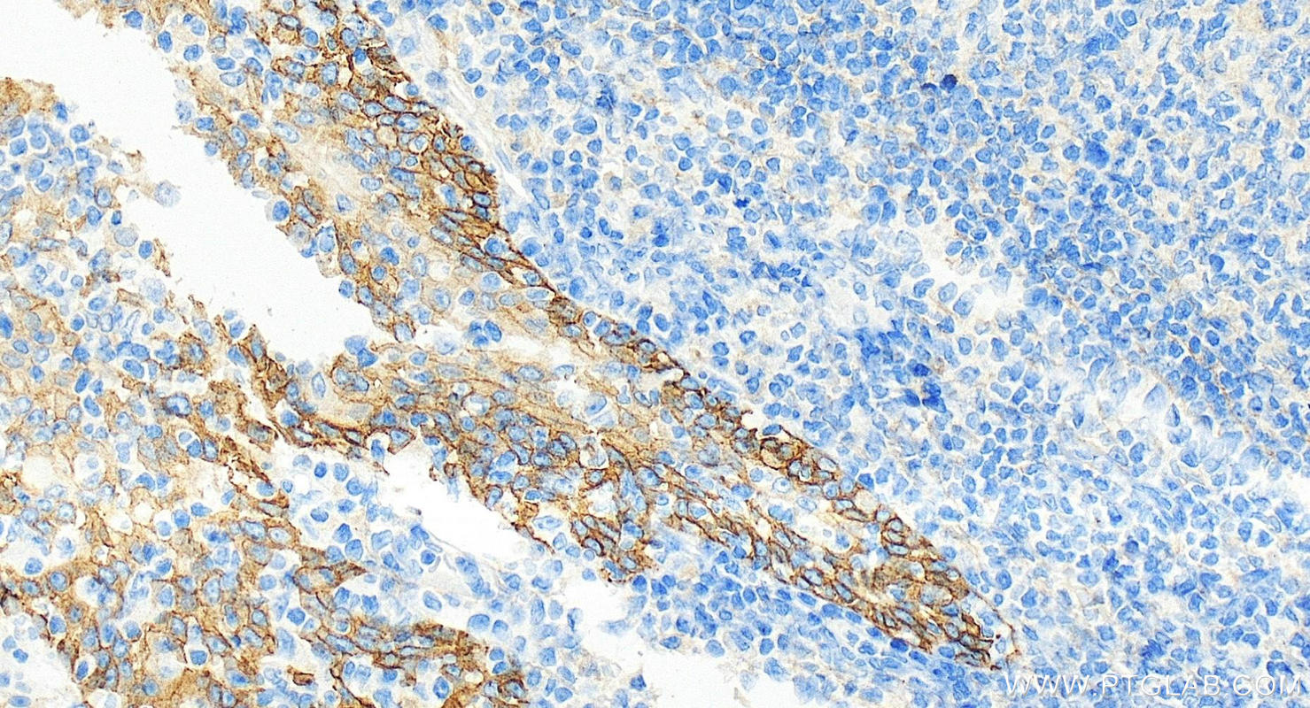
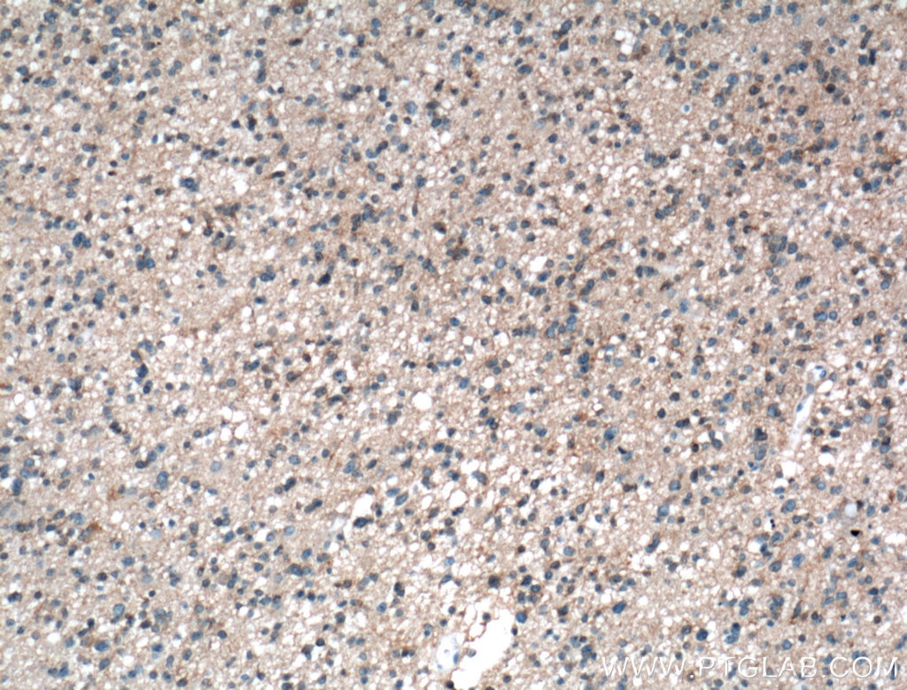
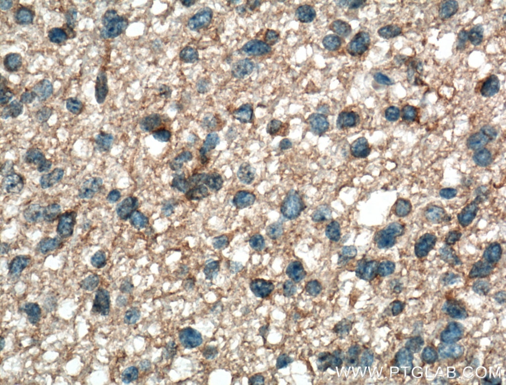
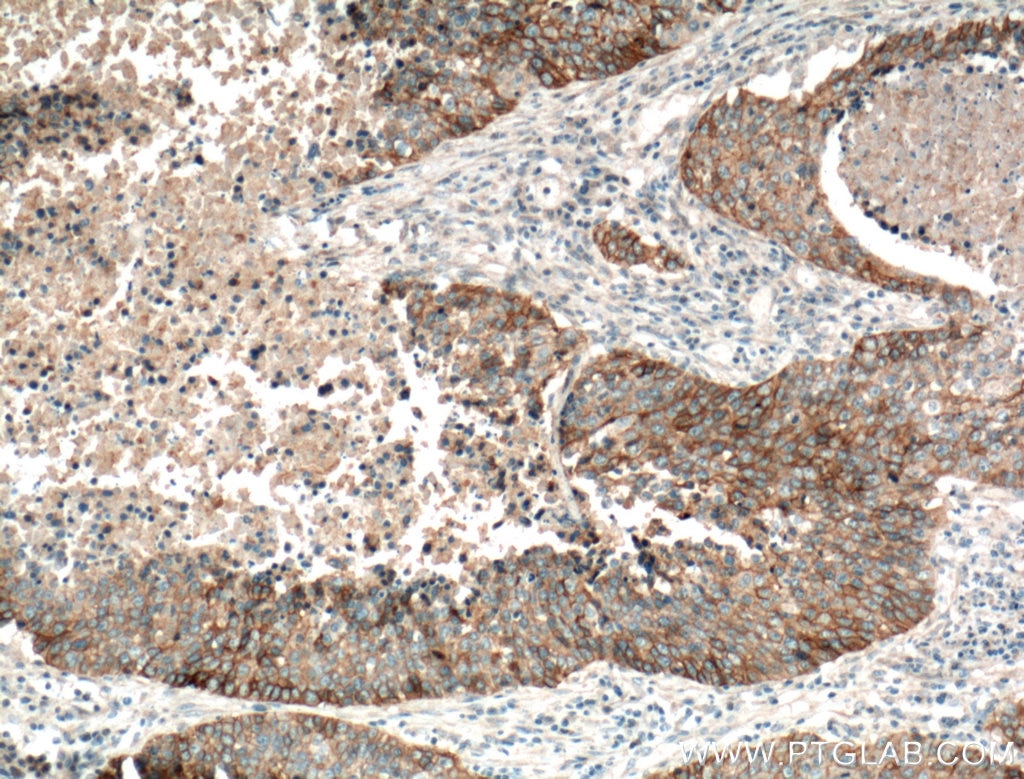
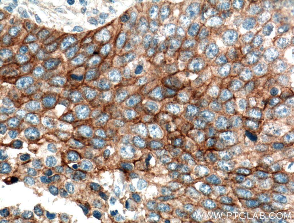
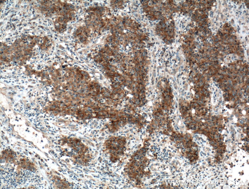
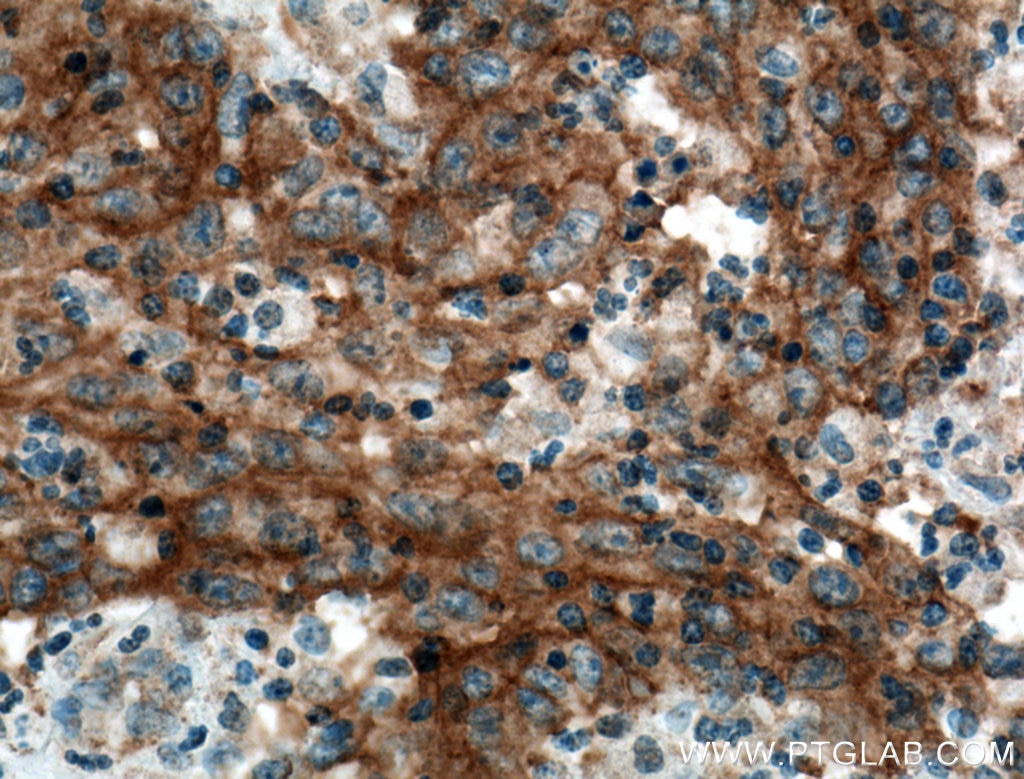
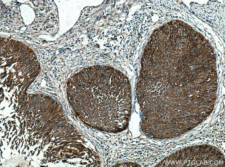
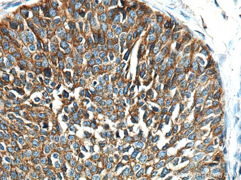
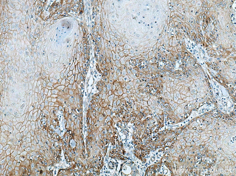
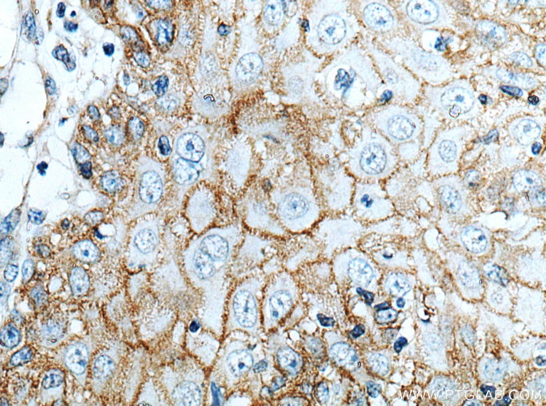
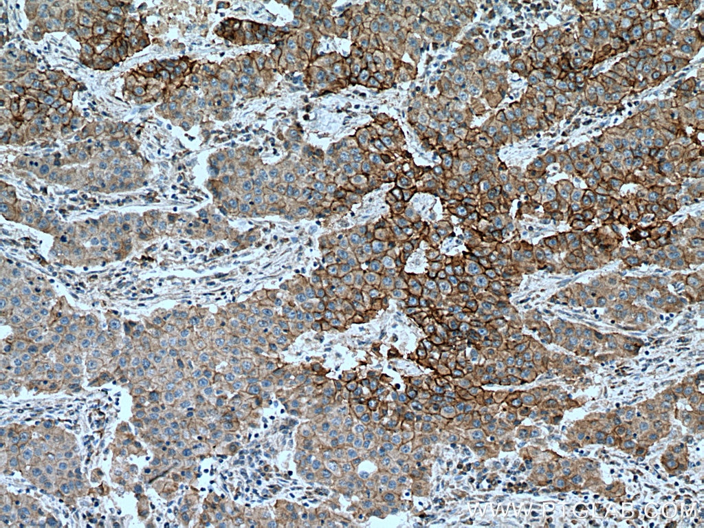
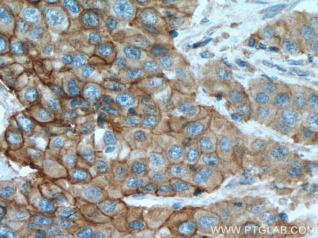
| Positive IHC detected in | human tonsillitis tissue, human breast cancer tissue, human cervical cancer tissue, human colon cancer tissue, human gliomas tissue, human lung cancer tissue, human placenta tissue, human skin cancer tissue Note: suggested antigen retrieval with TE buffer pH 9.0; (*) Alternatively, antigen retrieval may be performed with citrate buffer pH 6.0 |
Recommended dilution
| Application | Dilution |
|---|---|
| Western Blot (WB) | WB : 1:5000-1:50000 |
| Immunohistochemistry (IHC) | IHC : 1:2000-1:8000 |
| It is recommended that this reagent should be titrated in each testing system to obtain optimal results. | |
| Sample-dependent, Check data in validation data gallery. | |
Product Information
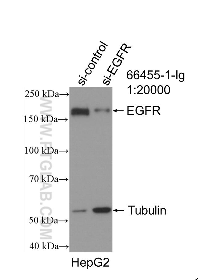
66455-1-Ig targets EGFR in WB, IHC, IF, CoIP, ELISA applications and shows reactivity with human samples.
| Tested Reactivity | human |
| Cited Reactivity | human |
| Host / Isotype | Mouse / IgG1 |
| Class | Monoclonal |
| Type | Antibody |
| Immunogen |
CatNo: Ag24947 Product name: Recombinant human EGFR protein Source: e coli.-derived, PET30a Tag: 6*His Domain: 291-534 aa of BC094761 Sequence: VCNGIGIGEFKDSLSINATNIKHFKNCTSISGDLHILPVAFRGDSFTHTPPLDPQELDILKTVKEITGFLLIQAWPENRTDLHAFENLEIIRGRTKQHGQFSLAVVSLNITSLGLRSLKEISDGDVIISGNKNLCYANTINWKKLFGTSGQKTKIISNRGENSCKATGQVCHALCSPEGCWGPEPRDCVSCRNVSRGRECVDKCNLLEGEPREFVENSECIQCHPECLPQAMNITCTGRGPDNC Predict reactive species |
| Full Name | epidermal growth factor receptor (erythroblastic leukemia viral (v-erb-b) oncogene homolog, avian) |
| Calculated Molecular Weight | 1210 aa, 134 kDa |
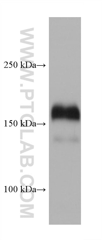
| Observed Molecular Weight | 145-180 kDa |
| GenBank Accession Number | BC094761 |
| Gene Symbol | EGFR |
| Gene ID (NCBI) | 1956 |
| RRID | AB_2881824 |
| Conjugate | Unconjugated |
| Form | Liquid |
| Purification Method | Protein G purification |
| UNIPROT ID | P00533 |
| Storage Buffer | PBS with 0.02% sodium azide and 50% glycerol, pH 7.3. |
| Storage Conditions | Store at -20°C. Stable for one year after shipment. Aliquoting is unnecessary for -20oC storage. 20ul sizes contain 0.1% BSA. |
Background Information
EGFR, also named as ERBB1, is a cell-surface receptor for members of the epidermal growth factor family (EGF-family) of extracellular protein ligands. Binding of the protein to a ligand induces receptor dimerization and tyrosine autophosphorylation and leads to cell proliferation. The gene resides on chromosome 7p12, encoding a 170 kDa membrane-associated glycoprotein. Recent studies have shown EGFR plays a critical role in cancer development and progression, including cell proliferation, apoptosis, angiogenesis, and metastatic spread. Mutations in this gene are associated with lung cancer.
Protocols
| Product Specific Protocols | |
|---|---|
| IHC protocol for EGFR antibody 66455-1-Ig | Download protocol |
| WB protocol for EGFR antibody 66455-1-Ig | Download protocol |
| Standard Protocols | |
|---|---|
| Click here to view our Standard Protocols |
Publications
What published studies show
Anti-EGFR antibody (Cat# 66455-1-Ig) has 141 total citations across over 90 journals, including Nature Communications, Cancer Research, Molecular Cell, Cell Death & Disease, Journal of Medicinal Chemistry, and Advanced Science. Key research fields include EGFR signaling in oncology (lung, liver, breast, colorectal, and brain cancers), drug resistance mechanisms, novel EGFR inhibitor and PROTAC development, network pharmacology with traditional Chinese medicine, exosome biology, ferroptosis, and glycosylation studies.
| Species | Application | Title |
|---|---|---|
Signal Transduct Target Ther Dual-targeting pharmacological UFMylation inhibition reprograms tumor and immune microenvironments to achieve long-term glioblastoma regression. | ||
Cancer Res Inhibition of EGFR Overcomes Acquired Lenvatinib Resistance Driven by STAT3-ABCB1 Signaling in Hepatocellular Carcinoma | ||
Cancer Res Adaptive Regulation of dNTP Homeostasis Confers Osimertinib Resistance in EGFR-Mutant Non-small Cell Lung Carcinoma. | ||
Nat Commun Self-reporting photodynamic nanobody conjugate for precise and sustainable large-volume tumor treatment | ||
Nat Commun EGFR core fucosylation, induced by hepatitis C virus, promotes TRIM40-mediated-RIG-I ubiquitination and suppresses interferon-I antiviral defenses | ||
Nat Commun Therapeutic potential of the secreted Kazal-type serine protease inhibitor SPINK4 in colitis |
Reviews
What customers say
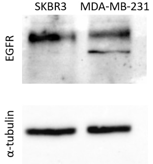
All four reviewers rated this antibody 4 out of 5 stars. Users report successful Western blot performance in human liver cells, breast cancer cell lines (SKBR3, MDA-MB-231), and ARPE-19 cells at 1:1000 dilution with overnight incubation at 4°C. One reviewer noted cross-reactivity with mouse liver proteins at 1:500–1:1000. A ~160 kDa band was detected as expected. One user also validated the antibody on an EDTA plasma antibody microarray. Overall, users find it reliable for detecting the target protein across multiple cell types and applications.
The reviews below have been submitted by verified Proteintech customers who received an incentive for providing their feedback.
k. (Verified Customer) (10-26-2023) | This antibody worked well for human cells and mouse liver cell proteins at 1:500 or 1:1000 concentrations at 4 °C over a night of incubation.
|
Christos (Verified Customer) (02-13-2023) | -35ug protein extract were loaded per well -Transfer was performed for 2hr at 400mA at 4oC, on a Nitrocellulose Blotting Membrane -Membrane blocking was performed in 5% non-fat milk in PBS-Tween20 at room temperature, under mild shacking -Antibodies dilutions were performed in 5% non-fat milk in PBS-Tween20. -Incubations with the primary antibodies were performed as followed: 1)EGFR: 1:1000 for 1.5hr at 4oC 2)tubulin (sc32293, Santa Cruz): 1:5000 for 1.5hr at 4oC -Incubations with the secondary antibodies were performed with Rb pAb to Ms IgG (HRP) (ab 6728, Abcam) at a 1:20000 dilution, for 1hr at 4oC.
 |
Guorong (Verified Customer) (03-31-2022) | A band of approximately 160 kDa was detected
 |
Carly (Verified Customer) (11-17-2020) | Tested using EDTA plasma on an antibody microarray
|


