Filter:
 at dilution of 1:10000 incubated at room temperature for 1.5 hours.")
 at dilution of 1:10000 incubated at room temperature for 1.5 hours.")
 at dilution of 1:10000 incubated at room temperature for 1.5 hours.")
 at dilution of 1:10000 incubated at room temperature for 1.5 hours.")
 with sh-Control and sh-DPEP1 transfected HepG2 cells.")
 at dilution of 1:1000 (under 10x lens). Heat mediated antigen retrieval with Tris-EDTA buffer (pH 9.0).")
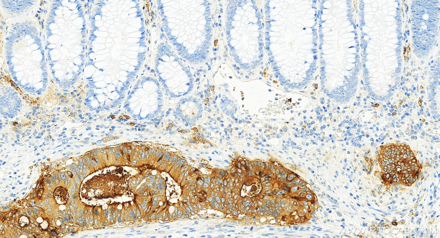
 at dilution of 1:1000 (under 40x lens). Heat mediated antigen retrieval with Tris-EDTA buffer (pH 9.0).")
 at dilution of 1:2000 (under 20x lens). Heat mediated antigen retrieval with Tris-EDTA buffer (pH 9.0).")
 fixed mouse kidney tissue using DPEP1 antibody (68081-1-Ig, Clone: 2B4F4 ) at dilution of 1:400 and CoraLite®488-Conjugated AffiniPure Goat Anti-Mouse IgG(H+L).")
Tested Applications
| Positive WB detected in | human testis tissue, rabbit kidney tissue, pig pancreas tissue, rat pancreas tissue, HepG2 cells |
| Positive IHC detected in | human kidney tissue, human colon cancer tissue Note: suggested antigen retrieval with TE buffer pH 9.0; (*) Alternatively, antigen retrieval may be performed with citrate buffer pH 6.0 |
| Positive IF-P detected in | mouse kidney tissue |
Recommended dilution
| Application | Dilution |
|---|---|
| Western Blot (WB) | WB : 1:5000-1:50000 |
| Immunohistochemistry (IHC) | IHC : 1:500-1:2000 |
| Immunofluorescence (IF)-P | IF-P : 1:200-1:800 |
| It is recommended that this reagent should be titrated in each testing system to obtain optimal results. | |
| Sample-dependent, Check data in validation data gallery. | |
Product Information
68081-1-Ig targets DPEP1 in WB, IHC, IF-P, ELISA applications and shows reactivity with human, mouse, rat, pig, rabbit samples.
| Tested Reactivity | human, mouse, rat, pig, rabbit |
| Host / Isotype | Mouse / IgG2a |
| Class | Monoclonal |
| Type | Antibody |
| Immunogen |
CatNo: Ag14591 Product name: Recombinant human DPEP1 protein Source: e coli.-derived, PET28a Tag: 6*His Domain: 1-385 aa of BC017023 Sequence: MWSGWWLWPLVAVCTADFFRDEAERIMRDSPVIDGHNDLPWQLLDMFNNRLQDERANLTTLAGTHTNIPKLRAGFVGGQFWSVYTPCDTQNKDAVRRTLEQMDVVHRMCRMYPETFLYVTSSAGIRQAFREGKVASLIGVEGGHSIDSSLGVLRALYQLGMRYLTLTHSCNTPWADNWLVDTGDSEPQSQGLSPFGQRVVKELNRLGVLIDLAHVSVATMKATLQLSRAPVIFSHSSAYSVCASRRNVPDDVLRLVKQTDSLVMVNFYNNYISCTNKANLSQVADHLDHIKEVAGARAVGFGGDFDGVPRVPEGLEDVSKYPDLIAELLRRNWTEAEVKGALADNLLRVFEAVEQASNLTQAPEEEPIPLDQLGGSCRTHYGYSS Predict reactive species |
| Full Name | dipeptidase 1 (renal) |
| Calculated Molecular Weight | 411 aa, 46 kDa |
| Observed Molecular Weight | 46 kDa |
| GenBank Accession Number | BC017023 |
| Gene Symbol | DPEP1 |
| Gene ID (NCBI) | 1800 |
| RRID | AB_2918818 |
| Conjugate | Unconjugated |
| Form | Liquid |
| Purification Method | Protein A purification |
| UNIPROT ID | P16444 |
| Storage Buffer | PBS with 0.02% sodium azide and 50% glycerol, pH 7.3. |
| Storage Conditions | Store at -20°C. Stable for one year after shipment. Aliquoting is unnecessary for -20oC storage. 20ul sizes contain 0.1% BSA. |
Protocols
| Product Specific Protocols | |
|---|---|
| IF protocol for DPEP1 antibody 68081-1-Ig | Download protocol |
| IHC protocol for DPEP1 antibody 68081-1-Ig | Download protocol |
| WB protocol for DPEP1 antibody 68081-1-Ig | Download protocol |
| Standard Protocols | |
|---|---|
| Click here to view our Standard Protocols |