Various lysates were subjected to SDS PAGE followed by western blot with 66735-1-Ig (Dystroglycan antibody) at dilution of 1:20000 incubated at room temperature for 1.5 hours.
Various lysates were subjected to SDS PAGE followed by western blot with 66735-1-Ig (Dystroglycan antibody) at dilution of 1:20000 incubated at room temperature for 1.5 hours.
IHC staining of mouse heart using 66735-1-Ig
Immunohistochemical analysis of paraffin-embedded mouse heart tissue slide using 66735-1-Ig (Dystroglycan antibody) at dilution of 1:4000 (under 10x lens). Heat mediated antigen retrieval with Tris-EDTA buffer (pH 9.0).
Immunohistochemical analysis of paraffin-embedded mouse skeletal muscle tissue slide using 66735-1-Ig (Dystroglycan antibody) at dilution of 1:4000 (under 40x lens). Heat mediated antigen retrieval with Tris-EDTA buffer (pH 9.0).
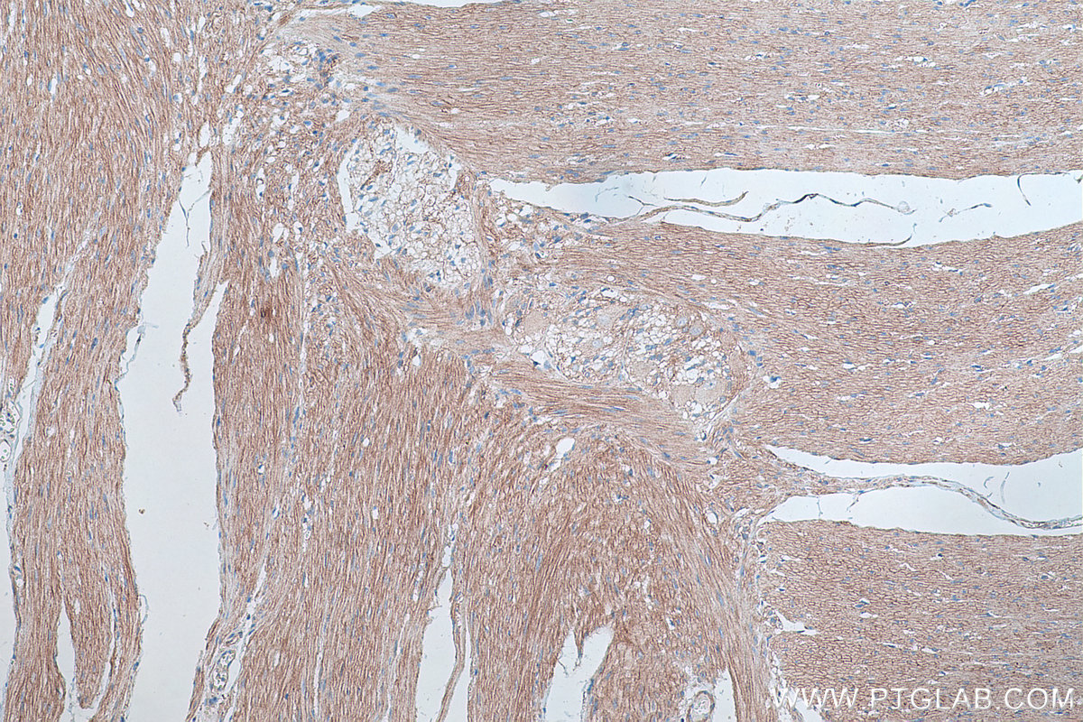
IHC staining of rat heart using 66735-1-Ig
Immunohistochemical analysis of paraffin-embedded rat heart tissue slide using 66735-1-Ig (Dystroglycan antibody) at dilution of 1:4000 (under 10x lens). Heat mediated antigen retrieval with Tris-EDTA buffer (pH 9.0).
Immunohistochemical analysis of paraffin-embedded rat heart tissue slide using 66735-1-Ig (Dystroglycan antibody) at dilution of 1:4000 (under 10x lens). Heat mediated antigen retrieval with Tris-EDTA buffer (pH 9.0).
IHC staining of rat heart using 66735-1-Ig
Immunohistochemical analysis of paraffin-embedded rat heart tissue slide using 66735-1-Ig (Dystroglycan antibody) at dilution of 1:4000 (under 40x lens). Heat mediated antigen retrieval with Tris-EDTA buffer (pH 9.0).
Immunohistochemical analysis of paraffin-embedded rat heart tissue slide using 66735-1-Ig (Dystroglycan antibody) at dilution of 1:4000 (under 40x lens). Heat mediated antigen retrieval with Tris-EDTA buffer (pH 9.0).
IHC staining of rat skeletal muscle using 66735-1-Ig
Immunohistochemical analysis of paraffin-embedded rat skeletal muscle tissue slide using 66735-1-Ig (Dystroglycan antibody) at dilution of 1:4000 (under 10x lens). Heat mediated antigen retrieval with Tris-EDTA buffer (pH 9.0).
Immunohistochemical analysis of paraffin-embedded rat skeletal muscle tissue slide using 66735-1-Ig (Dystroglycan antibody) at dilution of 1:4000 (under 10x lens). Heat mediated antigen retrieval with Tris-EDTA buffer (pH 9.0).
IHC staining of rat skeletal muscle using 66735-1-Ig
Immunohistochemical analysis of paraffin-embedded rat skeletal muscle tissue slide using 66735-1-Ig (Dystroglycan antibody) at dilution of 1:4000 (under 40x lens). Heat mediated antigen retrieval with Tris-EDTA buffer (pH 9.0).
Immunohistochemical analysis of paraffin-embedded rat skeletal muscle tissue slide using 66735-1-Ig (Dystroglycan antibody) at dilution of 1:4000 (under 40x lens). Heat mediated antigen retrieval with Tris-EDTA buffer (pH 9.0).
IHC staining of human colon using 66735-1-Ig
Immunohistochemical analysis of paraffin-embedded human colon tissue slide using 66735-1-Ig (Dystroglycan antibody) at dilution of 1:4000 (under 10x lens). Heat mediated antigen retrieval with Tris-EDTA buffer (pH 9.0).
Immunohistochemical analysis of paraffin-embedded human colon tissue slide using 66735-1-Ig (Dystroglycan antibody) at dilution of 1:4000 (under 10x lens). Heat mediated antigen retrieval with Tris-EDTA buffer (pH 9.0).
IF Staining of HeLa using 66735-1-Ig
Immunofluorescent analysis of (4% PFA) fixed HeLa cells using Dystroglycan antibody (66735-1-Ig, Clone: 2B1G12 ) at dilution of 1:800 and CoraLite®488-Conjugated AffiniPure Goat Anti-Mouse IgG(H+L).
Immunofluorescent analysis of (4% PFA) fixed HeLa cells using Dystroglycan antibody (66735-1-Ig, Clone: 2B1G12 ) at dilution of 1:800 and CoraLite®488-Conjugated AffiniPure Goat Anti-Mouse IgG(H+L).
IF Staining of A549 using 66735-1-Ig
Immunofluorescent analysis of (-20°C Ethanol) fixed A549 cells using Dystroglycan antibody (66735-1-Ig, Clone: 2B1G12 ) at dilution of 1:1000 and Multi-rAb CoraLite ® Plus 488-Goat Anti-Mouse Recombinant Secondary Antibody (H+L) (RGAM002).
The Proteintech guarantee covers Proteintech antibodies in any species and any application, including those not listed on the datasheet. If the antibody doesn’t perform, you can receive a hassle-free refund or credit note.
A549 cells, NCI-H1299 cells, HeLa cells, HepG2 cells, pig brain tissue, rat brain tissue, mouse brain
Positive IHC detected in
mouse heart tissue, human colon tissue, mouse skeletal muscle tissue, rat heart tissue, rat skeletal muscle tissue Note: suggested antigen retrieval with TE buffer pH 9.0; (*) Alternatively, antigen retrieval may be performed with citrate buffer pH 6.0
Positive IF/ICC detected in
HeLa cells, A549 cells
Recommended dilution
Application
Dilution
Western Blot (WB)
WB : 1:5000-1:50000
Immunohistochemistry (IHC)
IHC : 1:2000-1:8000
Immunofluorescence (IF)/ICC
IF/ICC : 1:400-1:1600
It is recommended that this reagent should be titrated in each testing system to obtain optimal results.
Sample-dependent, Check data in validation data gallery.
PBS with 0.02% sodium azide and 50% glycerol, pH 7.3.
Storage Conditions
Store at -20°C. Stable for one year after shipment. Aliquoting is unnecessary for -20oC storage. 20ul sizes contain 0.1% BSA.
Background Information
Dystroglycan, also known as DAG1 or DG, was originally isolated from skeletal muscle as an integral membrane component of the dystrophin-glycoprotein complex (DGC). In addition to skeletal muscle, dystroglycan is strongly expressed in heart and smooth muscle, as well as many non-muscle tissues including brain and peripheral nerve (PMID: 12556455). The dystroglycan is involved in a number of processes including laminin and basement membrane assembly, sarcolemmal stability, cell survival, peripheral nerve myelination, nodal structure, cell migration, and epithelial polarization. Dystroglycan consists of two subunits (alpha and beta), which are translated from a single mRNA as a propeptide that is proteolytically cleaved into two noncovalently associated proteins (PMID: 16410545). Alpha-dystroglycan is a 156-kDa extracellular peripheral glycoprotein, while beta-dystroglycan is a 43-kDa transmembrane protein (PMID: 9858474). The 43-kDa beta-dystroglycan can be cleaved into a ~30-kDa form (PMID: 14678802; 18458097; 17255331).
Various lysates were subjected to SDS PAGE followed by western blot with 66735-1-Ig (Dystroglycan antibody) at dilution of 1:20000 incubated at room temperature for 1.5 hours.
IHC Figures
IHC staining of mouse heart using 66735-1-Ig
Immunohistochemical analysis of paraffin-embedded mouse heart tissue slide using 66735-1-Ig (Dystroglycan antibody) at dilution of 1:4000 (under 10x lens). Heat mediated antigen retrieval with Tris-EDTA buffer (pH 9.0).
IHC staining of mouse heart using 66735-1-Ig
Immunohistochemical analysis of paraffin-embedded mouse heart tissue slide using 66735-1-Ig (Dystroglycan antibody) at dilution of 1:4000 (under 40x lens). Heat mediated antigen retrieval with Tris-EDTA buffer (pH 9.0).
IHC staining of mouse skeletal muscle using 66735-1-Ig
Immunohistochemical analysis of paraffin-embedded mouse skeletal muscle tissue slide using 66735-1-Ig (Dystroglycan antibody) at dilution of 1:4000 (under 10x lens). Heat mediated antigen retrieval with Tris-EDTA buffer (pH 9.0).
IHC staining of mouse skeletal muscle using 66735-1-Ig
Immunohistochemical analysis of paraffin-embedded mouse skeletal muscle tissue slide using 66735-1-Ig (Dystroglycan antibody) at dilution of 1:4000 (under 40x lens). Heat mediated antigen retrieval with Tris-EDTA buffer (pH 9.0).
IHC staining of rat heart using 66735-1-Ig
Immunohistochemical analysis of paraffin-embedded rat heart tissue slide using 66735-1-Ig (Dystroglycan antibody) at dilution of 1:4000 (under 10x lens). Heat mediated antigen retrieval with Tris-EDTA buffer (pH 9.0).
IHC staining of rat heart using 66735-1-Ig
Immunohistochemical analysis of paraffin-embedded rat heart tissue slide using 66735-1-Ig (Dystroglycan antibody) at dilution of 1:4000 (under 40x lens). Heat mediated antigen retrieval with Tris-EDTA buffer (pH 9.0).
IHC staining of rat skeletal muscle using 66735-1-Ig
Immunohistochemical analysis of paraffin-embedded rat skeletal muscle tissue slide using 66735-1-Ig (Dystroglycan antibody) at dilution of 1:4000 (under 10x lens). Heat mediated antigen retrieval with Tris-EDTA buffer (pH 9.0).
IHC staining of rat skeletal muscle using 66735-1-Ig
Immunohistochemical analysis of paraffin-embedded rat skeletal muscle tissue slide using 66735-1-Ig (Dystroglycan antibody) at dilution of 1:4000 (under 40x lens). Heat mediated antigen retrieval with Tris-EDTA buffer (pH 9.0).
IHC staining of human colon using 66735-1-Ig
Immunohistochemical analysis of paraffin-embedded human colon tissue slide using 66735-1-Ig (Dystroglycan antibody) at dilution of 1:4000 (under 10x lens). Heat mediated antigen retrieval with Tris-EDTA buffer (pH 9.0).
IF/ICC Figures
IF Staining of HeLa using 66735-1-Ig
Immunofluorescent analysis of (4% PFA) fixed HeLa cells using Dystroglycan antibody (66735-1-Ig, Clone: 2B1G12 ) at dilution of 1:800 and CoraLite®488-Conjugated AffiniPure Goat Anti-Mouse IgG(H+L).
IF Staining of A549 using 66735-1-Ig
Immunofluorescent analysis of (-20°C Ethanol) fixed A549 cells using Dystroglycan antibody (66735-1-Ig, Clone: 2B1G12 ) at dilution of 1:1000 and Multi-rAb CoraLite ® Plus 488-Goat Anti-Mouse Recombinant Secondary Antibody (H+L) (RGAM002).
The species listed in Tested Reactivity are in-house verified and applicable species. For unlisted species, please refer to the homology analysis of the immunogen sequence and related species. For rabbit polyclonal antibodies, homology >70% is recommended. For mouse monoclonal antibodies and rabbit recombinant antibodies, homology >90% is recommended. Generally, the higher the homology, the greater the applicability. However, there will be certain differences in protein expression in different species, tissues or cells. Therefore, the homology analysis results are for reference only and do not serve as a guarantee.
At Proteintech, we pride ourselves on our antibody quality, customer service and transparency. As such, we are comparing our antibodies with other vendors, enabling easy identification and comparisons of key data to help you choose the suitable antibody for your needs.
We have selected the top cited antibodies from these vendors for you to compare.
 at dilution of 1:20000 incubated at room temperature for 1.5 hours.")
 at dilution of 1:4000 (under 10x lens). Heat mediated antigen retrieval with Tris-EDTA buffer (pH 9.0).")
 at dilution of 1:4000 (under 40x lens). Heat mediated antigen retrieval with Tris-EDTA buffer (pH 9.0).")
 at dilution of 1:4000 (under 10x lens). Heat mediated antigen retrieval with Tris-EDTA buffer (pH 9.0).")
 at dilution of 1:4000 (under 40x lens). Heat mediated antigen retrieval with Tris-EDTA buffer (pH 9.0).")
 at dilution of 1:4000 (under 10x lens). Heat mediated antigen retrieval with Tris-EDTA buffer (pH 9.0).")
 at dilution of 1:4000 (under 40x lens). Heat mediated antigen retrieval with Tris-EDTA buffer (pH 9.0).")
 at dilution of 1:4000 (under 10x lens). Heat mediated antigen retrieval with Tris-EDTA buffer (pH 9.0).")
 at dilution of 1:4000 (under 40x lens). Heat mediated antigen retrieval with Tris-EDTA buffer (pH 9.0).")
 at dilution of 1:4000 (under 10x lens). Heat mediated antigen retrieval with Tris-EDTA buffer (pH 9.0).")
 fixed HeLa cells using Dystroglycan antibody (66735-1-Ig, Clone: 2B1G12 ) at dilution of 1:800 and CoraLite®488-Conjugated AffiniPure Goat Anti-Mouse IgG(H+L).")
 fixed A549 cells using Dystroglycan antibody (66735-1-Ig, Clone: 2B1G12 ) at dilution of 1:1000 and Multi-rAb CoraLite ® Plus 488-Goat Anti-Mouse Recombinant Secondary Antibody (H+L) (RGAM002).")