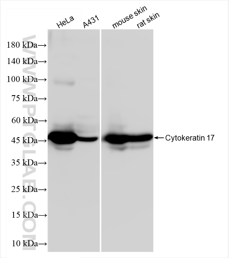
Various lysates were subjected to SDS PAGE followed by western blot with 84978-1-RR (KRT17-Specific antibody) at dilution of 1:10000 incubated at room temperature for 1.5 hours.
Various lysates were subjected to SDS PAGE followed by western blot with 84978-1-RR (KRT17-Specific antibody) at dilution of 1:10000 incubated at room temperature for 1.5 hours.
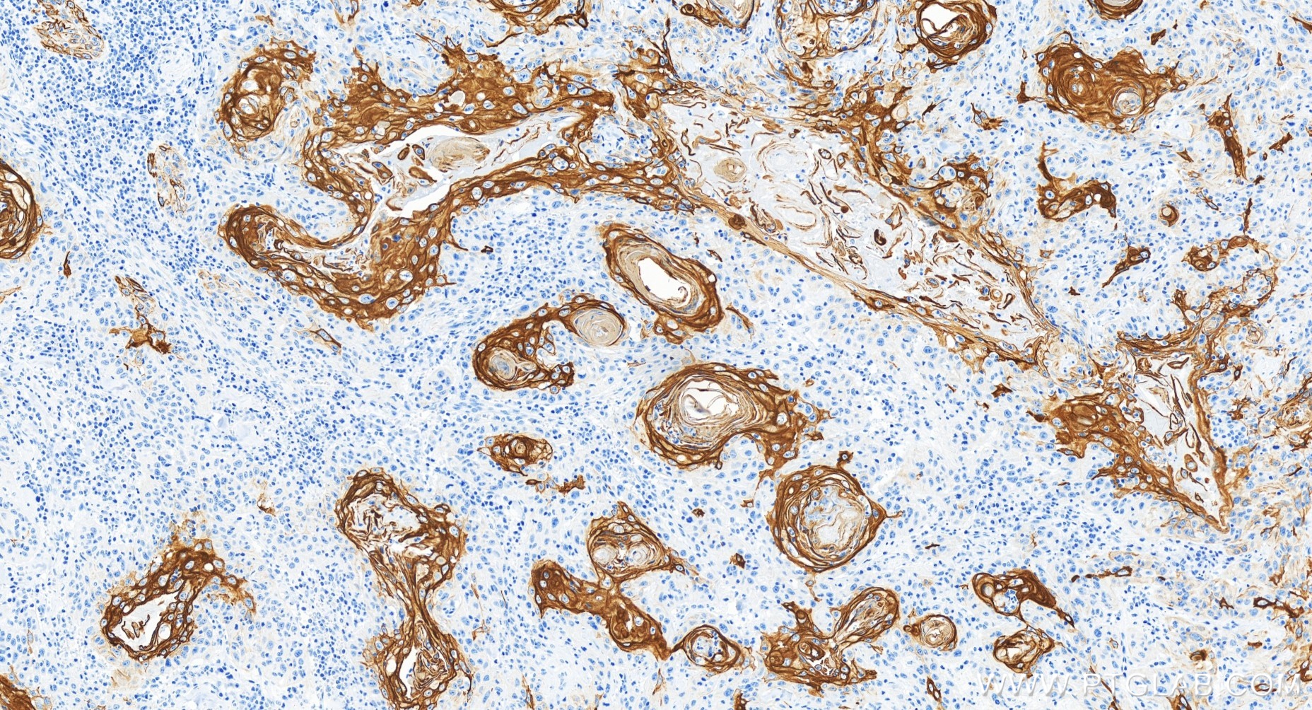
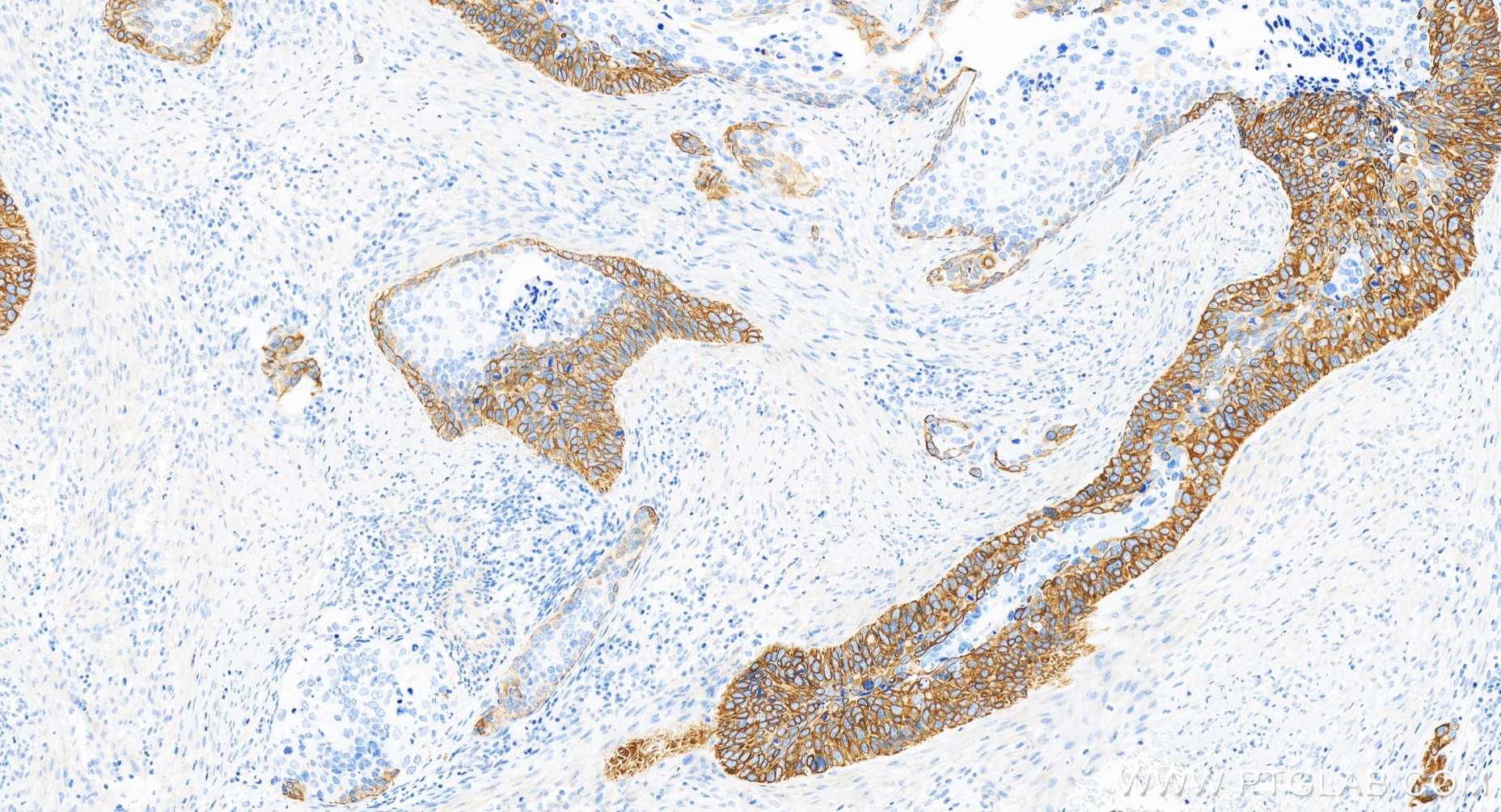
IHC staining of human skin cancer using 84978-1-RR
Immunohistochemical analysis of paraffin-embedded human skin cancer tissue slide using 84978-1-RR (Cytokeratin 17-specific antibody) at dilution of 1:6000 (under 20x lens). Heat mediated antigen retrieval with Tris-EDTA buffer (pH 9.0).
Immunohistochemical analysis of paraffin-embedded human skin cancer tissue slide using 84978-1-RR (Cytokeratin 17-specific antibody) at dilution of 1:6000 (under 20x lens). Heat mediated antigen retrieval with Tris-EDTA buffer (pH 9.0).
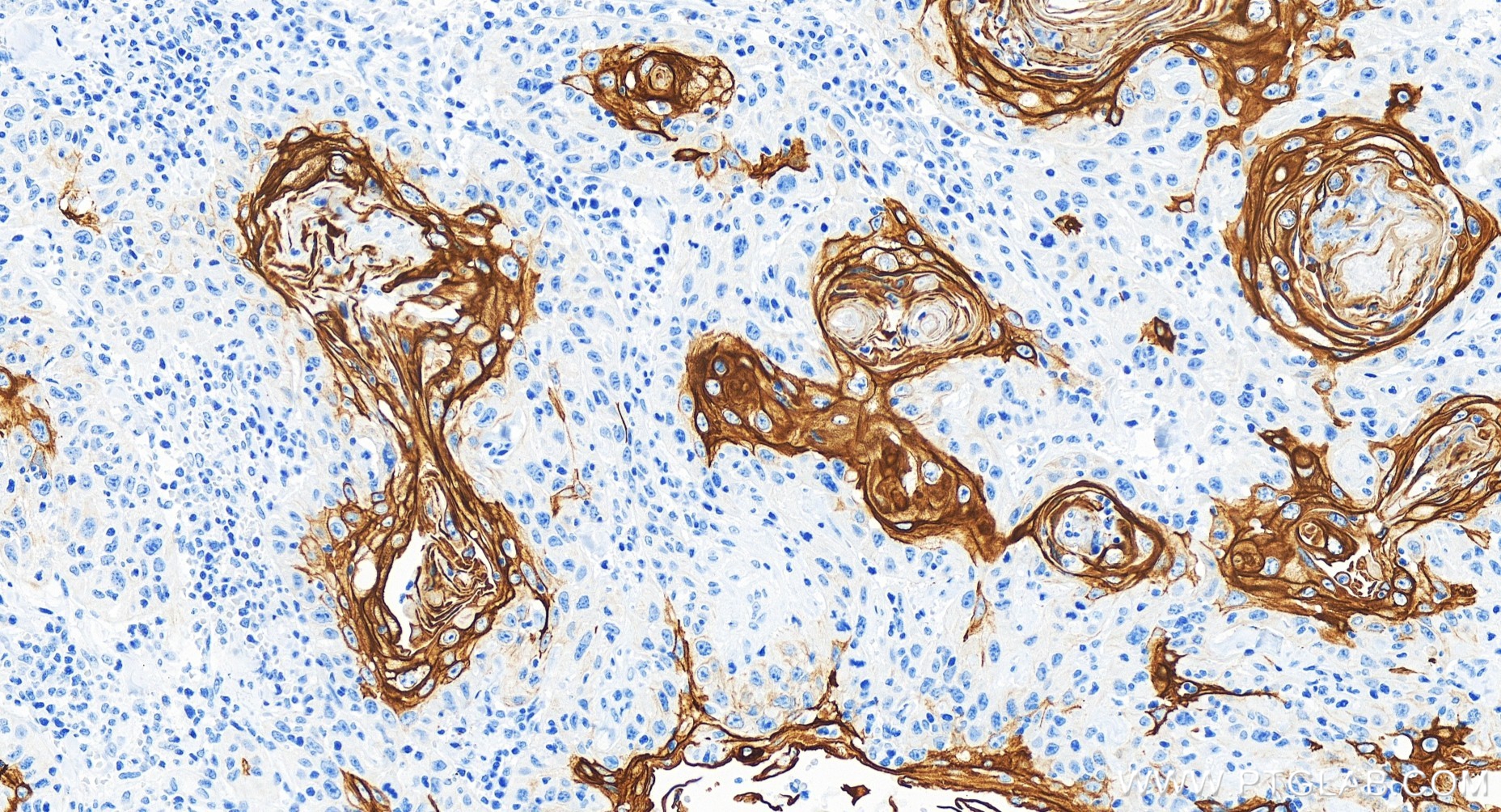
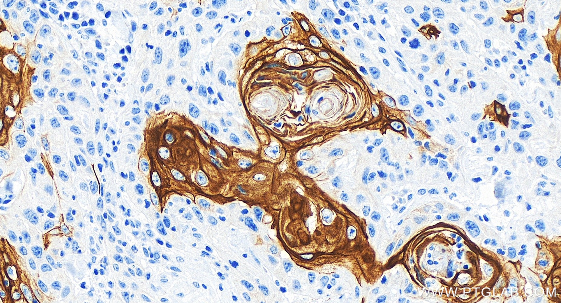
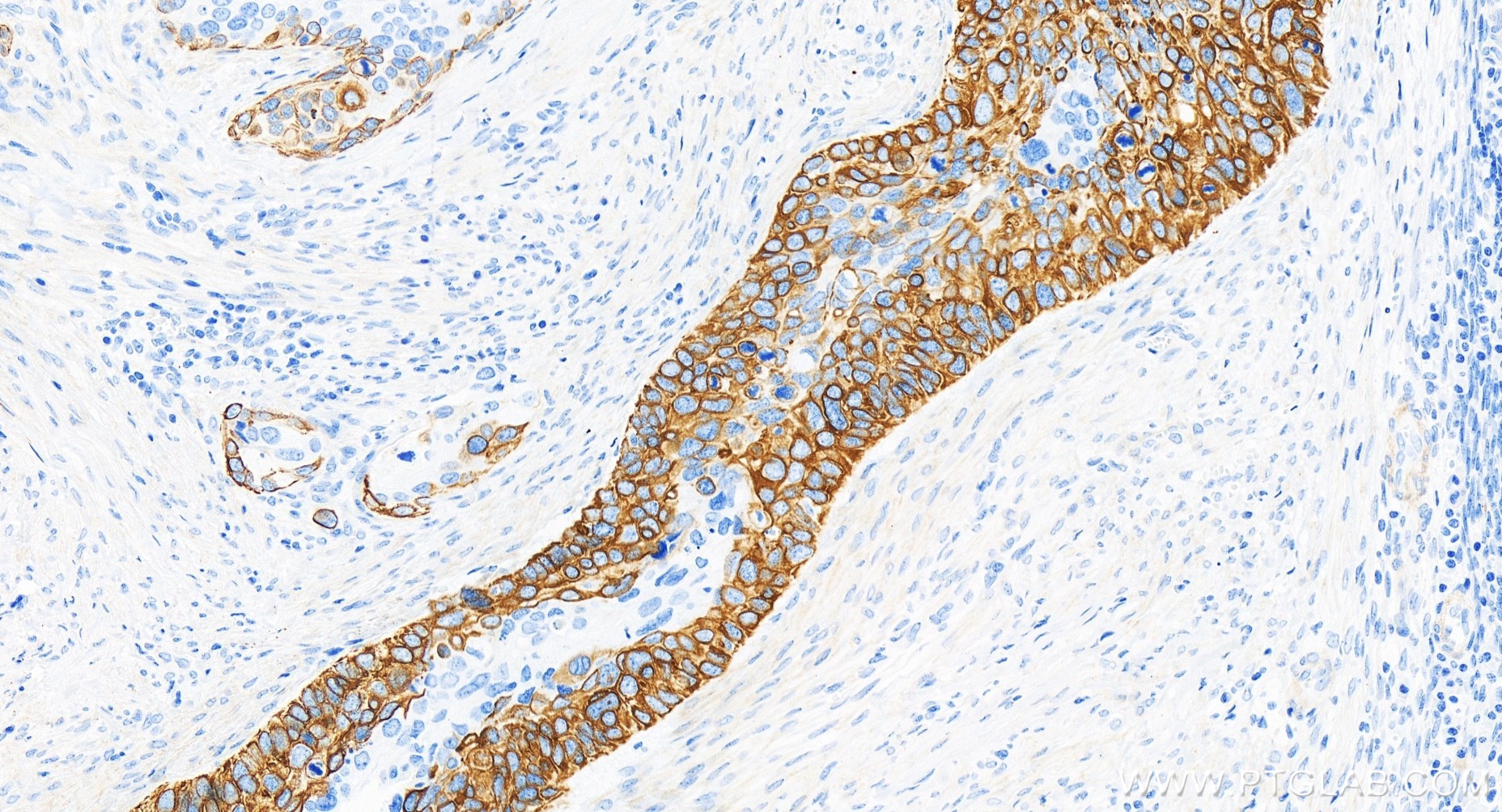
IHC staining of human cervical cancer using 84978-1-RR
Immunohistochemical analysis of paraffin-embedded human cervical cancer tissue slide using 84978-1-RR (Cytokeratin 17-specific antibody) at dilution of 1:6000 (under 20x lens). Heat mediated antigen retrieval with Tris-EDTA buffer (pH 9.0).
Immunohistochemical analysis of paraffin-embedded human cervical cancer tissue slide using 84978-1-RR (Cytokeratin 17-specific antibody) at dilution of 1:6000 (under 20x lens). Heat mediated antigen retrieval with Tris-EDTA buffer (pH 9.0).
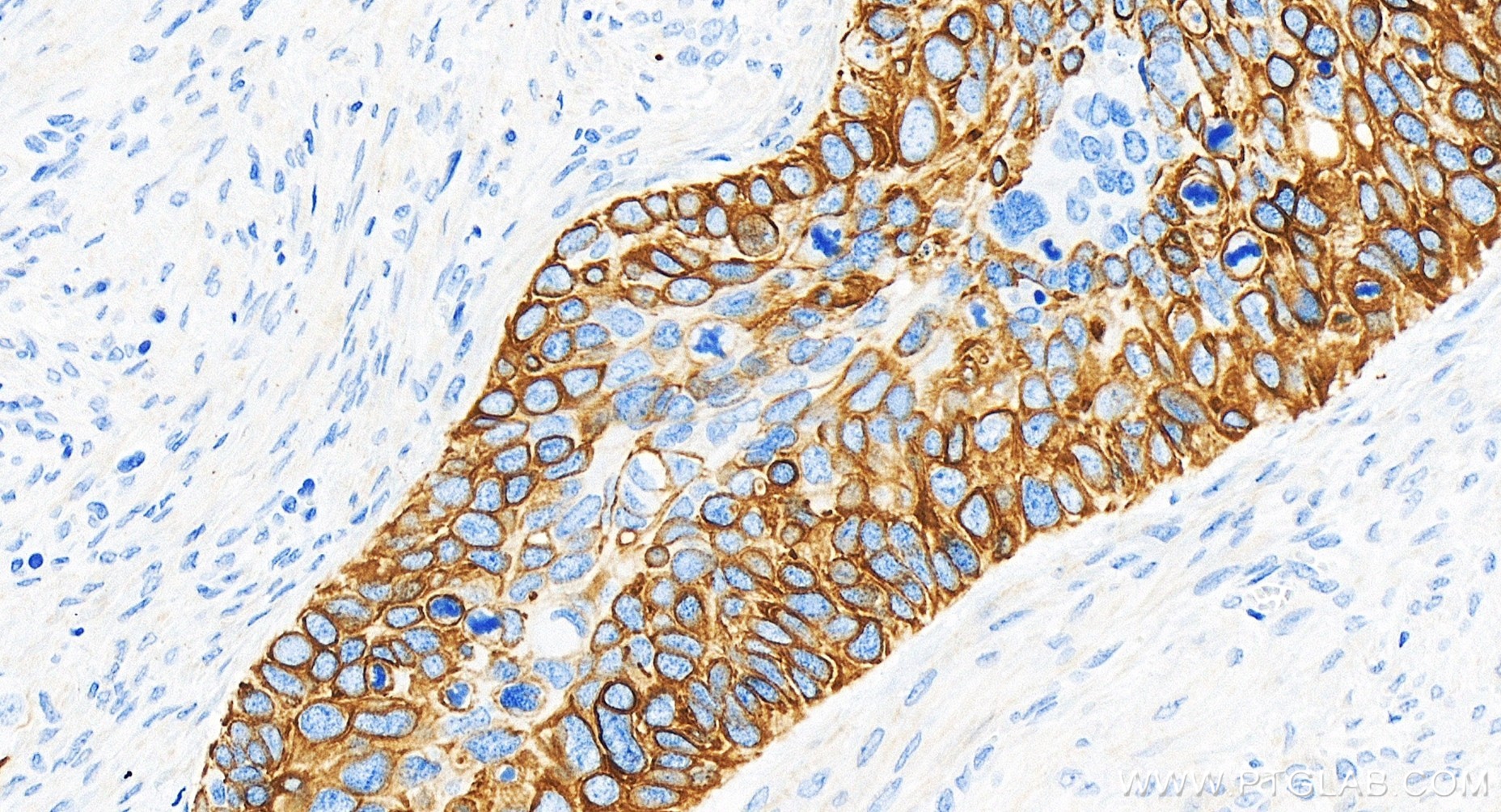
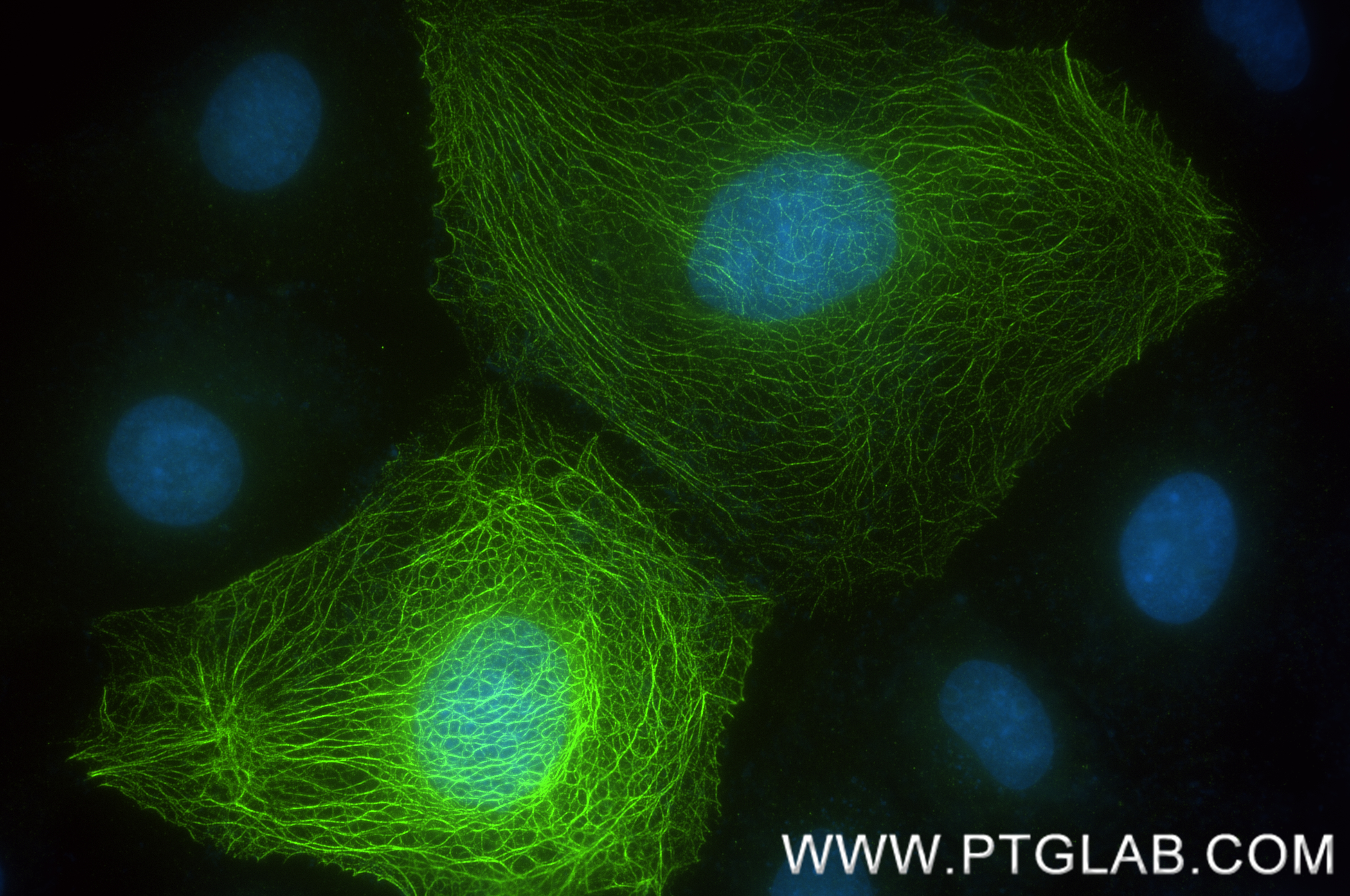
IF Staining of A431 using 84978-1-RR
Immunofluorescent analysis of (-20°C Methanol) fixed A431 cells using Cytokeratin 17-Specific antibody (84978-1-RR, Clone: 242240A8 ) at dilution of 1:1000 and CoraLite®488-Conjugated Goat Anti-Rabbit IgG(H+L) (SA00013-2).
Immunofluorescent analysis of (-20°C Methanol) fixed A431 cells using Cytokeratin 17-Specific antibody (84978-1-RR, Clone: 242240A8 ) at dilution of 1:1000 and CoraLite®488-Conjugated Goat Anti-Rabbit IgG(H+L) (SA00013-2).
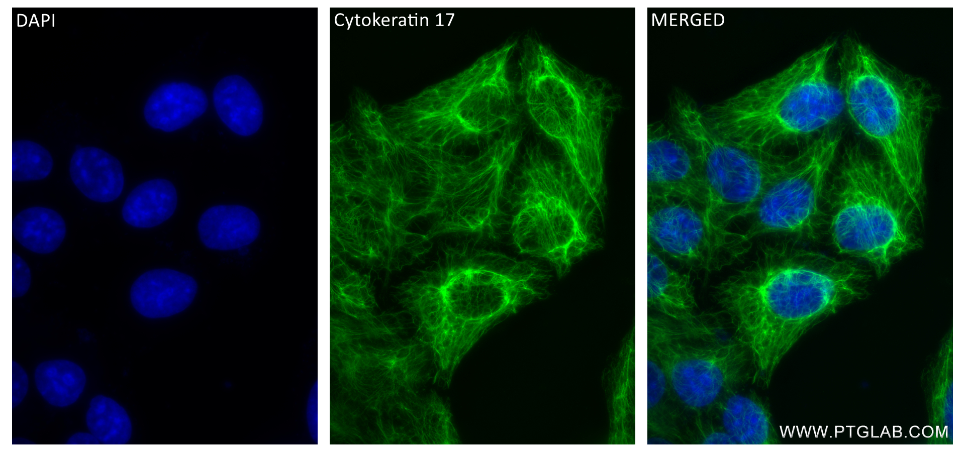
IF Staining of HeLa using 84978-1-RR
Immunofluorescent analysis of (-20°C Methanol) fixed HeLa cells using Cytokeratin 17-Specific antibody (84978-1-RR, Clone: 242240A8 ) at dilution of 1:500 and CoraLite®488-Conjugated Goat Anti-Rabbit IgG(H+L) (SA00013-2), CL594-Phalloidin (red).
Immunofluorescent analysis of (-20°C Methanol) fixed HeLa cells using Cytokeratin 17-Specific antibody (84978-1-RR, Clone: 242240A8 ) at dilution of 1:500 and CoraLite®488-Conjugated Goat Anti-Rabbit IgG(H+L) (SA00013-2), CL594-Phalloidin (red).
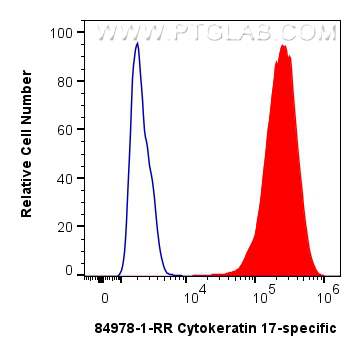
FC experiment of HeLa using 84978-1-RR
1x10^6 HeLa cells were intracellularly stained with 0.25 ug Cytokeratin 17-specific Recombinant antibody (84978-1-RR, Clone:242240A8) and CoraLite®488-Conjugated Goat Anti-Rabbit IgG(H+L) (SA00013-2)(red), or 0.25 ug Rabbit IgG Isotype Control RecAb (98136-1-RR, Clone: 240953C9) (blue). Cells were fixed with 4% PFA and permeabilized with Flow Cytometry Perm Buffer (PF00011-C).
1x10^6 HeLa cells were intracellularly stained with 0.25 ug Cytokeratin 17-specific Recombinant antibody (84978-1-RR, Clone:242240A8) and CoraLite®488-Conjugated Goat Anti-Rabbit IgG(H+L) (SA00013-2)(red), or 0.25 ug Rabbit IgG Isotype Control RecAb (98136-1-RR, Clone: 240953C9) (blue). Cells were fixed with 4% PFA and permeabilized with Flow Cytometry Perm Buffer (PF00011-C).
The Proteintech guarantee covers Proteintech antibodies in any species and any application, including those not listed on the datasheet. If the antibody doesn’t perform, you can receive a hassle-free refund or credit note.
HeLa cells, A431 cells, mouse skin tissue, rat skin tissue
Positive IHC detected in
human skin cancer tissue, human cervical cancer tissue Note: suggested antigen retrieval with TE buffer pH 9.0; (*) Alternatively, antigen retrieval may be performed with citrate buffer pH 6.0
Positive IF/ICC detected in
HeLa cells, A431 cells
Positive FC (Intra) detected in
HeLa cells
Recommended dilution
Application
Dilution
Western Blot (WB)
WB : 1:5000-1:50000
Immunohistochemistry (IHC)
IHC : 1:3000-1:12000
Immunofluorescence (IF)/ICC
IF/ICC : 1:250-1:1000
Flow Cytometry (FC) (INTRA)
FC (INTRA) : 0.25 ug per 10^6 cells in a 100 µl suspension
It is recommended that this reagent should be titrated in each testing system to obtain optimal results.
Sample-dependent, Check data in validation data gallery.
Product Information
84978-1-RR targets Cytokeratin 17-specific in WB, IHC, IF/ICC, FC (Intra), ELISA applications and shows reactivity with human, mouse, rat samples.
PBS with 0.02% sodium azide and 50% glycerol , pH 7.3
Storage Conditions
Store at -20°C. Stable for one year after shipment. Aliquoting is unnecessary for -20oC storage. 20ul sizes contain 0.1% BSA.
Background Information
Keratins are a large family of proteins that form the intermediate filaments that make up the cytoskeleton. Type I keratins are a group of acidic intermediate filament proteins and type II keratins are the basic or neutral counterparts. Keratin 17 is encoded by the KRT17 gene and is a type I cytokeratin found in nail beds, hair follicles, sebaceous glands, and epidermal appendages.The molecular weight of cytokeratin 17 is approximately 49 kDa.Cytokeratin 17 plays a role in the formation and maintenance of epidermal appendages, especially in determining the shape and orientation of hair. It is required for the maintenance of the anagen (growth) state of hair follicles by modulating the function of TNF-alpha for hair cycling and is also involved in tissue repair (PMID: 16702408).
Protocols
Product Specific Protocols
WB protocol for Cytokeratin 17-specific antibody 84978-1-RR
Various lysates were subjected to SDS PAGE followed by western blot with 84978-1-RR (KRT17-Specific antibody) at dilution of 1:10000 incubated at room temperature for 1.5 hours.
IHC Figures
IHC staining of human skin cancer using 84978-1-RR
Immunohistochemical analysis of paraffin-embedded human skin cancer tissue slide using 84978-1-RR (Cytokeratin 17-specific antibody) at dilution of 1:6000 (under 20x lens). Heat mediated antigen retrieval with Tris-EDTA buffer (pH 9.0).
IHC staining of human skin cancer using 84978-1-RR
Immunohistochemical analysis of paraffin-embedded human skin cancer tissue slide using 84978-1-RR (Cytokeratin 17-specific antibody) at dilution of 1:6000 (under 20x lens). Heat mediated antigen retrieval with Tris-EDTA buffer (pH 9.0).
IHC staining of human skin cancer using 84978-1-RR
Immunohistochemical analysis of paraffin-embedded human skin cancer tissue slide using 84978-1-RR (Cytokeratin 17-specific antibody) at dilution of 1:6000 (under 20x lens). Heat mediated antigen retrieval with Tris-EDTA buffer (pH 9.0).
IHC staining of human cervical cancer using 84978-1-RR
Immunohistochemical analysis of paraffin-embedded human cervical cancer tissue slide using 84978-1-RR (Cytokeratin 17-specific antibody) at dilution of 1:6000 (under 20x lens). Heat mediated antigen retrieval with Tris-EDTA buffer (pH 9.0).
IHC staining of human cervical cancer using 84978-1-RR
Immunohistochemical analysis of paraffin-embedded human cervical cancer tissue slide using 84978-1-RR (Cytokeratin 17-specific antibody) at dilution of 1:6000 (under 20x lens). Heat mediated antigen retrieval with Tris-EDTA buffer (pH 9.0).
IHC staining of human cervical cancer using 84978-1-RR
Immunohistochemical analysis of paraffin-embedded human cervical cancer tissue slide using 84978-1-RR (Cytokeratin 17-specific antibody) at dilution of 1:6000 (under 20x lens). Heat mediated antigen retrieval with Tris-EDTA buffer (pH 9.0).
IF/ICC Figures
IF Staining of A431 using 84978-1-RR
Immunofluorescent analysis of (-20°C Methanol) fixed A431 cells using Cytokeratin 17-Specific antibody (84978-1-RR, Clone: 242240A8 ) at dilution of 1:1000 and CoraLite®488-Conjugated Goat Anti-Rabbit IgG(H+L) (SA00013-2).
IF Staining of HeLa using 84978-1-RR
Immunofluorescent analysis of (-20°C Methanol) fixed HeLa cells using Cytokeratin 17-Specific antibody (84978-1-RR, Clone: 242240A8 ) at dilution of 1:500 and CoraLite®488-Conjugated Goat Anti-Rabbit IgG(H+L) (SA00013-2), CL594-Phalloidin (red).
FC (INTRA) Figures
FC experiment of HeLa using 84978-1-RR
1x10^6 HeLa cells were intracellularly stained with 0.25 ug Cytokeratin 17-specific Recombinant antibody (84978-1-RR, Clone:242240A8) and CoraLite®488-Conjugated Goat Anti-Rabbit IgG(H+L) (SA00013-2)(red), or 0.25 ug Rabbit IgG Isotype Control RecAb (98136-1-RR, Clone: 240953C9) (blue). Cells were fixed with 4% PFA and permeabilized with Flow Cytometry Perm Buffer (PF00011-C).
The species listed in Tested Reactivity are in-house verified and applicable species. For unlisted species, please refer to the homology analysis of the immunogen sequence and related species. For rabbit polyclonal antibodies, homology >70% is recommended. For mouse monoclonal antibodies and rabbit recombinant antibodies, homology >90% is recommended. Generally, the higher the homology, the greater the applicability. However, there will be certain differences in protein expression in different species, tissues or cells. Therefore, the homology analysis results are for reference only and do not serve as a guarantee.
At Proteintech, we pride ourselves on our antibody quality, customer service and transparency. As such, we are comparing our antibodies with other vendors, enabling easy identification and comparisons of key data to help you choose the suitable antibody for your needs.
We have selected the top cited antibodies from these vendors for you to compare.
Proteintech
Cytokeratin 17-specific Recombinant antibody
Catalog Number
84978-1-RR
Citations
-
Dilutions
WB : 1:5000-1:50000 IHC : 1:3000-1:12000 IF/ICC : 1:250-1:1000 FC (INTRA) : 0.25 ug per 10^6 cells in a 100 µl suspension
Applications
WB, IHC, IF/ICC, FC (Intra), ELISA
Reactivity
human, mouse, rat
Product Guarantee
Covers any species including not listed on datasheet
Covers any applications including not listed on datasheet
 at dilution of 1:10000 incubated at room temperature for 1.5 hours.")
 at dilution of 1:6000 (under 20x lens). Heat mediated antigen retrieval with Tris-EDTA buffer (pH 9.0).")
 at dilution of 1:6000 (under 20x lens). Heat mediated antigen retrieval with Tris-EDTA buffer (pH 9.0).")
 at dilution of 1:6000 (under 20x lens). Heat mediated antigen retrieval with Tris-EDTA buffer (pH 9.0).")
 at dilution of 1:6000 (under 20x lens). Heat mediated antigen retrieval with Tris-EDTA buffer (pH 9.0).")
 at dilution of 1:6000 (under 20x lens). Heat mediated antigen retrieval with Tris-EDTA buffer (pH 9.0).")
 at dilution of 1:6000 (under 20x lens). Heat mediated antigen retrieval with Tris-EDTA buffer (pH 9.0).")
 fixed A431 cells using Cytokeratin 17-Specific antibody (84978-1-RR, Clone: 242240A8 ) at dilution of 1:1000 and CoraLite®488-Conjugated Goat Anti-Rabbit IgG(H+L) (SA00013-2).")
 fixed HeLa cells using Cytokeratin 17-Specific antibody (84978-1-RR, Clone: 242240A8 ) at dilution of 1:500 and CoraLite®488-Conjugated Goat Anti-Rabbit IgG(H+L) (SA00013-2), CL594-Phalloidin (red).")
 and CoraLite®488-Conjugated Goat Anti-Rabbit IgG(H+L) (SA00013-2)(red), or 0.25 ug Rabbit IgG Isotype Control RecAb (98136-1-RR, Clone: 240953C9) (blue). Cells were fixed with 4% PFA and permeabilized with Flow Cytometry Perm Buffer (PF00011-C).")