IHC Figures
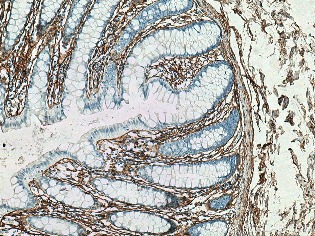
IHC staining of human colon using 80009-1-RR (same clone as 80009-1-PBS)
Immunohistochemical analysis of paraffin-embedded human colon tissue slide using 80009-1-RR (COL3A1 antibody) at dilution of 1:4000 (under 10x lens). Heat mediated antigen retrieval with Tris-EDTA buffer (pH 9.0). This data was developed using the same antibody clone with 80009-1-PBS in a different storage buffer formulation.
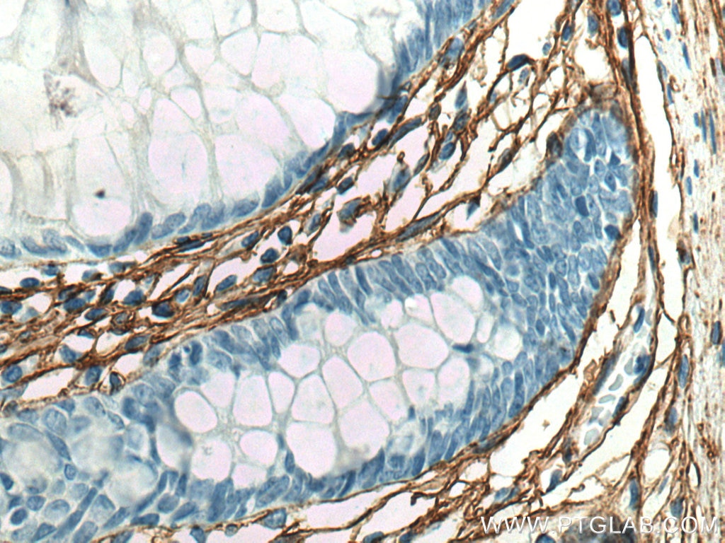
IHC staining of human colon using 80009-1-RR (same clone as 80009-1-PBS)
Immunohistochemical analysis of paraffin-embedded human colon tissue slide using 80009-1-RR (COL3A1 antibody) at dilution of 1:4000 (under 40x lens). Heat mediated antigen retrieval with Tris-EDTA buffer (pH 9.0). This data was developed using the same antibody clone with 80009-1-PBS in a different storage buffer formulation.
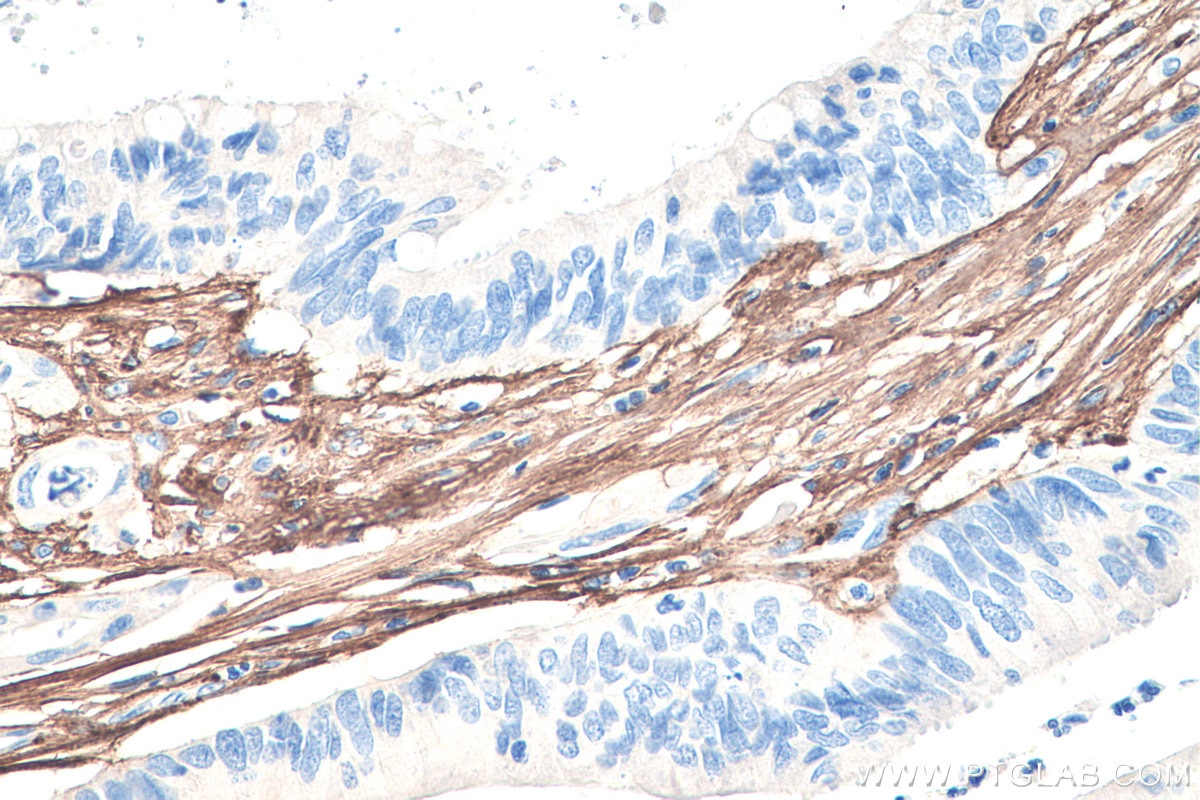
IHC staining of human colon cancer using 80009-1-RR (same clone as 80009-1-PBS)
Immunohistochemical analysis of paraffin-embedded human colon cancer tissue slide using 80009-1-RR (Collagen Type III (N-terminal) antibody) at dilution of 1:8000 (under 40x lens). Heat mediated antigen retrieval with Tris-EDTA buffer (pH 9.0). This data was developed using the same antibody clone with 80009-1-PBS in a different storage buffer formulation.
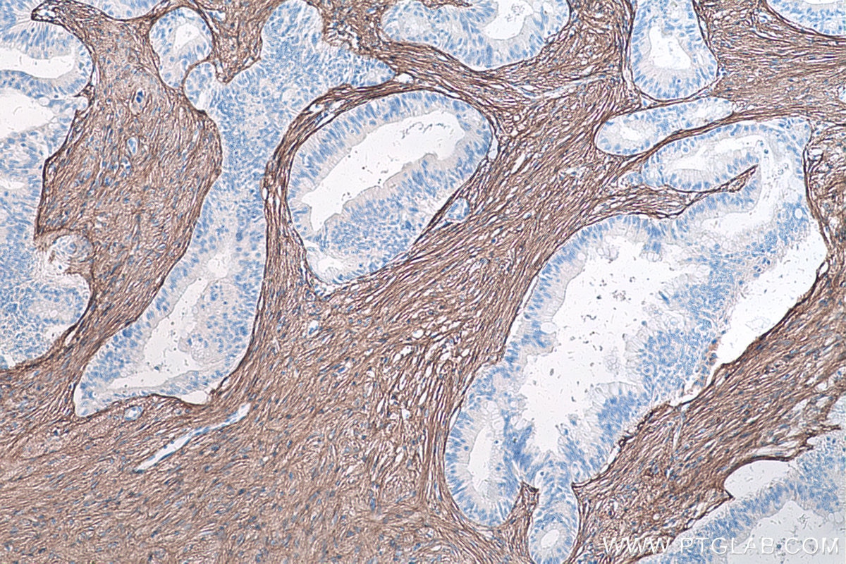
IHC staining of human ovary tumor using 80009-1-RR (same clone as 80009-1-PBS)
Immunohistochemical analysis of paraffin-embedded human ovary tumor tissue slide using 80009-1-RR (Collagen Type III (N-terminal) antibody) at dilution of 1:8000 (under 10x lens). Heat mediated antigen retrieval with Tris-EDTA buffer (pH 9.0). This data was developed using the same antibody clone with 80009-1-PBS in a different storage buffer formulation.
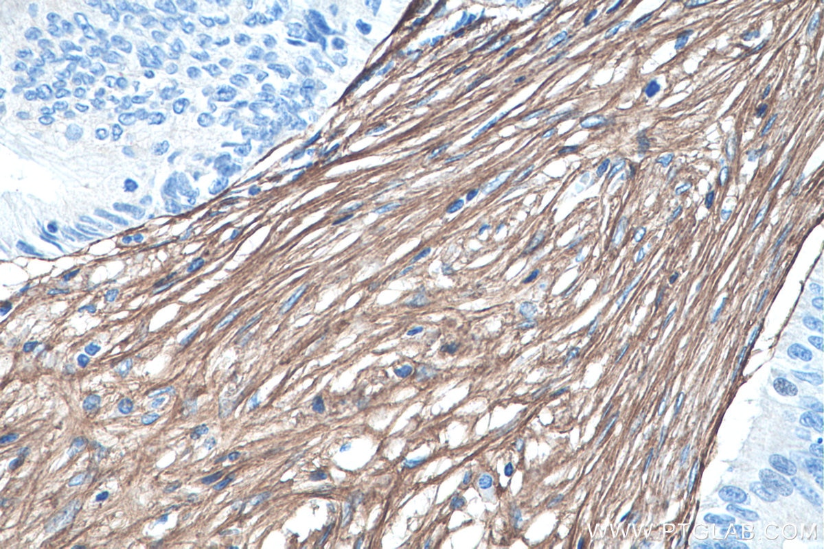
IHC staining of human ovary tumor using 80009-1-RR (same clone as 80009-1-PBS)
Immunohistochemical analysis of paraffin-embedded human ovary tumor tissue slide using 80009-1-RR (Collagen Type III (N-terminal) antibody) at dilution of 1:8000 (under 40x lens). Heat mediated antigen retrieval with Tris-EDTA buffer (pH 9.0). This data was developed using the same antibody clone with 80009-1-PBS in a different storage buffer formulation.
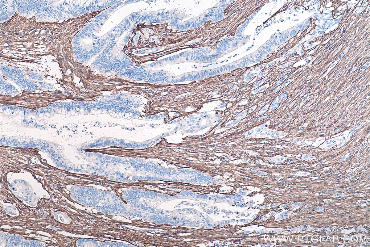
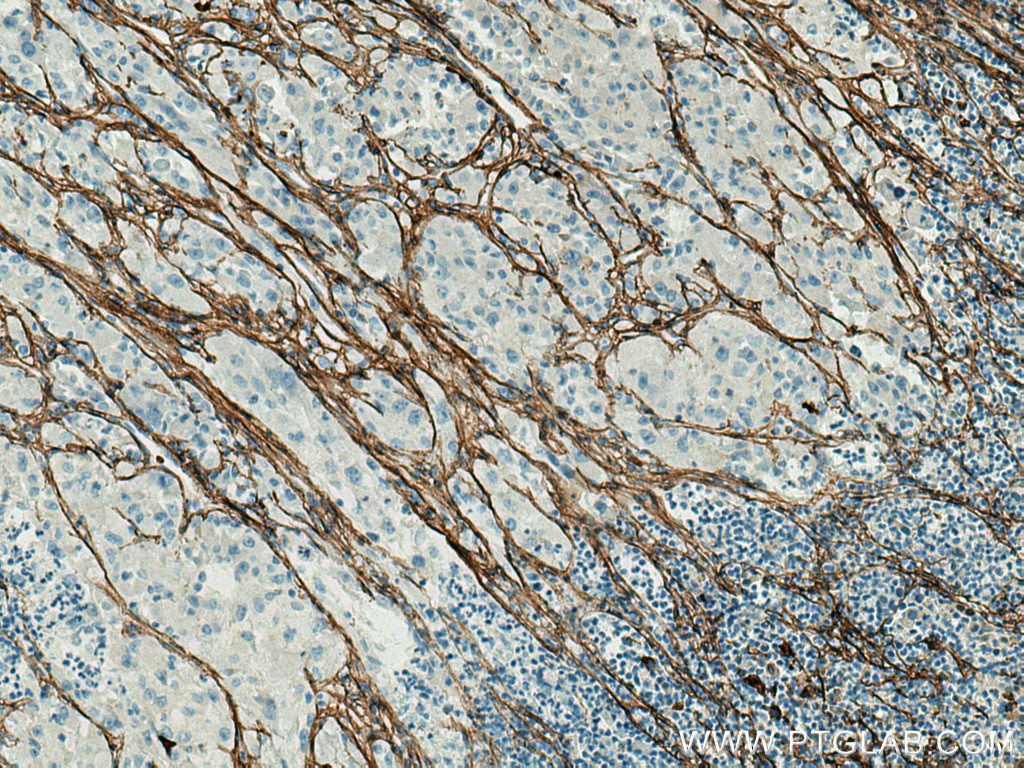
IHC staining of human pancreas cancer using 80009-1-RR (same clone as 80009-1-PBS)
Immunohistochemical analysis of paraffin-embedded human pancreas cancer tissue slide using 80009-1-RR (Collagen Type III (N-terminal) antibody) at dilution of 1:8000 (under 10x lens). Heat mediated antigen retrieval with Tris-EDTA buffer (pH 9.0). This data was developed using the same antibody clone with 80009-1-PBS in a different storage buffer formulation.
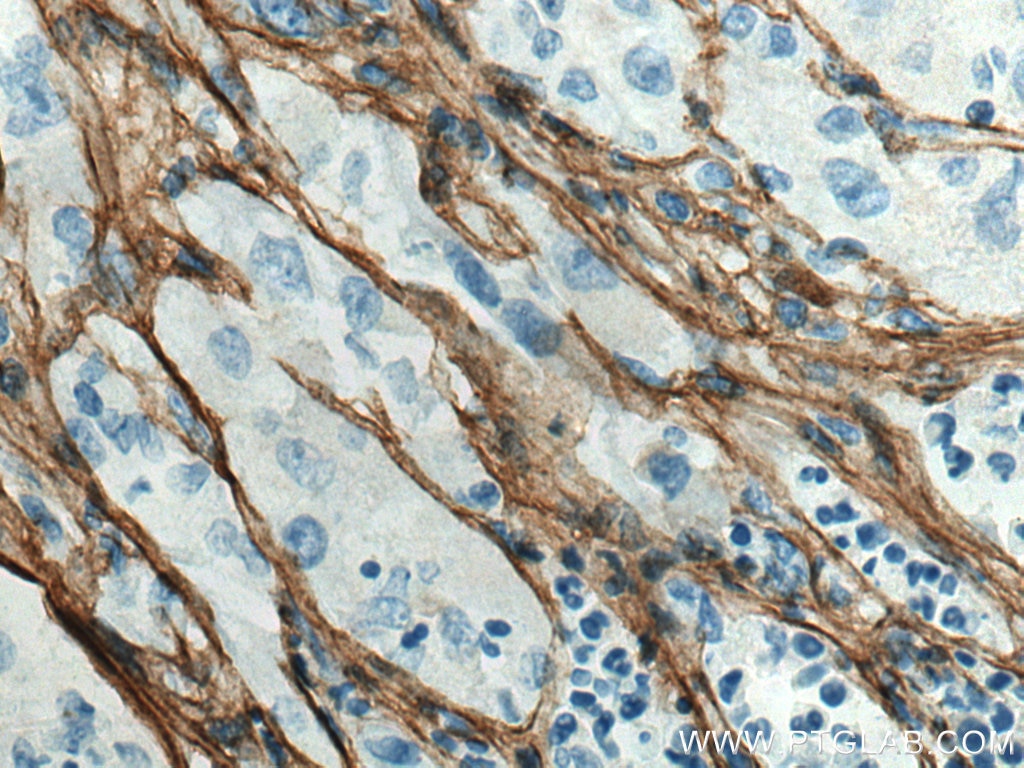
IHC staining of human pancreas cancer using 80009-1-RR (same clone as 80009-1-PBS)
Immunohistochemical analysis of paraffin-embedded human pancreas cancer tissue slide using 80009-1-RR (Collagen Type III (N-terminal) antibody) at dilution of 1:8000 (under 40x lens). Heat mediated antigen retrieval with Tris-EDTA buffer (pH 9.0). This data was developed using the same antibody clone with 80009-1-PBS in a different storage buffer formulation.
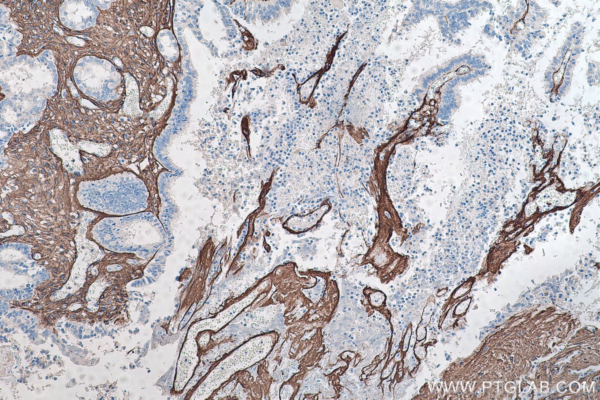
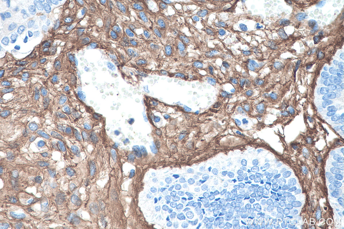
IHC staining of human colon cancer using 80009-1-RR (same clone as 80009-1-PBS)
Immunohistochemical analysis of paraffin-embedded human colon cancer tissue slide using 80009-1-RR (Collagen Type III (N-terminal) antibody) at dilution of 1:8000 (under 10x lens). Heat mediated antigen retrieval with Tris-EDTA buffer (pH 9.0). This data was developed using the same antibody clone with 80009-1-PBS in a different storage buffer formulation.
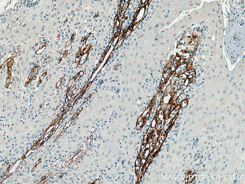
IHC staining of human skin cancer using 80009-1-RR (same clone as 80009-1-PBS)
Immunohistochemical analysis of paraffin-embedded human skin cancer tissue slide using 80009-1-RR (COL3A1 antibody) at dilution of 1:4000 (under 10x lens). Heat mediated antigen retrieval with Tris-EDTA buffer (pH 9.0). This data was developed using the same antibody clone with 80009-1-PBS in a different storage buffer formulation.
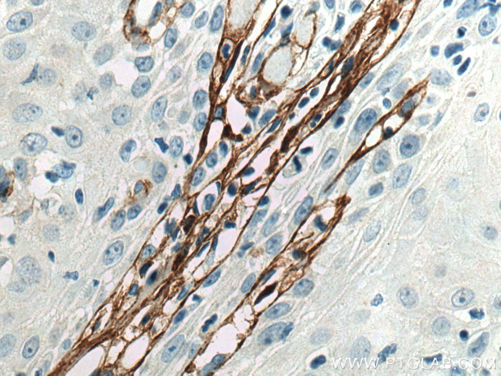
IHC staining of human skin cancer using 80009-1-RR (same clone as 80009-1-PBS)
Immunohistochemical analysis of paraffin-embedded human skin cancer tissue slide using 80009-1-RR (COL3A1 antibody) at dilution of 1:4000 (under 40x lens). Heat mediated antigen retrieval with Tris-EDTA buffer (pH 9.0). This data was developed using the same antibody clone with 80009-1-PBS in a different storage buffer formulation.
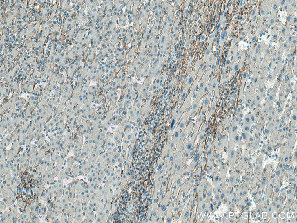
IHC staining of human liver cancer using 80009-1-RR (same clone as 80009-1-PBS)
Immunohistochemical analysis of paraffin-embedded human liver cancer tissue slide using 80009-1-RR (COL3A1 antibody) at dilution of 1:4000 (under 10x lens). Heat mediated antigen retrieval with Tris-EDTA buffer (pH 9.0). This data was developed using the same antibody clone with 80009-1-PBS in a different storage buffer formulation.
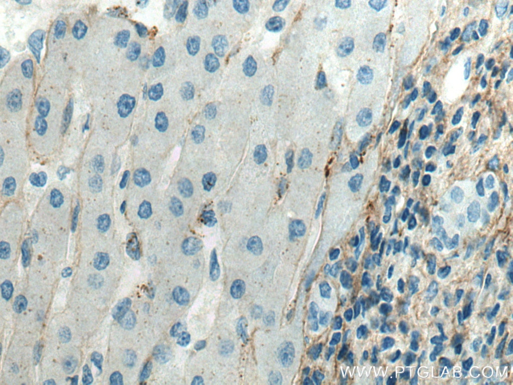
IHC staining of human liver cancer using 80009-1-RR (same clone as 80009-1-PBS)
Immunohistochemical analysis of paraffin-embedded human liver cancer tissue slide using 80009-1-RR (COL3A1 antibody) at dilution of 1:4000 (under 40x lens). Heat mediated antigen retrieval with Tris-EDTA buffer (pH 9.0). This data was developed using the same antibody clone with 80009-1-PBS in a different storage buffer formulation.
IHC staining of human liver cancer using 80009-1-RR (same clone as 80009-1-PBS)
Immunohistochemical analysis of paraffin-embedded human liver cancer tissue slide using 80009-1-RR (COL3A1 antibody) at dilution of 1:4000 (under 10x lens). Heat mediated antigen retrieval with Tris-EDTA buffer (pH 9.0). This data was developed using the same antibody clone with 80009-1-PBS in a different storage buffer formulation.
IHC staining of human liver cancer using 80009-1-RR (same clone as 80009-1-PBS)
Immunohistochemical analysis of paraffin-embedded human liver cancer tissue slide using 80009-1-RR (COL3A1 antibody) at dilution of 1:4000 (under 40x lens). Heat mediated antigen retrieval with Tris-EDTA buffer (pH 9.0). This data was developed using the same antibody clone with 80009-1-PBS in a different storage buffer formulation.
IF-P Figures
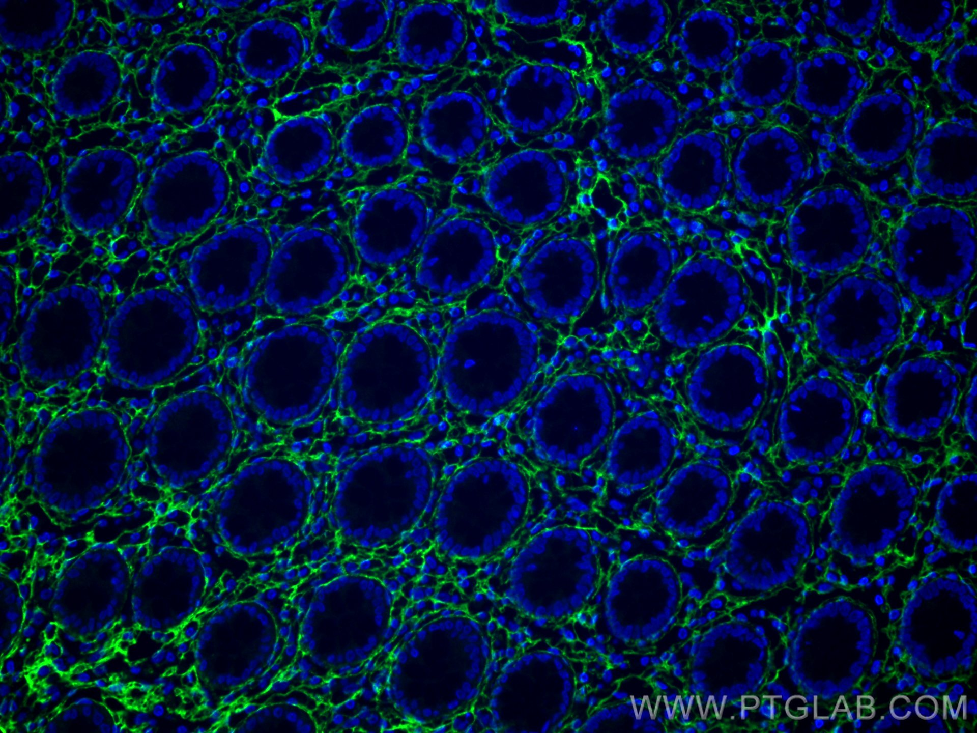
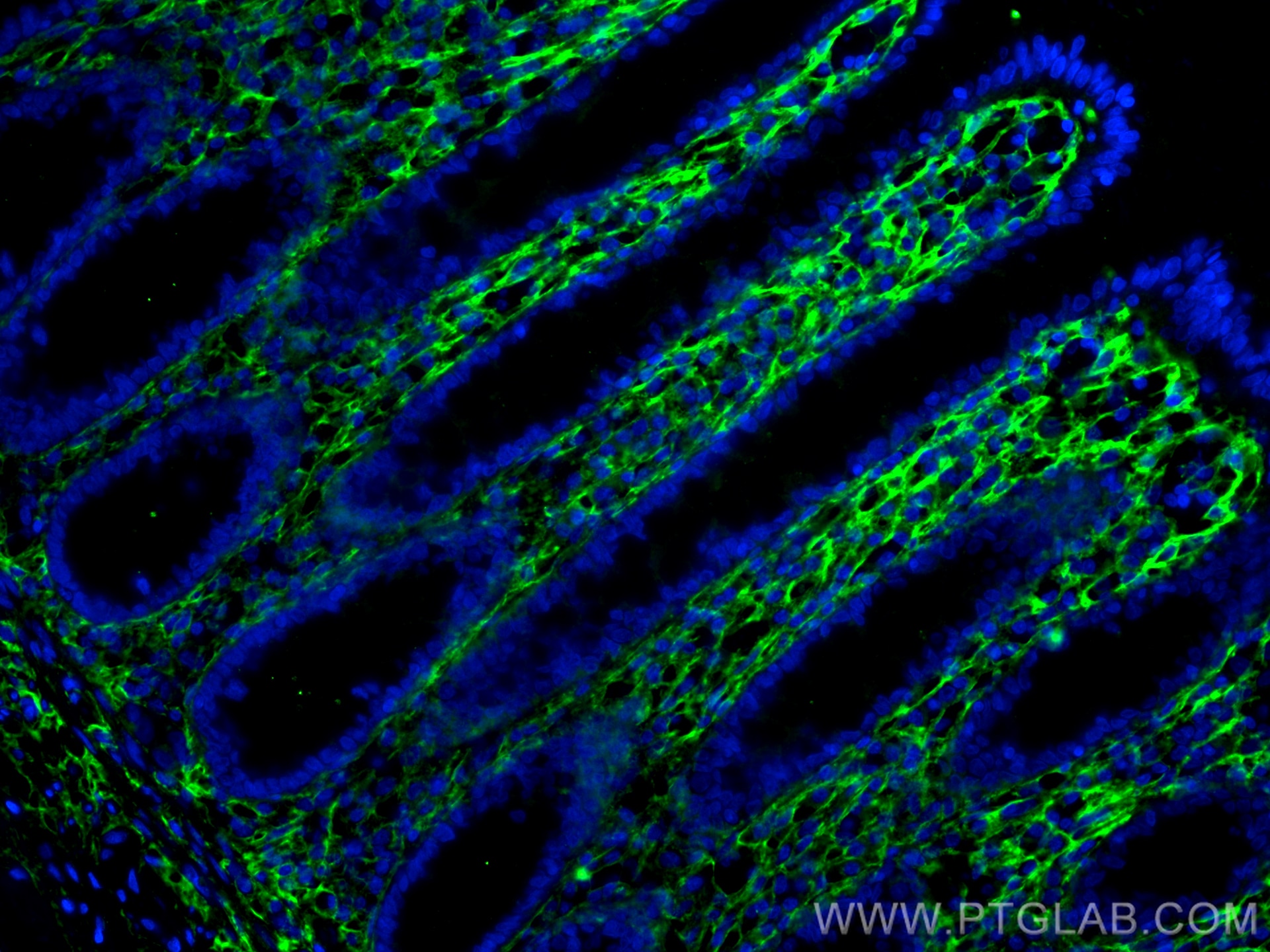
IF Staining of human colon using 80009-1-RR (same clone as 80009-1-PBS)
Immunofluorescent analysis of (4% PFA) fixed human colon tissue using Collagen Type III (N-terminal) antibody (80009-1-RR, Clone: 2O1 ) at dilution of 1:200 and CoraLite®488-Conjugated AffiniPure Goat Anti-Rabbit IgG(H+L). This data was developed using the same antibody clone with 80009-1-PBS in a different storage buffer formulation.
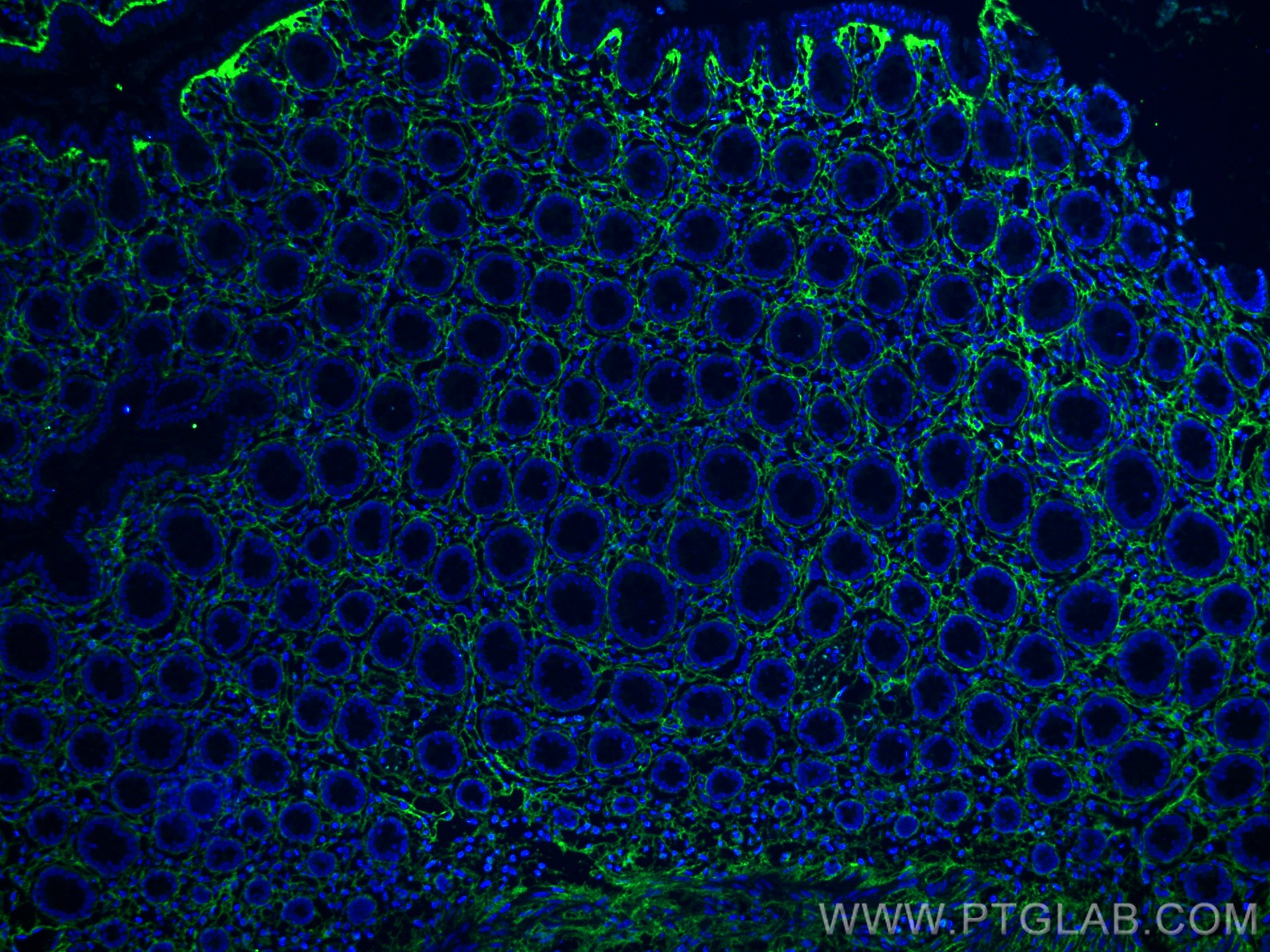
IF Staining of human colon using 80009-1-RR (same clone as 80009-1-PBS)
Immunofluorescent analysis of (4% PFA) fixed paraffin-embedded human colon tissue using Collagen Type III (N-terminal) antibody (80009-1-RR, Clone: 2O1 ) at dilution of 1:400 and Multi-rAb CoraLite ® Plus 488-Goat Anti-Rabbit Recombinant Secondary Antibody (H+L) (RGAR002). Heat mediated antigen retrieval with Tris-EDTA buffer (pH 9.0). This data was developed using the same antibody clone with 80009-1-PBS in a different storage buffer formulation.

