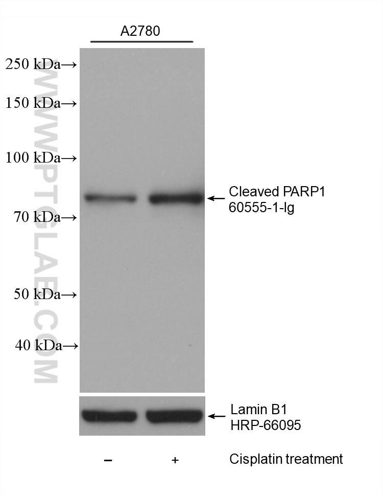
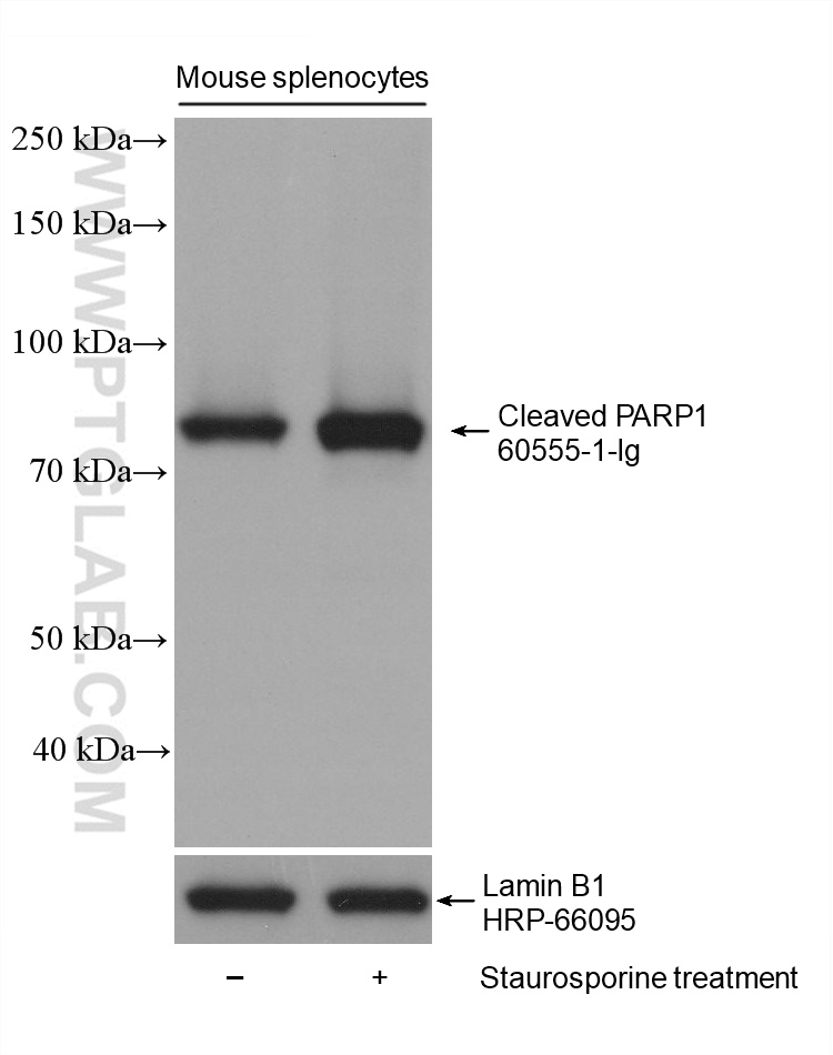
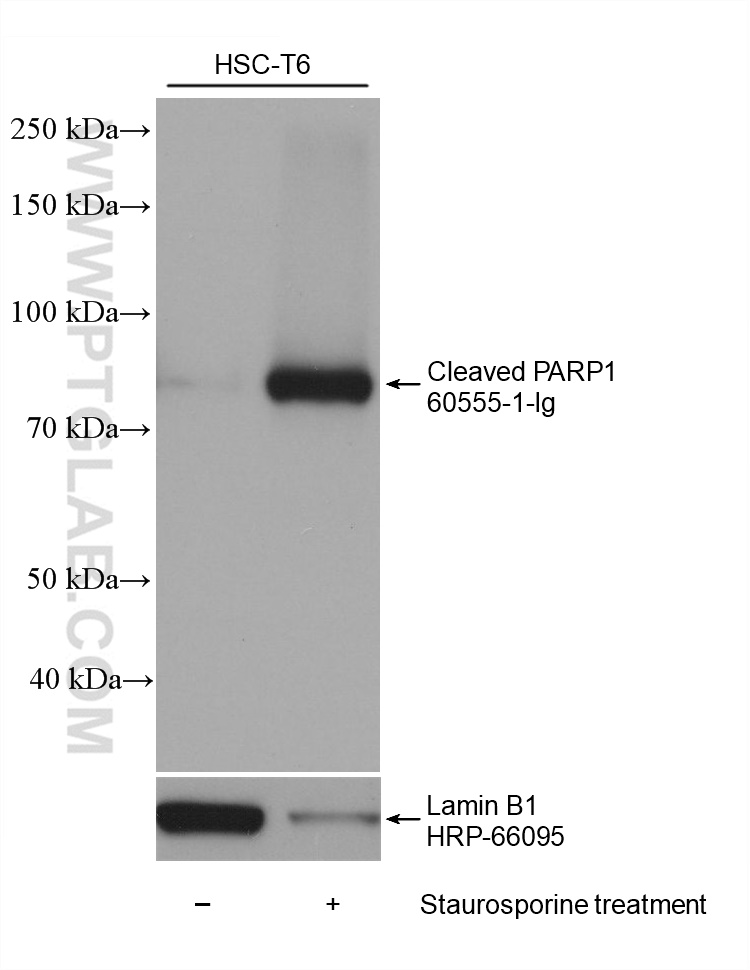
at dilution of 1:20000 incubated at room temperature for 1.5 hours. The membrane was stripped and re-blotted with HRP-conjugated Lamin B1 (HRP-66095) antibody as a loading control.")
 at dilution of 1:20000 incubated at room temperature for 1.5 hours. The membrane was stripped and re-blotted with HRP-conjugated Lamin B1 (HRP-66095) antibody as a loading control.")
 at dilution of 1:20000 incubated at room temperature for 1.5 hours. The membrane was stripped and re-blotted with HRP-conjugated Lamin B1 (HRP-66095) antibody as a loading control.")
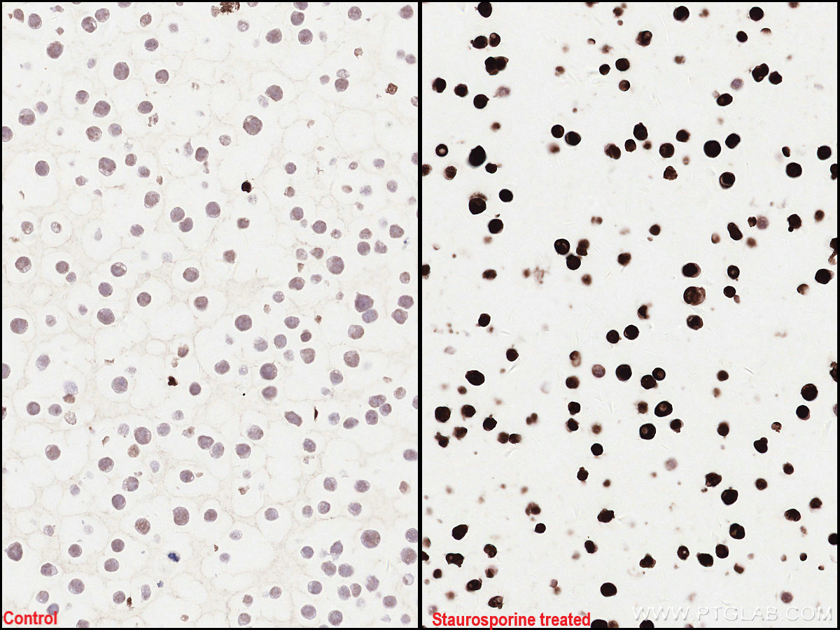
 and Staurosporine treated Jurkat (right) cells slide using 60555-1-Ig (Cleaved PARP1 antibody) at dilution of 1:2000 (under 40x lens). Heat mediated antigen retrieval with Tris-EDTA buffer (pH 9.0).")
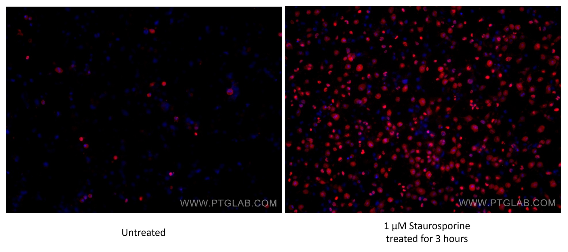
 fixed untreated and 1 μM Staurosporine (3 hours) treated HSC-T6 cells using Cleaved PARP1 antibody (60555-1-Ig, Clone: 4G4C8 ) at dilution of 1:1000 and Multi-rAb CoraLite® Plus 594-Goat Anti-Mouse Recombinant Secondary Antibody (H+L) (Cat.NO. RGAM004).")
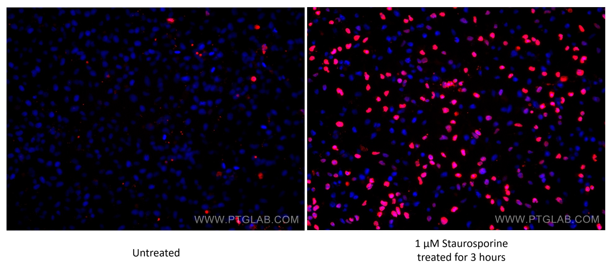
 fixed untreated and 1 μM Staurosporine (3 hours) treated HeLa cells using Cleaved PARP1 antibody (60555-1-Ig, Clone: 4G4C8 ) at dilution of 1:366 and Multi-rAb CoraLite® Plus 594-Goat Anti-Mouse Recombinant Secondary Antibody (H+L) (Cat.NO. RGAM004 ).")
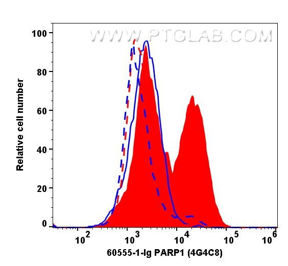
 and 1 μM Staurosporine (3 hours) treated HSC-T6 cells (full lines) were intracellularly stained with 0.4 μg Cleaved PARP1 Monoclonal Antibody (60555-1-Ig, Clone:4G4C8, red) and CoraLite® Plus 647-Goat Anti-Mouse Recombinant Secondary Antibody (H+L)(Cat.NO.RGAM005). Mouse IgG1 isotype control (66360-1-Ig, Clone: 1F8D3, blue) was parallel stained as control. Cells were fixed with 4% PFA.")
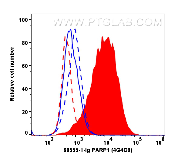
 and 1 μM Staurosporine (3 hours) treated HeLa cells (full lines) were intracellularly stained with 0.1 μg Cleaved PARP1 Monoclonal Antibody (60555-1-Ig, Clone:4G4C8, red) and CoraLite® Plus 647-Goat Anti-Mouse Recombinant Secondary Antibody (H+L)(Cat.NO.RGAM005). Mouse IgG1 isotype control (66360-1-Ig, Clone: 1F8D3, blue) was parallel stained as control. Cells were fixed with 4% PFA .")
Tested Applications
| Positive WB detected in | A2780 cells, HSC-T6 cells, mouse splenocytes, Staurosporine treated A2780 cells, Staurosporine treated HSC-T6 cells, Staurosporine treated mouse splenocytes |
| Positive IHC detected in | Jurkat cells Note: suggested antigen retrieval with TE buffer pH 9.0; (*) Alternatively, antigen retrieval may be performed with citrate buffer pH 6.0 |
| Positive IF/ICC detected in | 1 μM Staurosporine (3 hours) treated HSC-T6 cells, 1 μM Staurosporine (3 hours) treated HeLa cells |
| Positive FC (Intra) detected in | 1 μM Staurosporine (3 hours) treated HSC-T6 cells, 1 μM Staurosporine (3 hours) treated HeLa cells |
Recommended dilution
| Application | Dilution |
|---|---|
| Western Blot (WB) | WB : 1:5000-1:50000 |
| Immunohistochemistry (IHC) | IHC : 1:1000-1:4000 |
| Immunofluorescence (IF)/ICC | IF/ICC : 1:500-1:2000 |
| Flow Cytometry (FC) (INTRA) | FC (INTRA) : 0.40 ug per 10^6 cells in a 100 µl suspension |
| It is recommended that this reagent should be titrated in each testing system to obtain optimal results. | |
| Sample-dependent, Check data in validation data gallery. | |
Published Applications
| WB | See 6 publications below |
Product Information
60555-1-Ig targets Cleaved PARP1 in WB, IHC, IF/ICC, FC (Intra), ELISA applications and shows reactivity with human, mouse, rat samples.
| Tested Reactivity | human, mouse, rat |
| Cited Reactivity | human, mouse |
| Host / Isotype | Mouse / IgG1 |
| Class | Monoclonal |
| Type | Antibody |
| Immunogen |
Peptide Predict reactive species |
| Full Name | poly (ADP-ribose) polymerase 1 |
| Calculated Molecular Weight | 1014 aa, 113 kDa |
| Observed Molecular Weight | 89 kDa |
| GenBank Accession Number | BC037545 |
| Gene Symbol | PARP1 |
| Gene ID (NCBI) | 142 |
| Conjugate | Unconjugated |
| Form | Liquid |
| Purification Method | Protein G purification |
| UNIPROT ID | P09874 |
| Storage Buffer | PBS with 0.02% sodium azide and 50% glycerol, pH 7.3. |
| Storage Conditions | Store at -20°C. Stable for one year after shipment. Aliquoting is unnecessary for -20oC storage. 20ul sizes contain 0.1% BSA. |
Background Information
PARP1 (poly(ADP-ribose) polymerase 1) is a nuclear enzyme catalyzing the poly(ADP-ribosyl)ation of many key proteins in vivo. The normal function of PARP1 is the routine repair of DNA damage. Activated by DNA strand breaks, the PARP1 is cleaved into an 85 to 89-kDa COOH-terminal fragment and a 24 kDa NH2-terminal peptide by caspases during the apoptotic process. The appearance of PARP fragments is commonly considered an important biomarker of apoptosis. In addition to caspases, other proteases like calpains, cathepsins, granzymes, and matrix metalloproteinases (MMPs) have also been reported to cleave PARP1 and give rise to fragments ranging from 42-89 kDa.
This antibody only recognizes the cleaved form of PAPR1 but not full-length PARP1.
Protocols
| Product Specific Protocols | |
|---|---|
| IF protocol for Cleaved PARP1 antibody 60555-1-Ig | Download protocol |
| IHC protocol for Cleaved PARP1 antibody 60555-1-Ig | Download protocol |
| WB protocol for Cleaved PARP1 antibody 60555-1-Ig | Download protocol |
| Standard Protocols | |
|---|---|
| Click here to view our Standard Protocols |
Publications
| Species | Application | Title |
|---|---|---|
Acta Pharm Sin B Design, synthesis, and antitumor activity of novel thioheterocyclic nucleoside derivatives by suppressing the c-MYC pathway | ||
Oncogene Vimentin intermediate filaments orchestrate DNA nonhomologous end joining repair and lipolysis after DNA damage | ||
Front Immunol An anoikis-related signature predicts prognosis and immunotherapy response in gastrointestinal cancers | ||
Front Endocrinol (Lausanne) Bioinformatics analysis combined with experimental validation reveals the novel mechanisms of multi-targets of dapagliflozin attenuating diabetic liver injury | ||
Bioorg Chem Synthesis and biological evaluation of novel isatin-phenol hybrids as potential antitumor agents | ||
Int J Mol Med RBM8A promotes gastric cancer progression by binding with UPF3B to induce BBC3 mRNA degradation |