at dilution of 1:20000 incubated at room temperature for 1.5 hours.")
 at dilution of 1:4000 incubated at room temperature for 1.5 hours.")
 at dilution of 1:20000 incubated at room temperature for 1.5 hours.")
 at dilution of 1:20000 incubated at room temperature for 1.5 hours.")
 at dilution of 1:20000 incubated at room temperature for 1.5 hours.")
 at dilution of 1:600 (under 10x lens). Heat mediated antigen retrieval with Tris-EDTA buffer (pH 9.0).")
 at dilution of 1:600 (under 40x lens). Heat mediated antigen retrieval with Tris-EDTA buffer (pH 9.0).")
 at dilution of 1:600 (under 10x lens). Heat mediated antigen retrieval with Tris-EDTA buffer (pH 9.0).")
 at dilution of 1:600 (under 40x lens). Heat mediated antigen retrieval with Tris-EDTA buffer (pH 9.0).")
 fixed mouse liver tissue using CYP3A4 antibody (67110-1-Ig, Clone: 2F11C4 ) at dilution of 1:400 and CoraLite®488-Conjugated AffiniPure Goat Anti-Mouse IgG(H+L).")
 fixed mouse liver tissue using CYP3A4 antibody (67110-1-Ig, Clone: 2F11C4 ) at dilution of 1:400 and CoraLite®488-Conjugated AffiniPure Goat Anti-Mouse IgG(H+L).")
 fixed rat liver tissue using CYP3A4 antibody (67110-1-Ig, Clone: 2F11C4 ) at dilution of 1:1000 and CoraLite®488-Conjugated AffiniPure Goat Anti-Mouse IgG(H+L).")
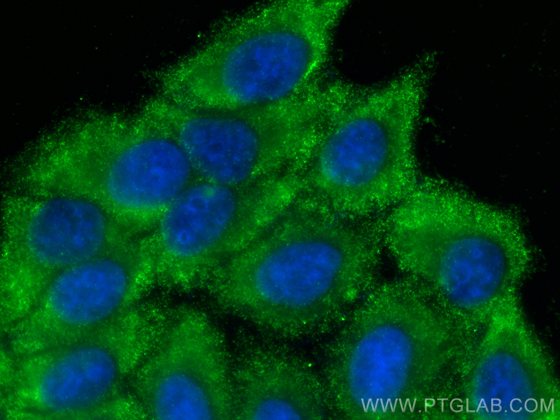
 fixed HepG2 cells using CYP3A4 antibody (67110-1-Ig, Clone: 2F11C4 ) at dilution of 1:800 and CoraLite®488-Conjugated Goat Anti-Mouse IgG(H+L) (SA00013-1).")
Tested Applications
| Positive WB detected in | mouse liver tissue, pig liver tissue, pig gall bladder tissue, mouse lung tissue, rat liver tissue |
| Positive IHC detected in | mouse liver tissue, human liver cancer tissue Note: suggested antigen retrieval with TE buffer pH 9.0; (*) Alternatively, antigen retrieval may be performed with citrate buffer pH 6.0 |
| Positive IF-P detected in | mouse liver tissue, rat liver tissue |
| Positive IF/ICC detected in | HepG2 cells |
Recommended dilution
| Application | Dilution |
|---|---|
| Western Blot (WB) | WB : 1:5000-1:50000 |
| Immunohistochemistry (IHC) | IHC : 1:300-1:1200 |
| Immunofluorescence (IF)-P | IF-P : 1:200-1:800 |
| Immunofluorescence (IF)/ICC | IF/ICC : 1:400-1:1600 |
| It is recommended that this reagent should be titrated in each testing system to obtain optimal results. | |
| Sample-dependent, Check data in validation data gallery. | |
Published Applications
| WB | See 2 publications below |
| IF | See 4 publications below |
Product Information
67110-1-Ig targets CYP3A4 in WB, IHC, IF/ICC, IF-P, ELISA applications and shows reactivity with human, mouse, rat, pig samples.
| Tested Reactivity | human, mouse, rat, pig |
| Cited Reactivity | human, mouse, rat |
| Host / Isotype | Mouse / IgG1 |
| Class | Monoclonal |
| Type | Antibody |
| Immunogen |
CatNo: Ag13074 Product name: Recombinant human CYP3A4 protein Source: e coli.-derived, T-HIS Tag: 6*His Domain: 154-503 aa of BC101631 Sequence: DVLVRNLRREAETGKPVTLKDVFGAYSMDVITSTSFGVNIDSLNNPQDPFVENTKKLLRFDFLDPFFLSITVFPFLIPILEVLNICVFPREVTNFLRKSVKRMKESRLEDTQKHRVDFLQLMIDSQNSKETESHKALSDLELVAQSIIFIFAGYETTSSVLSFIMYELATHPDVQQKLQEEIDAVLPNKAPPTYDTVLQMEYLDMVVNETLRLFPIAMRLERVCKKDVEINGMFIPKGVVVMIPSYALHRDPKYWTEPEKFLPERFSKKNKDNIDPYIYTPFGSGPRNCIGMRFALMNMKLALIRVLQNFSFKPCKETQIPLKLSLGGLLQPEKPVVLKVESRDGTVSGA Predict reactive species |
| Full Name | cytochrome P450, family 3, subfamily A, polypeptide 4 |
| Calculated Molecular Weight | 503 aa, 57 kDa |
| Observed Molecular Weight | 51-57 kDa |
| GenBank Accession Number | BC101631 |
| Gene Symbol | CYP3A4 |
| Gene ID (NCBI) | 1576 |
| RRID | AB_2882414 |
| Conjugate | Unconjugated |
| Form | Liquid |
| Purification Method | Protein G purification |
| UNIPROT ID | P08684 |
| Storage Buffer | PBS with 0.02% sodium azide and 50% glycerol, pH 7.3. |
| Storage Conditions | Store at -20°C. Stable for one year after shipment. Aliquoting is unnecessary for -20oC storage. 20ul sizes contain 0.1% BSA. |
Protocols
| Product Specific Protocols | |
|---|---|
| IF protocol for CYP3A4 antibody 67110-1-Ig | Download protocol |
| IHC protocol for CYP3A4 antibody 67110-1-Ig | Download protocol |
| WB protocol for CYP3A4 antibody 67110-1-Ig | Download protocol |
| Standard Protocols | |
|---|---|
| Click here to view our Standard Protocols |
Publications
| Species | Application | Title |
|---|---|---|
Biomaterials Hepatic endoplasmic reticulum-derived nanodiscs for broad-spectrum drug detoxification | ||
Biomed Rep Phenotypical, functional and transcriptomic comparison of two modified methods of hepatocyte differentiation from human induced pluripotent stem cells. | ||
Methods Mol Biol Homogeneous Differentiation of Functional Hepatocytes from Human Induced Pluripotent Stem Cells. | ||
Eur J Pharmacol Investigating the 'RT-PK' phenomenon: Effects of X-ray radiation on cabozantinib metabolism | ||
Cell Rep Identifying the target, mechanism, and agonist of α-ketoglutaric acid in delaying mesenchymal stem cell senescence | ||
Acta Biomater Hepar-on-a-sensor-platform with hybridization chain reaction amplification strategy to intuitively monitor the hepatoxicity of natural compounds |