at dilution of 1:400 incubated at room temperature for 1.5 hours.")
 at dilution of 1:2000 incubated at room temperature for 1.5 hours.")
 at dilution of 1:300 incubated at room temperature for 1.5 hours.")
 at dilution of 1:2000 incubated at room temperature for 1.5 hours.")
 at dilution of 1:200 (under 40x lens. Heat mediated antigen retrieval with Tris-EDTA buffer (pH 9.0).")
 at dilution of 1:200 (under 10x lens. Heat mediated antigen retrieval with Tris-EDTA buffer (pH 9.0).")
 fixed human gliomas tissue using Mu Crystallin antibody (12495-1-AP) at dilution of 1:200 and CoraLite®488-Conjugated AffiniPure Goat Anti-Rabbit IgG(H+L).")
 fixed human gliomas tissue using Mu Crystallin antibody (12495-1-AP) at dilution of 1:600 and CoraLite®488-Conjugated AffiniPure Goat Anti-Rabbit IgG(H+L).")
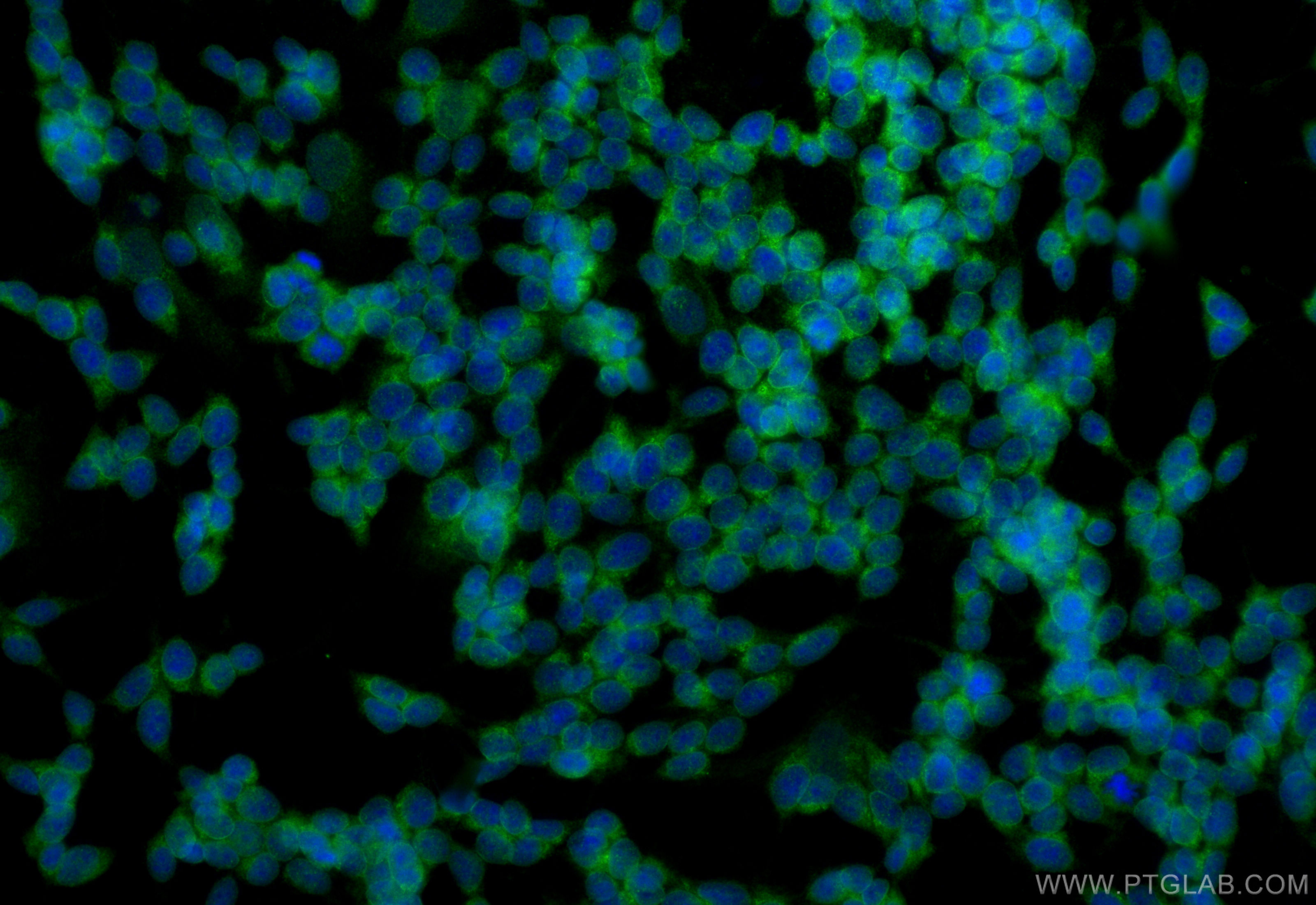
 fixed SH-SY5Y cells using Mu Crystallin antibody (12495-1-AP) at dilution of 1:200 and CoraLite®488-Conjugated Goat Anti-Rabbit IgG(H+L) (SA00013-2).")
 and CoraLite®488-Conjugated AffiniPure Goat Anti-Rabbit IgG(H+L) at dilution 1:1000 (red), or 0.2 ug Control Antibody. Cells were fixed with 4% PFA and permeabilized with Flow Cytometry Perm Buffer.")
Tested Applications
| Positive WB detected in | Y79 cells, Jurkat cells, human kidney tissue, human heart tissue |
| Positive IHC detected in | human gliomas tissue Note: suggested antigen retrieval with TE buffer pH 9.0; (*) Alternatively, antigen retrieval may be performed with citrate buffer pH 6.0 |
| Positive IF-P detected in | human gliomas tissue |
| Positive IF/ICC detected in | SH-SY5Y cells |
| Positive FC (Intra) detected in | SH-SY5Y cells |
Recommended dilution
| Application | Dilution |
|---|---|
| Western Blot (WB) | WB : 1:1000-1:4000 |
| Immunohistochemistry (IHC) | IHC : 1:50-1:500 |
| Immunofluorescence (IF)-P | IF-P : 1:50-1:500 |
| Immunofluorescence (IF)/ICC | IF/ICC : 1:50-1:500 |
| Flow Cytometry (FC) (INTRA) | FC (INTRA) : 0.20 ug per 10^6 cells in a 100 µl suspension |
| It is recommended that this reagent should be titrated in each testing system to obtain optimal results. | |
| Sample-dependent, Check data in validation data gallery. | |
Published Applications
| WB | See 2 publications below |
| IHC | See 1 publications below |
| IF | See 2 publications below |
Product Information
12495-1-AP targets Mu Crystallin in WB, IHC, IF/ICC, IF-P, FC (Intra), ELISA applications and shows reactivity with human, mouse, rat samples.
| Tested Reactivity | human, mouse, rat |
| Cited Reactivity | mouse, rat |
| Host / Isotype | Rabbit / IgG |
| Class | Polyclonal |
| Type | Antibody |
| Immunogen |
CatNo: Ag3161 Product name: Recombinant human CRYM protein Source: e coli.-derived, PGEX-4T Tag: GST Domain: 1-314 aa of BC018061 Sequence: MSRVPAFLSAAEVEEHLRSSSLLIPPLETALANFSSGPEGGVMQPVRTVVPVTKHRGYLGVMPAYSAAEDALTTKLVTFYEDRGITSVVPSHQATVLLFEPSNGTLLAVMDGNVITAKRTAAVSAIATKFLKPPSSEVLCILGAGVQAYSHYEIFTEQFSFKEVRIWNRTKENAEKFADTVQGEVRVCSSVQEAVAGADVIITVTLATEPILFGEWVKPGAHINAVGASRPDWRELDDELMKEAVLYVDSQEAALKESGDVLLSGAEIFAELGEVIKGVKPAHCEKTTVFKSLGMAVEDTVAAKLIYDSWSSGK Predict reactive species |
| Full Name | crystallin, mu |
| Calculated Molecular Weight | 314 aa, 34 kDa |
| Observed Molecular Weight | 34 kDa |
| GenBank Accession Number | BC018061 |
| Gene Symbol | Mu Crystallin |
| Gene ID (NCBI) | 1428 |
| RRID | AB_2084620 |
| Conjugate | Unconjugated |
| Form | Liquid |
| Purification Method | Antigen affinity purification |
| UNIPROT ID | Q14894 |
| Storage Buffer | PBS with 0.02% sodium azide and 50% glycerol, pH 7.3. |
| Storage Conditions | Store at -20°C. Stable for one year after shipment. Aliquoting is unnecessary for -20oC storage. 20ul sizes contain 0.1% BSA. |
Background Information
mu Crystallin(thiomorpholine-carboxylate dehydrogenase) is also named as THBP, CRYM, ketimine reductase and belongs to the ornithine cyclodeaminase family. This protein catalyzes the reduction of imine bonds in brain substrates that may include cystathionine ketimine (CysK) and lanthionine ketimine (LK). It is also involved in the regulation of the free intracellular concentration of triiodothyronine and access to its nuclear receptor.
Protocols
| Product Specific Protocols | |
|---|---|
| FC protocol for Mu Crystallin antibody 12495-1-AP | Download protocol |
| IF protocol for Mu Crystallin antibody 12495-1-AP | Download protocol |
| IHC protocol for Mu Crystallin antibody 12495-1-AP | Download protocol |
| WB protocol for Mu Crystallin antibody 12495-1-AP | Download protocol |
| Standard Protocols | |
|---|---|
| Click here to view our Standard Protocols |
Publications
| Species | Application | Title |
|---|---|---|
Dev Cell Single-Cell Transcriptomic Analyses of the Developing Meninges Reveal Meningeal Fibroblast Diversity and Function. | ||
Endocr J Follicular thyroglobulin enhances gene expression necessary for thyroid hormone secretion. | ||
Neuropathology Expression of CRYM in different rat organs during development and its decreased expression in degenerating pyramidal tracts in amyotrophic lateral sclerosis. |