at dilution of 1:10000 incubated at room temperature for 1.5 hours.")
 at dilution of 1:10000 incubated at room temperature for 1.5 hours.")
 at dilution of 1:10000 incubated at room temperature for 1.5 hours.")
 at dilution of 1:10000 incubated at room temperature for 1.5 hours.")
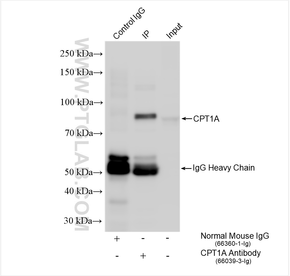
 with MCF-7 cells lysate 1520 ug.")
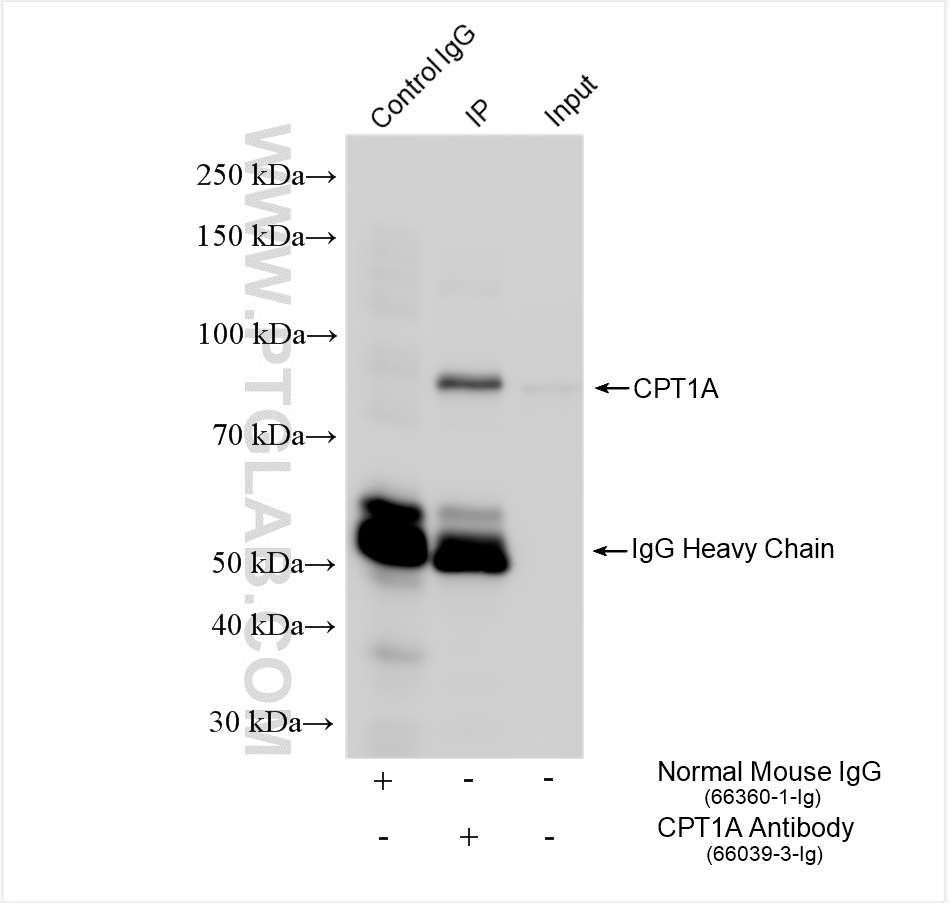
 with HepG2 cells lysate 1800 ug.")
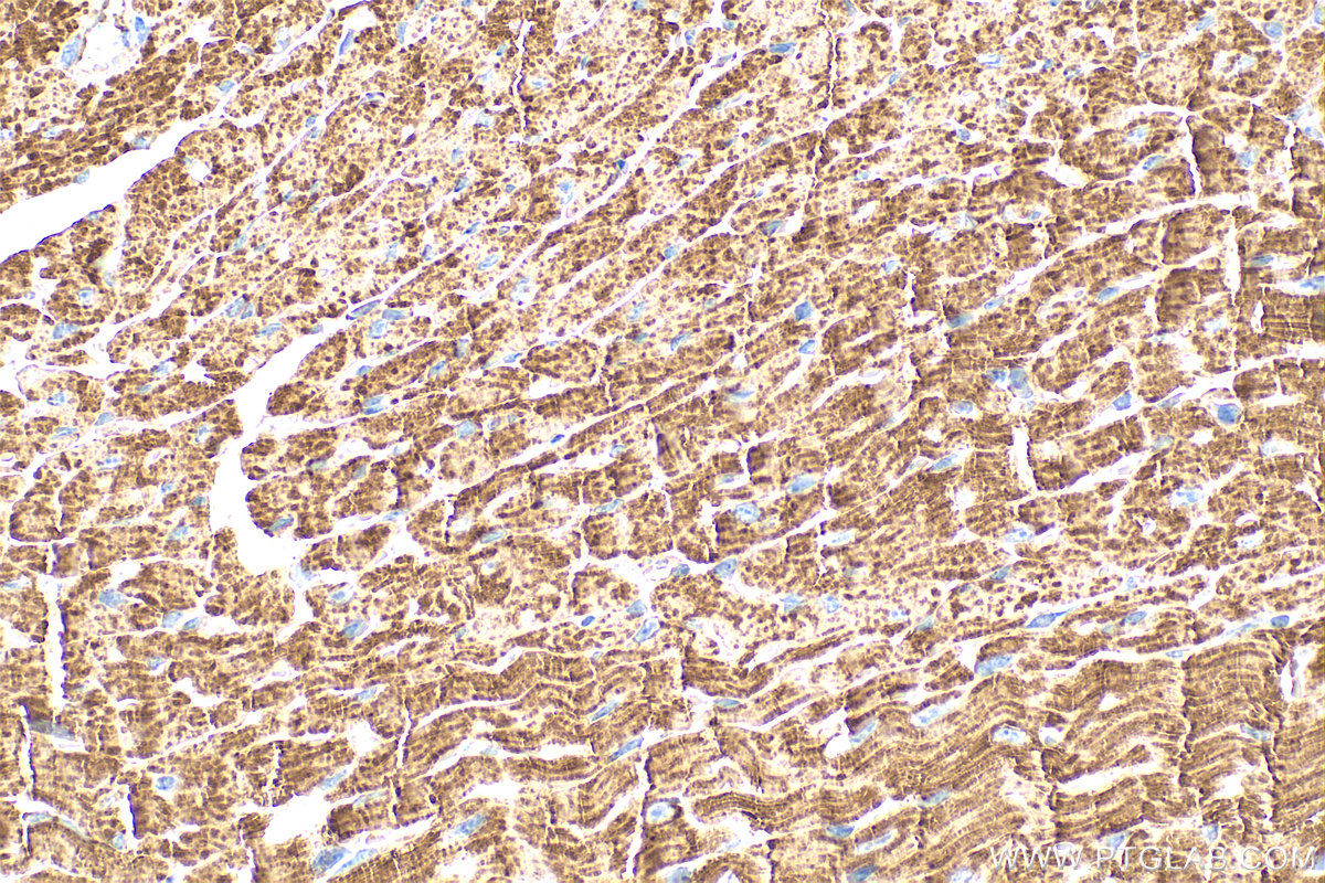
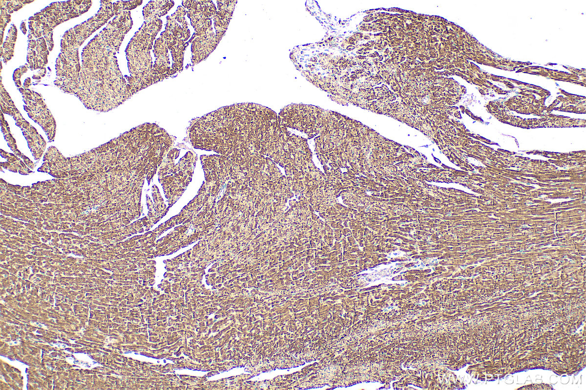
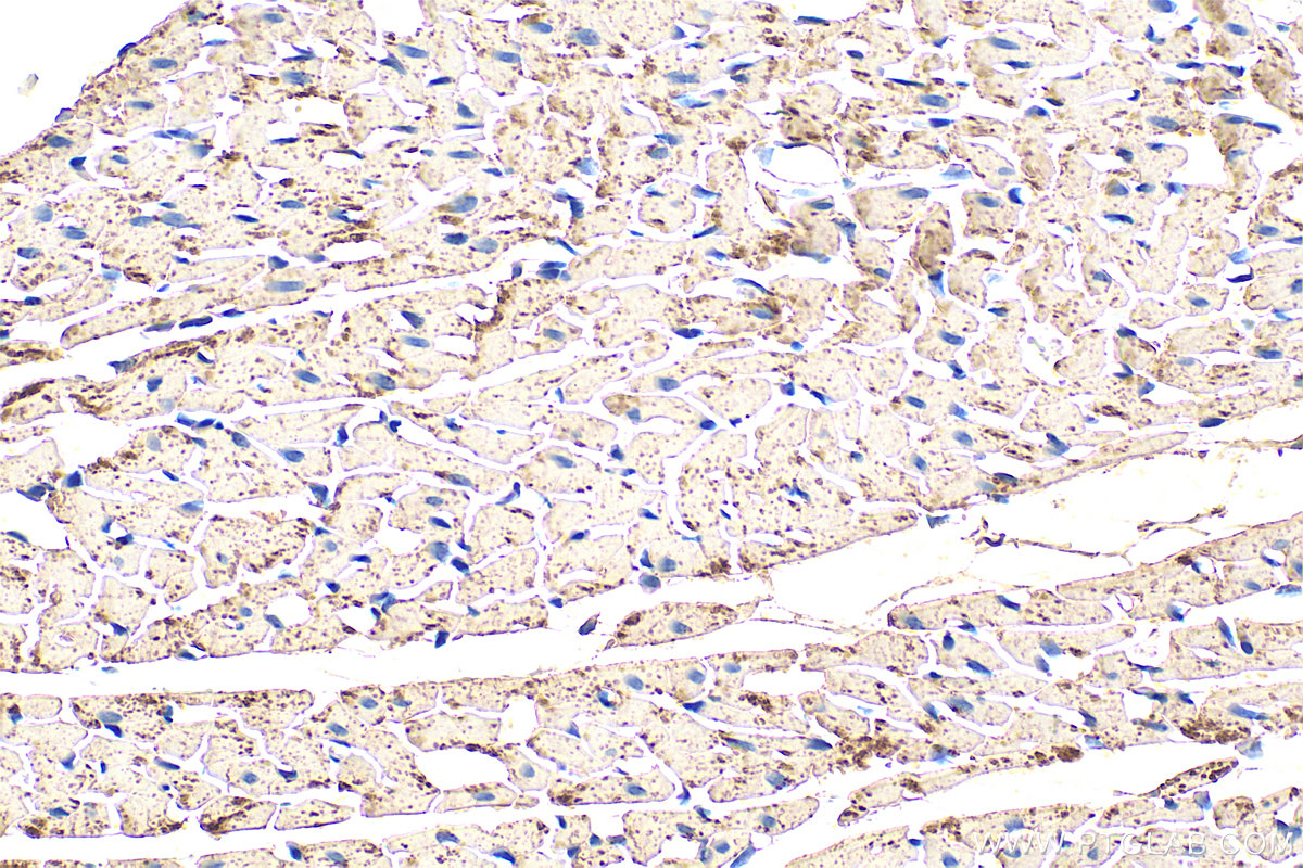
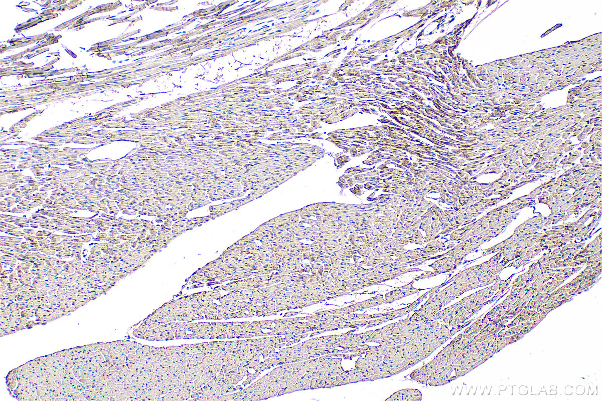
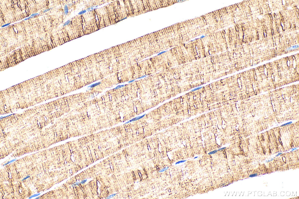
 at dilution of 1:1000 (under 40x lens). Heat mediated antigen retrieval with Tris-EDTA buffer (pH 9.0).")
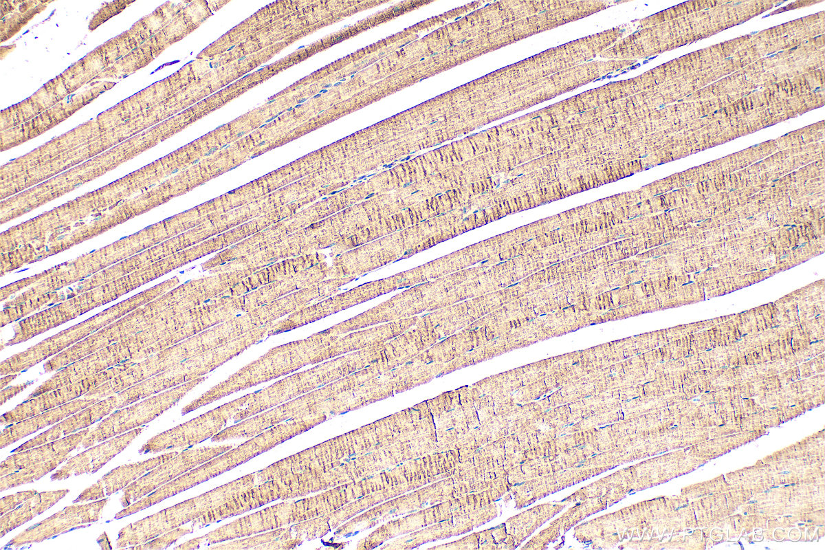
 at dilution of 1:1000 (under 10x lens). Heat mediated antigen retrieval with Tris-EDTA buffer (pH 9.0).")
 at dilution of 1:1000 (under 40x lens). Heat mediated antigen retrieval with Tris-EDTA buffer (pH 9.0).")
 at dilution of 1:1000 (under 10x lens). Heat mediated antigen retrieval with Tris-EDTA buffer (pH 9.0).")
 at dilution of 1:1000 (under 10x lens). Heat mediated antigen retrieval with Tris-EDTA buffer (pH 9.0).")
 at dilution of 1:1000 (under 40x lens). Heat mediated antigen retrieval with Tris-EDTA buffer (pH 9.0).")
Tested Applications
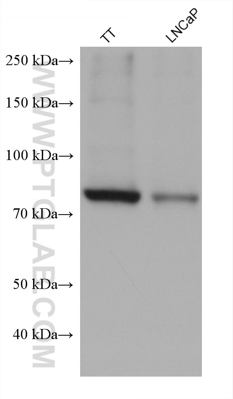
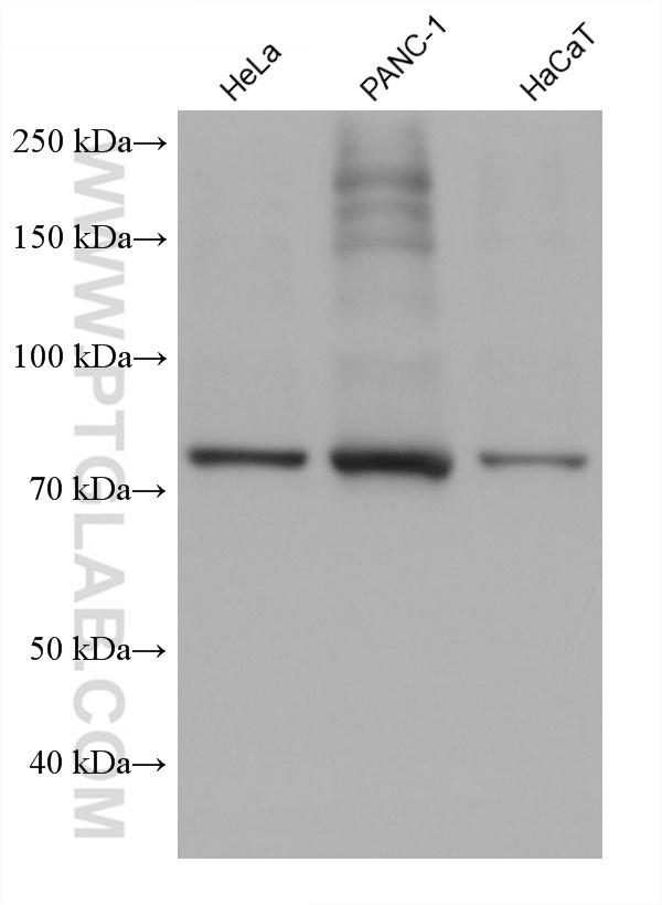
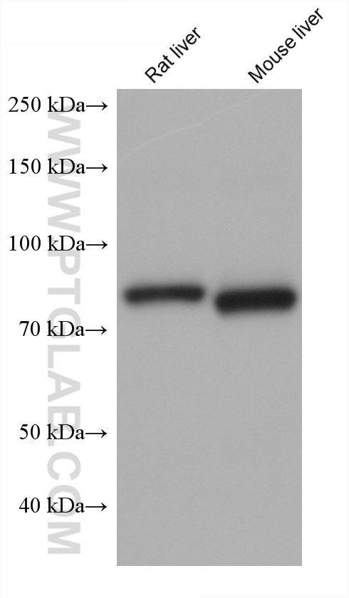
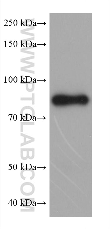
| Positive WB detected in | TT cells, HeLa cells, rat liver tissue, mouse liver tissue, LNCaP cells, PANC-1 cells, HaCaT cells |
| Positive IP detected in | MCF-7 cells, HepG2 cells |
| Positive IHC detected in | mouse heart tissue, mouse skeletal muscle tissue, rat heart tissue Note: suggested antigen retrieval with TE buffer pH 9.0; (*) Alternatively, antigen retrieval may be performed with citrate buffer pH 6.0 |
Recommended dilution
| Application | Dilution |
|---|---|
| Western Blot (WB) | WB : 1:5000-1:50000 |
| Immunoprecipitation (IP) | IP : 0.5-4.0 ug for 1.0-3.0 mg of total protein lysate |
| Immunohistochemistry (IHC) | IHC : 1:500-1:2000 |
| It is recommended that this reagent should be titrated in each testing system to obtain optimal results. | |
| Sample-dependent, Check data in validation data gallery. | |
Product Information
66039-3-Ig targets CPT1A in WB, IHC, IP, ELISA applications and shows reactivity with human, mouse, rat samples.
| Tested Reactivity | human, mouse, rat |
| Host / Isotype | Mouse / IgG1 |
| Class | Monoclonal |
| Type | Antibody |
| Immunogen | CPT1A fusion protein Ag7745 Predict reactive species |
| Full Name | carnitine palmitoyltransferase 1A (liver) |
| Calculated Molecular Weight | 88 kDa |
| Observed Molecular Weight | 86 kDa |
| GenBank Accession Number | BC000185 |
| Gene Symbol | CPT1A |
| Gene ID (NCBI) | 1374 |
| RRID | AB_3670374 |
| Conjugate | Unconjugated |
| Form | Liquid |
| Purification Method | Protein G purification |
| UNIPROT ID | P50416 |
| Storage Buffer | PBS with 0.02% sodium azide and 50% glycerol, pH 7.3. |
| Storage Conditions | Store at -20°C. Stable for one year after shipment. Aliquoting is unnecessary for -20oC storage. 20ul sizes contain 0.1% BSA. |
Background Information
CPT1A, also named as CPT1, CPT1-L and L-CPTI, belongs to the carnitine/choline acetyltransferase family. Carnitine palmitoyltransferase (CPT) deficiencies are common disorders of mitochondrial fatty acid oxidation. The CPT system is made up of two separate proteins located in the outer (CPT1) and inner (CPT2) mitochondrial membranes. CPT1A is an active forms of related liver-type carnitine palmitoyltransferase I. (PMID: 11001805). CPT1A deficiency presents as recurrent attacks of fasting hypoketotic hypoglycemia. (PMID: 15363638). This antibody can bind the close sequences genes.
Protocols
| Product Specific Protocols | |
|---|---|
| WB protocol for CPT1A antibody 66039-3-Ig | Download protocol |
| IHC protocol for CPT1A antibody 66039-3-Ig | Download protocol |
| IP protocol for CPT1A antibody 66039-3-Ig | Download protocol |
| Standard Protocols | |
|---|---|
| Click here to view our Standard Protocols |