Various lysates were subjected to SDS PAGE followed by western blot with 68259-1-Ig (CHCHD3 antibody) at dilution of 1:20000 incubated at room temperature for 1.5 hours.
Various lysates were subjected to SDS PAGE followed by western blot with 68259-1-Ig (CHCHD3 antibody) at dilution of 1:20000 incubated at room temperature for 1.5 hours.
WB analysis using 68259-1-Ig
Various lysates were subjected to SDS PAGE followed by western blot with 68259-1-Ig (CHCHD3 antibody) at dilution of 1:20000 incubated at room temperature for 1.5 hours.
Various lysates were subjected to SDS PAGE followed by western blot with 68259-1-Ig (CHCHD3 antibody) at dilution of 1:20000 incubated at room temperature for 1.5 hours.
WB analysis of HeLa using 68259-1-Ig
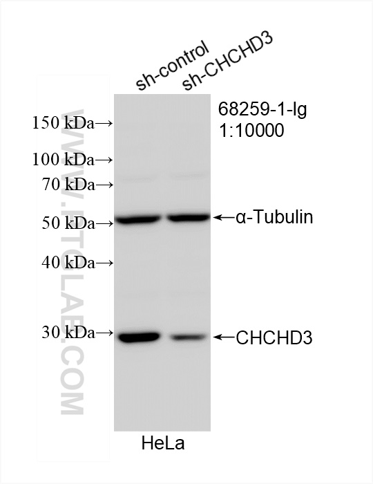
WB result of CHCHD3 antibody (68259-1-Ig; 1:10000; incubated at room temperature for 1.5 hours) with sh-Control and sh-CHCHD3 transfected HeLa cells.
WB result of CHCHD3 antibody (68259-1-Ig; 1:10000; incubated at room temperature for 1.5 hours) with sh-Control and sh-CHCHD3 transfected HeLa cells.
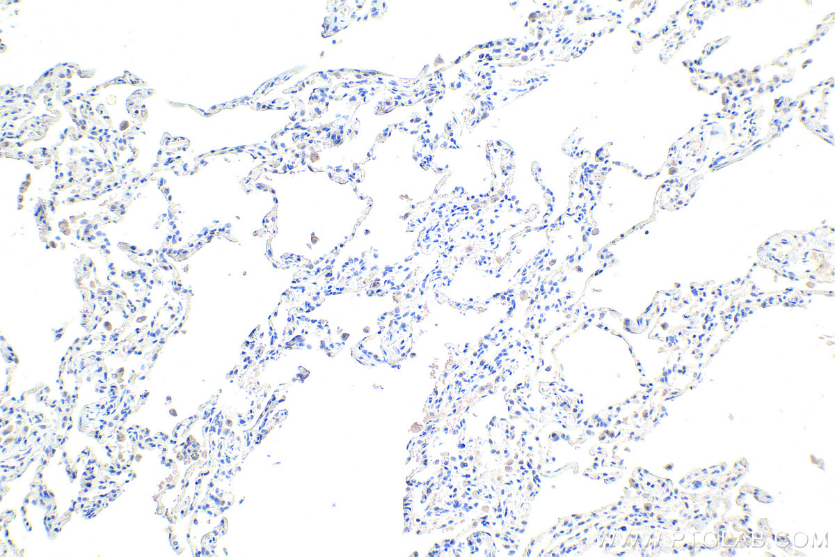
IHC staining of human lung using 68259-1-Ig
Immunohistochemical analysis of paraffin-embedded human lung tissue slide using 68259-1-Ig (CHCHD3 antibody) at dilution of 1:1000 (under 10x lens). Heat mediated antigen retrieval with Tris-EDTA buffer (pH 9.0).
Immunohistochemical analysis of paraffin-embedded human lung tissue slide using 68259-1-Ig (CHCHD3 antibody) at dilution of 1:1000 (under 10x lens). Heat mediated antigen retrieval with Tris-EDTA buffer (pH 9.0).
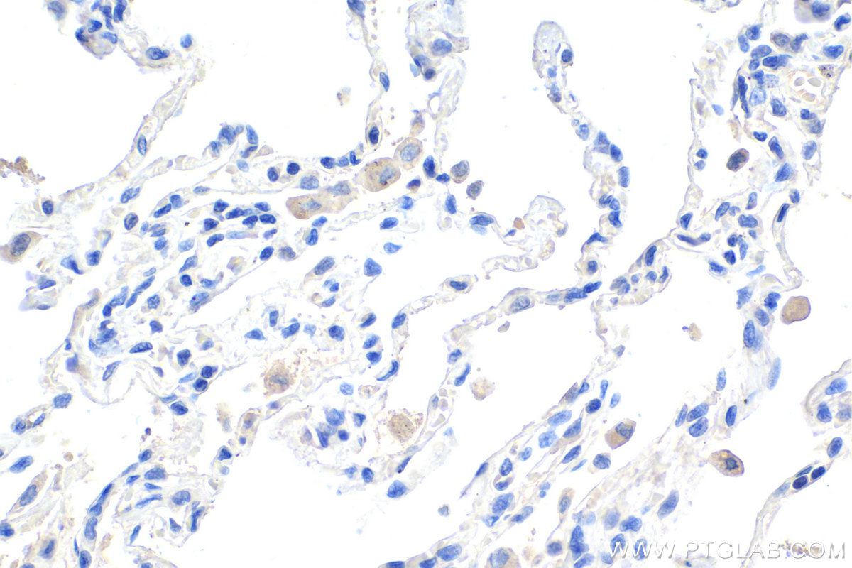
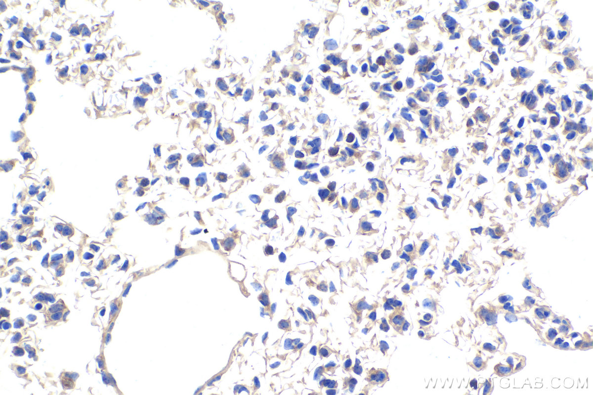
IHC staining of human lung using 68259-1-Ig
Immunohistochemical analysis of paraffin-embedded human lung tissue slide using 68259-1-Ig (CHCHD3 antibody) at dilution of 1:1000 (under 40x lens). Heat mediated antigen retrieval with Tris-EDTA buffer (pH 9.0).
Immunohistochemical analysis of paraffin-embedded human lung tissue slide using 68259-1-Ig (CHCHD3 antibody) at dilution of 1:1000 (under 40x lens). Heat mediated antigen retrieval with Tris-EDTA buffer (pH 9.0).
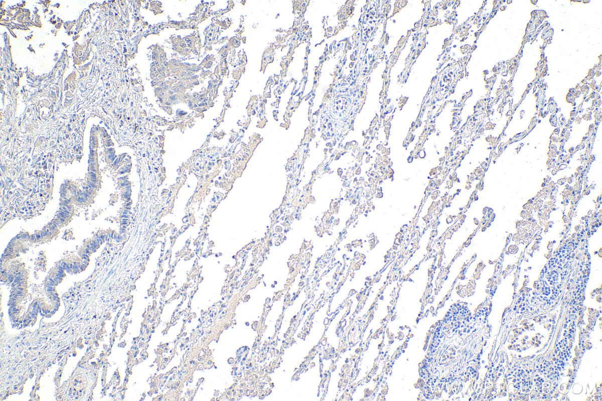
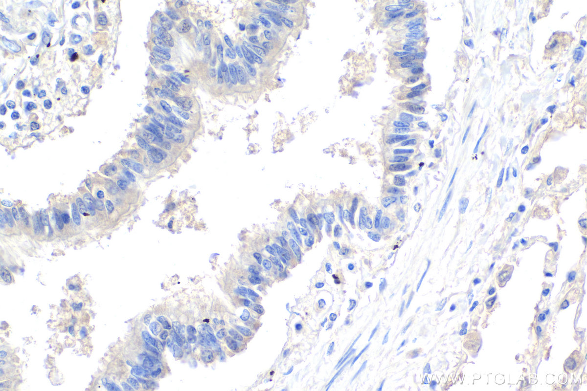
IHC staining of human lung cancer using 68259-1-Ig
Immunohistochemical analysis of paraffin-embedded human lung cancer tissue slide using 68259-1-Ig (CHCHD3 antibody) at dilution of 1:1000 (under 10x lens). Heat mediated antigen retrieval with Tris-EDTA buffer (pH 9.0).
Immunohistochemical analysis of paraffin-embedded human lung cancer tissue slide using 68259-1-Ig (CHCHD3 antibody) at dilution of 1:1000 (under 10x lens). Heat mediated antigen retrieval with Tris-EDTA buffer (pH 9.0).
IHC staining of human lung cancer using 68259-1-Ig
Immunohistochemical analysis of paraffin-embedded human lung cancer tissue slide using 68259-1-Ig (CHCHD3 antibody) at dilution of 1:1000 (under 40x lens). Heat mediated antigen retrieval with Tris-EDTA buffer (pH 9.0).
Immunohistochemical analysis of paraffin-embedded human lung cancer tissue slide using 68259-1-Ig (CHCHD3 antibody) at dilution of 1:1000 (under 40x lens). Heat mediated antigen retrieval with Tris-EDTA buffer (pH 9.0).
IHC staining of mouse cerebellum using 68259-1-Ig
Immunohistochemical analysis of paraffin-embedded mouse cerebellum tissue slide using 68259-1-Ig (CHCHD3 antibody) at dilution of 1:1000 (under 10x lens). Heat mediated antigen retrieval with Tris-EDTA buffer (pH 9.0).
Immunohistochemical analysis of paraffin-embedded mouse lung tissue slide using 68259-1-Ig (CHCHD3 antibody) at dilution of 1:1000 (under 40x lens). Heat mediated antigen retrieval with Tris-EDTA buffer (pH 9.0).
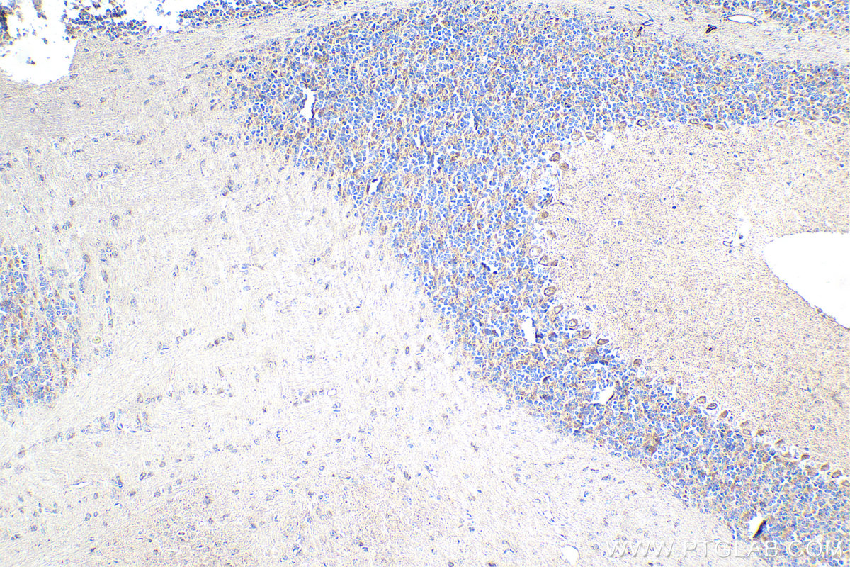
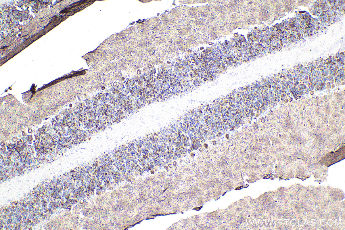
IHC staining of rat cerebellum using 68259-1-Ig
Immunohistochemical analysis of paraffin-embedded rat cerebellum tissue slide using 68259-1-Ig (CHCHD3 antibody) at dilution of 1:1000 (under 10x lens). Heat mediated antigen retrieval with Tris-EDTA buffer (pH 9.0).
Immunohistochemical analysis of paraffin-embedded rat cerebellum tissue slide using 68259-1-Ig (CHCHD3 antibody) at dilution of 1:1000 (under 10x lens). Heat mediated antigen retrieval with Tris-EDTA buffer (pH 9.0).
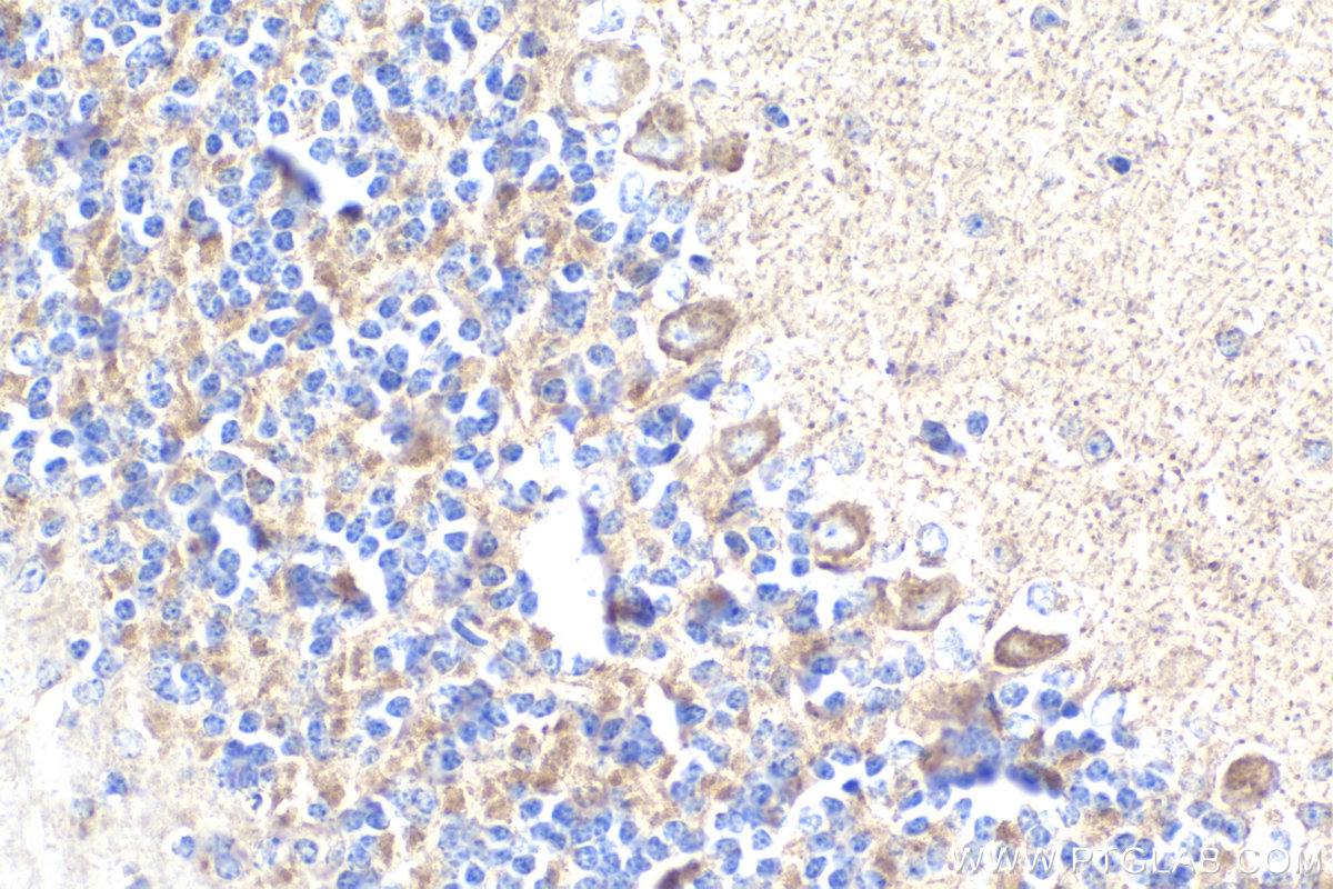
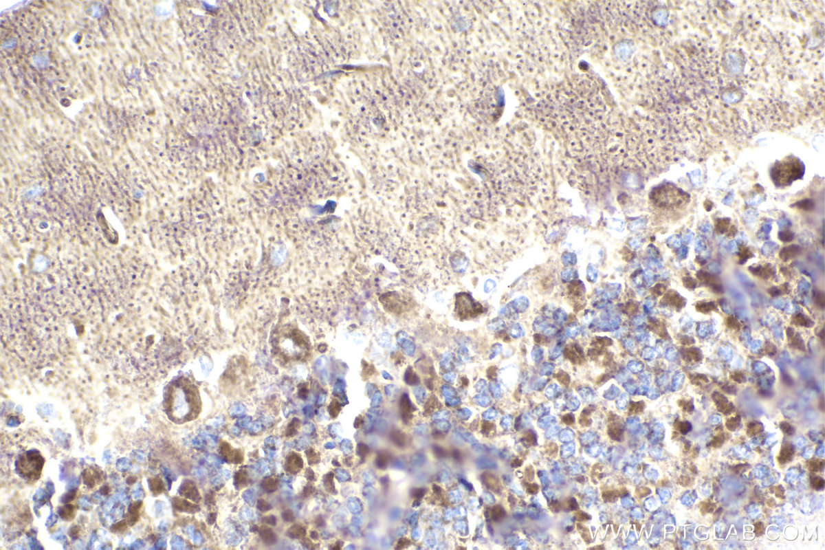
IHC staining of rat cerebellum using 68259-1-Ig
Immunohistochemical analysis of paraffin-embedded rat cerebellum tissue slide using 68259-1-Ig (CHCHD3 antibody) at dilution of 1:1000 (under 40x lens). Heat mediated antigen retrieval with Tris-EDTA buffer (pH 9.0).
Immunohistochemical analysis of paraffin-embedded rat cerebellum tissue slide using 68259-1-Ig (CHCHD3 antibody) at dilution of 1:1000 (under 40x lens). Heat mediated antigen retrieval with Tris-EDTA buffer (pH 9.0).
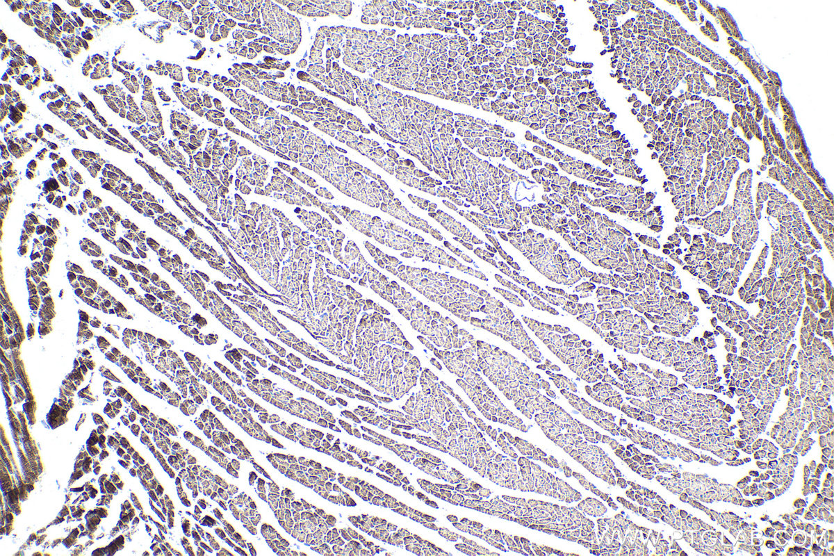
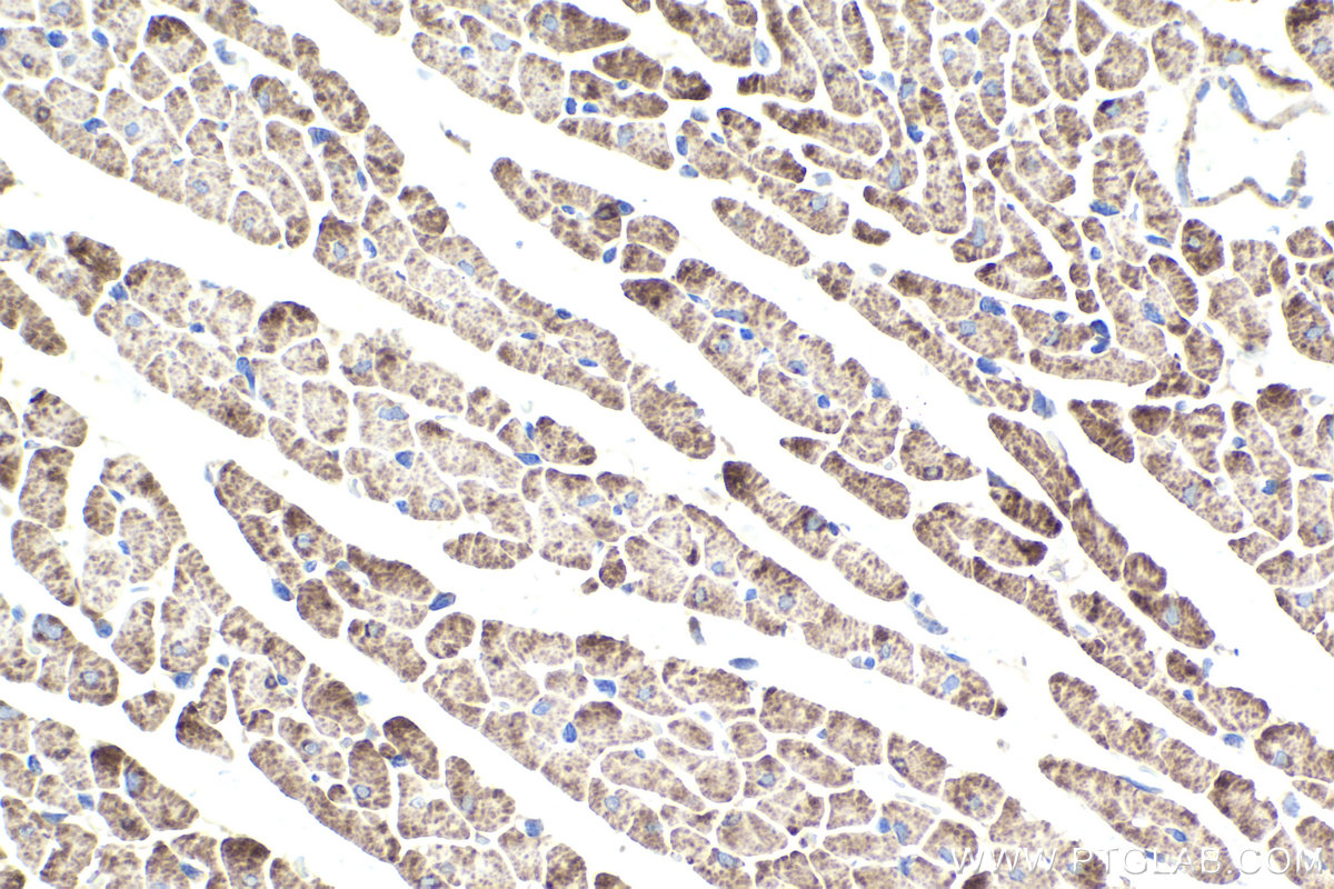
IHC staining of rat heart using 68259-1-Ig
Immunohistochemical analysis of paraffin-embedded rat heart tissue slide using 68259-1-Ig (CHCHD3 antibody) at dilution of 1:1000 (under 10x lens). Heat mediated antigen retrieval with Tris-EDTA buffer (pH 9.0).
Immunohistochemical analysis of paraffin-embedded rat heart tissue slide using 68259-1-Ig (CHCHD3 antibody) at dilution of 1:1000 (under 10x lens). Heat mediated antigen retrieval with Tris-EDTA buffer (pH 9.0).
IHC staining of rat heart using 68259-1-Ig
Immunohistochemical analysis of paraffin-embedded rat heart tissue slide using 68259-1-Ig (CHCHD3 antibody) at dilution of 1:1000 (under 40x lens). Heat mediated antigen retrieval with Tris-EDTA buffer (pH 9.0).
Immunohistochemical analysis of paraffin-embedded rat heart tissue slide using 68259-1-Ig (CHCHD3 antibody) at dilution of 1:1000 (under 40x lens). Heat mediated antigen retrieval with Tris-EDTA buffer (pH 9.0).
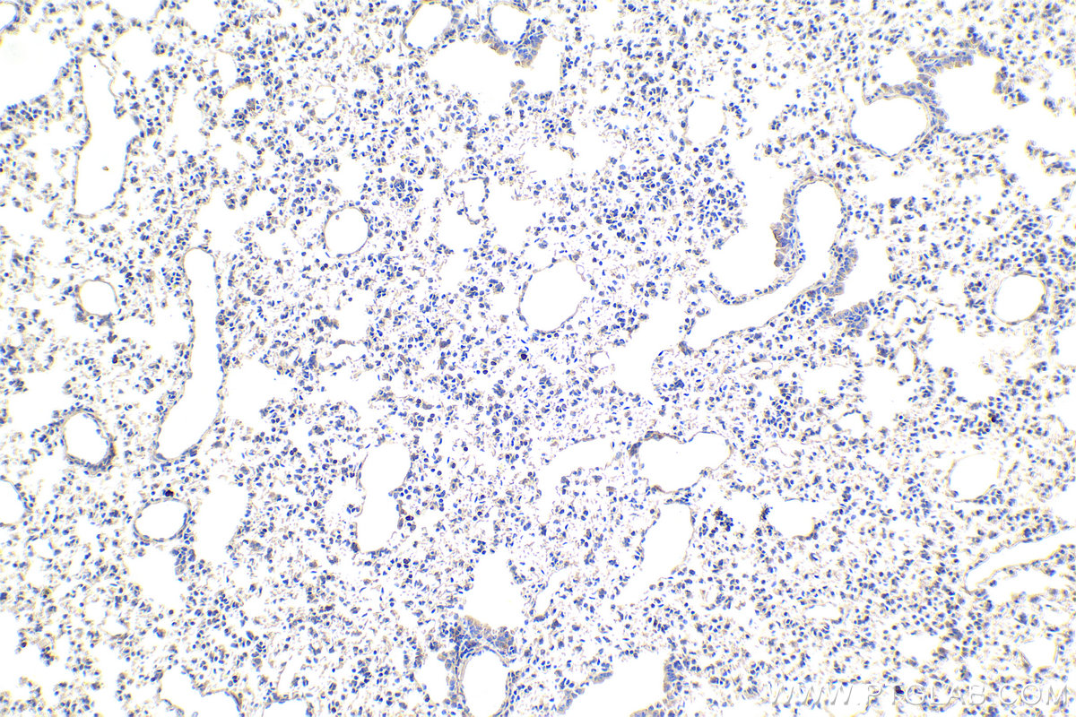
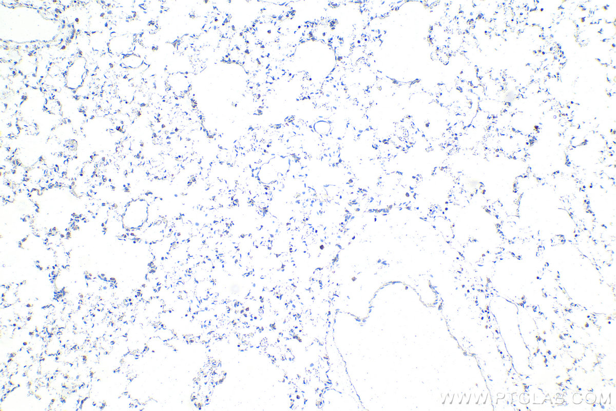
IHC staining of rat lung using 68259-1-Ig
Immunohistochemical analysis of paraffin-embedded rat lung tissue slide using 68259-1-Ig (CHCHD3 antibody) at dilution of 1:1000 (under 10x lens). Heat mediated antigen retrieval with Tris-EDTA buffer (pH 9.0).
Immunohistochemical analysis of paraffin-embedded rat lung tissue slide using 68259-1-Ig (CHCHD3 antibody) at dilution of 1:1000 (under 10x lens). Heat mediated antigen retrieval with Tris-EDTA buffer (pH 9.0).
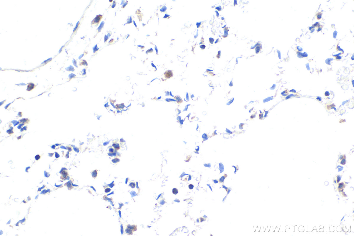
IHC staining of rat lung using 68259-1-Ig
Immunohistochemical analysis of paraffin-embedded rat lung tissue slide using 68259-1-Ig (CHCHD3 antibody) at dilution of 1:1000 (under 40x lens). Heat mediated antigen retrieval with Tris-EDTA buffer (pH 9.0).
Immunohistochemical analysis of paraffin-embedded rat lung tissue slide using 68259-1-Ig (CHCHD3 antibody) at dilution of 1:1000 (under 40x lens). Heat mediated antigen retrieval with Tris-EDTA buffer (pH 9.0).
IF Staining of MCF-7 using 68259-1-Ig
Immunofluorescent analysis of (-20°C Methanol) fixed MCF-7 cells using CHCHD3 antibody (68259-1-Ig, Clone: 1C8H12 ) at dilution of 1:800 and CoraLite®488-Conjugated AffiniPure Goat Anti-Mouse IgG(H+L).
Immunofluorescent analysis of (-20°C Methanol) fixed MCF-7 cells using CHCHD3 antibody (68259-1-Ig, Clone: 1C8H12 ) at dilution of 1:800 and CoraLite®488-Conjugated AffiniPure Goat Anti-Mouse IgG(H+L).
The Proteintech guarantee covers Proteintech antibodies in any species and any application, including those not listed on the datasheet. If the antibody doesn’t perform, you can receive a hassle-free refund or credit note.
pig heart tissue, A549 cells, HeLa cells, rabbit heart tissue, rat heart tissue, mouse heart tissue, rat brain tissue
Positive IHC detected in
human lung tissue, human lung cancer tissue, mouse cerebellum tissue, mouse lung tissue, rat cerebellum tissue, rat heart tissue, rat lung tissue Note: suggested antigen retrieval with TE buffer pH 9.0; (*) Alternatively, antigen retrieval may be performed with citrate buffer pH 6.0
Positive IF/ICC detected in
MCF-7 cells
Recommended dilution
Application
Dilution
Western Blot (WB)
WB : 1:5000-1:50000
Immunohistochemistry (IHC)
IHC : 1:500-1:2000
Immunofluorescence (IF)/ICC
IF/ICC : 1:400-1:1600
It is recommended that this reagent should be titrated in each testing system to obtain optimal results.
Sample-dependent, Check data in validation data gallery.
PBS with 0.02% sodium azide and 50% glycerol, pH 7.3.
Storage Conditions
Store at -20°C. Stable for one year after shipment. Aliquoting is unnecessary for -20oC storage. 20ul sizes contain 0.1% BSA.
Background Information
CHCHD3, initially identified as a substrate for cAMP-dependent protein kinase (PKA), is a ubiquitous protein in the mitochondria and plays a prominent role in maintaining cristae integrity and mitochondrial function. In mitochondria, ChChd3 is predominantly localized to the inner membrane (IM), facing toward the intermembrane space (IMS), and is part of the large protein complex now called as MINOS (mitochondrial inner membrane organizing system), or MIB (mitochondrial intermembrane space bridging) CHCHD3 is highly conserved in mammals with human and mouse protein sharing ∼92% sequence similarity.
Various lysates were subjected to SDS PAGE followed by western blot with 68259-1-Ig (CHCHD3 antibody) at dilution of 1:20000 incubated at room temperature for 1.5 hours.
WB analysis using 68259-1-Ig
Various lysates were subjected to SDS PAGE followed by western blot with 68259-1-Ig (CHCHD3 antibody) at dilution of 1:20000 incubated at room temperature for 1.5 hours.
WB analysis of HeLa using 68259-1-Ig
WB result of CHCHD3 antibody (68259-1-Ig; 1:10000; incubated at room temperature for 1.5 hours) with sh-Control and sh-CHCHD3 transfected HeLa cells.
IHC Figures
IHC staining of human lung using 68259-1-Ig
Immunohistochemical analysis of paraffin-embedded human lung tissue slide using 68259-1-Ig (CHCHD3 antibody) at dilution of 1:1000 (under 10x lens). Heat mediated antigen retrieval with Tris-EDTA buffer (pH 9.0).
IHC staining of human lung using 68259-1-Ig
Immunohistochemical analysis of paraffin-embedded human lung tissue slide using 68259-1-Ig (CHCHD3 antibody) at dilution of 1:1000 (under 40x lens). Heat mediated antigen retrieval with Tris-EDTA buffer (pH 9.0).
IHC staining of human lung cancer using 68259-1-Ig
Immunohistochemical analysis of paraffin-embedded human lung cancer tissue slide using 68259-1-Ig (CHCHD3 antibody) at dilution of 1:1000 (under 10x lens). Heat mediated antigen retrieval with Tris-EDTA buffer (pH 9.0).
IHC staining of human lung cancer using 68259-1-Ig
Immunohistochemical analysis of paraffin-embedded human lung cancer tissue slide using 68259-1-Ig (CHCHD3 antibody) at dilution of 1:1000 (under 40x lens). Heat mediated antigen retrieval with Tris-EDTA buffer (pH 9.0).
IHC staining of mouse cerebellum using 68259-1-Ig
Immunohistochemical analysis of paraffin-embedded mouse cerebellum tissue slide using 68259-1-Ig (CHCHD3 antibody) at dilution of 1:1000 (under 10x lens). Heat mediated antigen retrieval with Tris-EDTA buffer (pH 9.0).
IHC staining of mouse cerebellum using 68259-1-Ig
Immunohistochemical analysis of paraffin-embedded mouse cerebellum tissue slide using 68259-1-Ig (CHCHD3 antibody) at dilution of 1:1000 (under 40x lens). Heat mediated antigen retrieval with Tris-EDTA buffer (pH 9.0).
IHC staining of mouse lung using 68259-1-Ig
Immunohistochemical analysis of paraffin-embedded mouse lung tissue slide using 68259-1-Ig (CHCHD3 antibody) at dilution of 1:1000 (under 10x lens). Heat mediated antigen retrieval with Tris-EDTA buffer (pH 9.0).
IHC staining of mouse lung using 68259-1-Ig
Immunohistochemical analysis of paraffin-embedded mouse lung tissue slide using 68259-1-Ig (CHCHD3 antibody) at dilution of 1:1000 (under 40x lens). Heat mediated antigen retrieval with Tris-EDTA buffer (pH 9.0).
IHC staining of rat cerebellum using 68259-1-Ig
Immunohistochemical analysis of paraffin-embedded rat cerebellum tissue slide using 68259-1-Ig (CHCHD3 antibody) at dilution of 1:1000 (under 10x lens). Heat mediated antigen retrieval with Tris-EDTA buffer (pH 9.0).
IHC staining of rat cerebellum using 68259-1-Ig
Immunohistochemical analysis of paraffin-embedded rat cerebellum tissue slide using 68259-1-Ig (CHCHD3 antibody) at dilution of 1:1000 (under 40x lens). Heat mediated antigen retrieval with Tris-EDTA buffer (pH 9.0).
IHC staining of rat heart using 68259-1-Ig
Immunohistochemical analysis of paraffin-embedded rat heart tissue slide using 68259-1-Ig (CHCHD3 antibody) at dilution of 1:1000 (under 10x lens). Heat mediated antigen retrieval with Tris-EDTA buffer (pH 9.0).
IHC staining of rat heart using 68259-1-Ig
Immunohistochemical analysis of paraffin-embedded rat heart tissue slide using 68259-1-Ig (CHCHD3 antibody) at dilution of 1:1000 (under 40x lens). Heat mediated antigen retrieval with Tris-EDTA buffer (pH 9.0).
IHC staining of rat lung using 68259-1-Ig
Immunohistochemical analysis of paraffin-embedded rat lung tissue slide using 68259-1-Ig (CHCHD3 antibody) at dilution of 1:1000 (under 10x lens). Heat mediated antigen retrieval with Tris-EDTA buffer (pH 9.0).
IHC staining of rat lung using 68259-1-Ig
Immunohistochemical analysis of paraffin-embedded rat lung tissue slide using 68259-1-Ig (CHCHD3 antibody) at dilution of 1:1000 (under 40x lens). Heat mediated antigen retrieval with Tris-EDTA buffer (pH 9.0).
IF/ICC Figures
IF Staining of MCF-7 using 68259-1-Ig
Immunofluorescent analysis of (-20°C Methanol) fixed MCF-7 cells using CHCHD3 antibody (68259-1-Ig, Clone: 1C8H12 ) at dilution of 1:800 and CoraLite®488-Conjugated AffiniPure Goat Anti-Mouse IgG(H+L).
The species listed in Tested Reactivity are in-house verified and applicable species. For unlisted species, please refer to the homology analysis of the immunogen sequence and related species. For rabbit polyclonal antibodies, homology >70% is recommended. For mouse monoclonal antibodies and rabbit recombinant antibodies, homology >90% is recommended. Generally, the higher the homology, the greater the applicability. However, there will be certain differences in protein expression in different species, tissues or cells. Therefore, the homology analysis results are for reference only and do not serve as a guarantee.
At Proteintech, we pride ourselves on our antibody quality, customer service and transparency. As such, we are comparing our antibodies with other vendors, enabling easy identification and comparisons of key data to help you choose the suitable antibody for your needs.
We have selected the top cited antibodies from these vendors for you to compare.
 at dilution of 1:20000 incubated at room temperature for 1.5 hours.")
 at dilution of 1:20000 incubated at room temperature for 1.5 hours.")
 with sh-Control and sh-CHCHD3 transfected HeLa cells.")
 at dilution of 1:1000 (under 10x lens). Heat mediated antigen retrieval with Tris-EDTA buffer (pH 9.0).")
 at dilution of 1:1000 (under 40x lens). Heat mediated antigen retrieval with Tris-EDTA buffer (pH 9.0).")
 at dilution of 1:1000 (under 10x lens). Heat mediated antigen retrieval with Tris-EDTA buffer (pH 9.0).")
 at dilution of 1:1000 (under 40x lens). Heat mediated antigen retrieval with Tris-EDTA buffer (pH 9.0).")
 at dilution of 1:1000 (under 10x lens). Heat mediated antigen retrieval with Tris-EDTA buffer (pH 9.0).")
 at dilution of 1:1000 (under 40x lens). Heat mediated antigen retrieval with Tris-EDTA buffer (pH 9.0).")
 at dilution of 1:1000 (under 10x lens). Heat mediated antigen retrieval with Tris-EDTA buffer (pH 9.0).")
 at dilution of 1:1000 (under 40x lens). Heat mediated antigen retrieval with Tris-EDTA buffer (pH 9.0).")
 at dilution of 1:1000 (under 10x lens). Heat mediated antigen retrieval with Tris-EDTA buffer (pH 9.0).")
 at dilution of 1:1000 (under 40x lens). Heat mediated antigen retrieval with Tris-EDTA buffer (pH 9.0).")
 at dilution of 1:1000 (under 10x lens). Heat mediated antigen retrieval with Tris-EDTA buffer (pH 9.0).")
 at dilution of 1:1000 (under 40x lens). Heat mediated antigen retrieval with Tris-EDTA buffer (pH 9.0).")
 at dilution of 1:1000 (under 10x lens). Heat mediated antigen retrieval with Tris-EDTA buffer (pH 9.0).")
 at dilution of 1:1000 (under 40x lens). Heat mediated antigen retrieval with Tris-EDTA buffer (pH 9.0).")
 fixed MCF-7 cells using CHCHD3 antibody (68259-1-Ig, Clone: 1C8H12 ) at dilution of 1:800 and CoraLite®488-Conjugated AffiniPure Goat Anti-Mouse IgG(H+L).")