Tested Applications
| Positive WB detected in | THP-1 cells, U-937 cells |
| Positive IHC detected in | human liver tissue, human tonsillitis tissue Note: suggested antigen retrieval with TE buffer pH 9.0; (*) Alternatively, antigen retrieval may be performed with citrate buffer pH 6.0 |
| Positive IF-P detected in | human colon tissue, human tonsillitis tissue, human appendicitis tissue |
Recommended dilution
| Application | Dilution |
|---|---|
| Western Blot (WB) | WB : 1:1000-1:8000 |
| Immunohistochemistry (IHC) | IHC : 1:2000-1:8000 |
| Immunofluorescence (IF)-P | IF-P : 1:50-1:500 |
| It is recommended that this reagent should be titrated in each testing system to obtain optimal results. | |
| Sample-dependent, Check data in validation data gallery. | |
Published Applications
| WB | See 15 publications below |
| IHC | See 47 publications below |
| IF | See 54 publications below |
Product Information
25747-1-AP targets CD68 in WB, IHC, IF-P, ELISA applications and shows reactivity with human samples.
| Tested Reactivity | human |
| Cited Reactivity | human, pig, canine, zebrafish |
| Host / Isotype | Rabbit / IgG |
| Class | Polyclonal |
| Type | Antibody |
| Immunogen |
CatNo: Ag22815 Product name: Recombinant human CD68 protein Source: e coli.-derived, PET28a Tag: 6*His Domain: 29-319 aa of BC015557 Sequence: SATLLPSFTVTPTVTESTGTTSHRTTKSHKTTTHRTTTTGTTSHGPTTATHNPTTTSHGNVTVHPTSNSTATSQGPSTATHSPATTSHGNATVHPTSNSTATSPGFTSSAHPEPPPPSPSPSPTSKETIGDYTWTNGSQPCVHLQAQIQIRVMYTTQGGGEAWGISVLNPNKTKVQGSCEGAHPHLLLSFPYGHLSFGFMQDLQQKVVYLSYMAVEYNVSFPHAAQWTFSAQNASLRDLQAPLGQSFSCSNSSIILSPAVHLDLLSLRLQAAQLPHTGVFGQSFSCPSDRS Predict reactive species |
| Full Name | CD68 molecule |
| Calculated Molecular Weight | 37 kDa |
| Observed Molecular Weight | 60-70 kDa |
| GenBank Accession Number | BC015557 |
| Gene Symbol | CD68 |
| Gene ID (NCBI) | 968 |
| RRID | AB_2721140 |
| Conjugate | Unconjugated |
| Form | Liquid |
| Purification Method | Antigen affinity purification |
| UNIPROT ID | P34810 |
| Storage Buffer | PBS with 0.02% sodium azide and 50% glycerol, pH 7.3. |
| Storage Conditions | Store at -20°C. Stable for one year after shipment. Aliquoting is unnecessary for -20oC storage. 20ul sizes contain 0.1% BSA. |
Background Information
CD68 is a type I transmembrane glycoprotein that is highly expressed by human monocytes and tissue macrophages. It belongs to the lysosomal/endosomal-associated membrane glycoprotein (LAMP) family and primarily localizes to lysosomes and endosomes with a smaller fraction circulating to the cell surface. CD68 is also a member of the scavenger receptor family. It may play a role in phagocytic activities of tissue macrophages. The apparent molecular weight of CD68 is larger than calculated molecular weight due to post-translation modification.
Protocols
| Product Specific Protocols | |
|---|---|
| IF protocol for CD68 antibody 25747-1-AP | Download protocol |
| IHC protocol for CD68 antibody 25747-1-AP | Download protocol |
| WB protocol for CD68 antibody 25747-1-AP | Download protocol |
| Standard Protocols | |
|---|---|
| Click here to view our Standard Protocols |
Publications
| Species | Application | Title |
|---|---|---|
Cell Metab Dual impacts of serine/glycine-free diet in enhancing antitumor immunity and promoting evasion via PD-L1 lactylation | ||
Cell Metab Pharmacological inhibition of arachidonate 12-lipoxygenase ameliorates myocardial ischemia-reperfusion injury in multiple species. | ||
Theranostics Platelets promote CRC by activating the C5a/C5aR1 axis via PSGL-1/JNK/STAT1 signaling in tumor-associated macrophages | ||
Nat Commun The ubiquitin ligase ZNRF1 promotes caveolin-1 ubiquitination and degradation to modulate inflammation. | ||
Aging Cell Inhibition of DNA methyltransferase aberrations reinstates antioxidant aging suppressors and ameliorates renal aging. |
Reviews
The reviews below have been submitted by verified Proteintech customers who received an incentive for providing their feedback.
FH Zach (Verified Customer) (09-12-2025) | The antibody works perfectly with liver samples of MASH mice, and pancreas samples of chronic pancreatitis mice.
|
FH Emma (Verified Customer) (11-29-2021) | Works well by IF on FFPE tissue with a Tris-EDTA antigen retrieval. Also works by IHC.
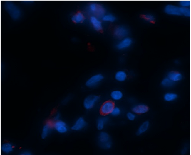 |
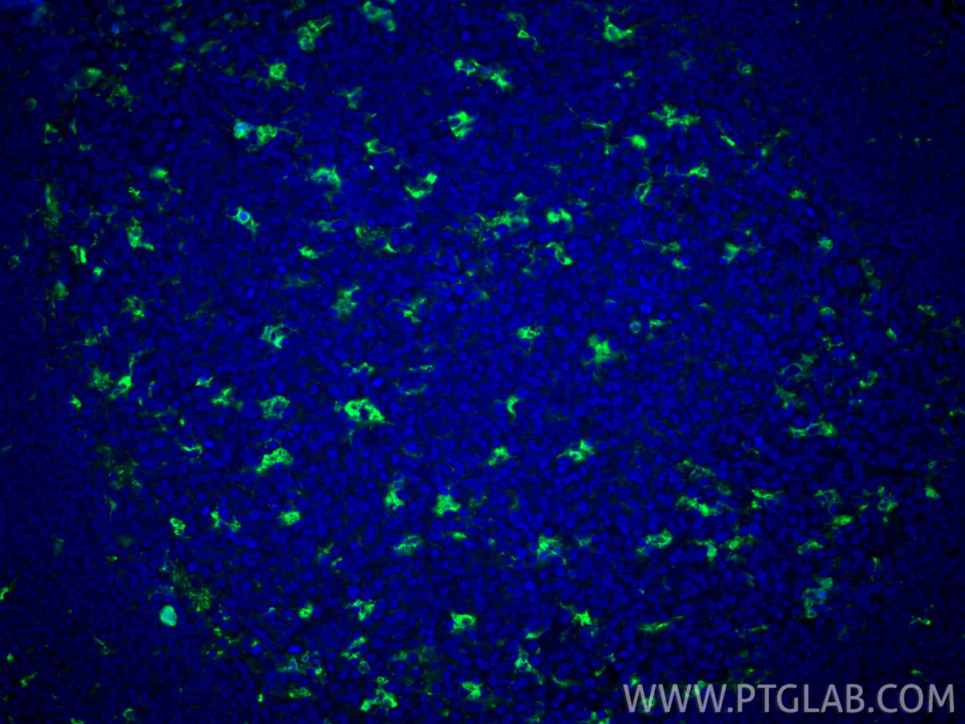
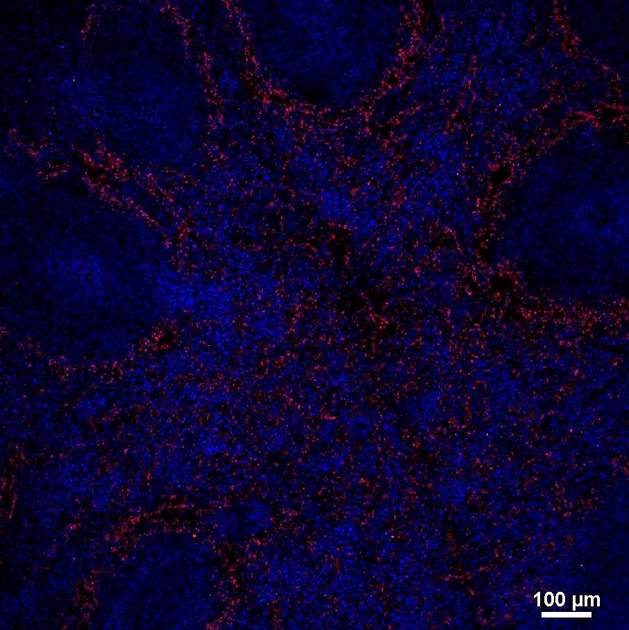
FH Fabio Henrique (Verified Customer) (01-17-2019) | Figure 1. IF of CD68 (red) in human spleen. Formalin-fixed paraffin-embedded human spleen tissue was used as positive control to probe for CD68. Heat-induced antigen retrieval was performed in sodium citrate buffer pH 6.0 + Tween20 at 0.5%. Tissue incubated at 96*C for 20 min inside the buffer. Permeabilization was done with washes of TBSTritonX 0.25% for 3x 5min and blocking was done in TBS 10% Goat serum, 1% BSA, for 1 hr at RT. Anti-CD68 was used at 1:200 in blocking buffer, overnight incubation at 4*C. Secondary antibody AlexaFluor 488 was used at 1:500 for 1 hr at RT. Imaged using a Nikon A1 confocal microscope. Figure 2: CD68 (red) and CD206 (green) in human primary macrophages polarized to M2-phenotype, encapsulated in 3D hydrogel (hyaluronic acid and collagen type 1). Staining was performed as described above, except primary (CD68) was used at 1:100 and secondary antibodies were used at 1:500.
 |