with sh-Control and sh-CD3 transfected Jurkat cells.")
 at dilution of 1:20000 incubated at room temperature for 1.5 hours.")
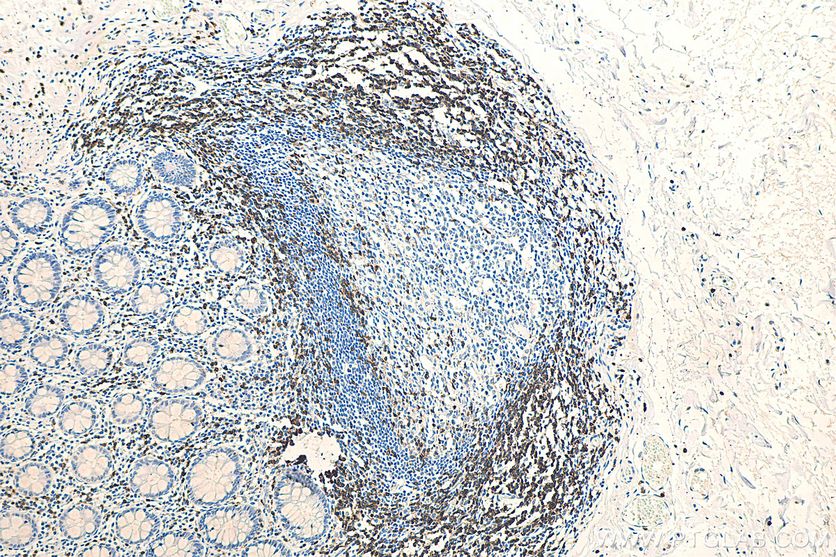
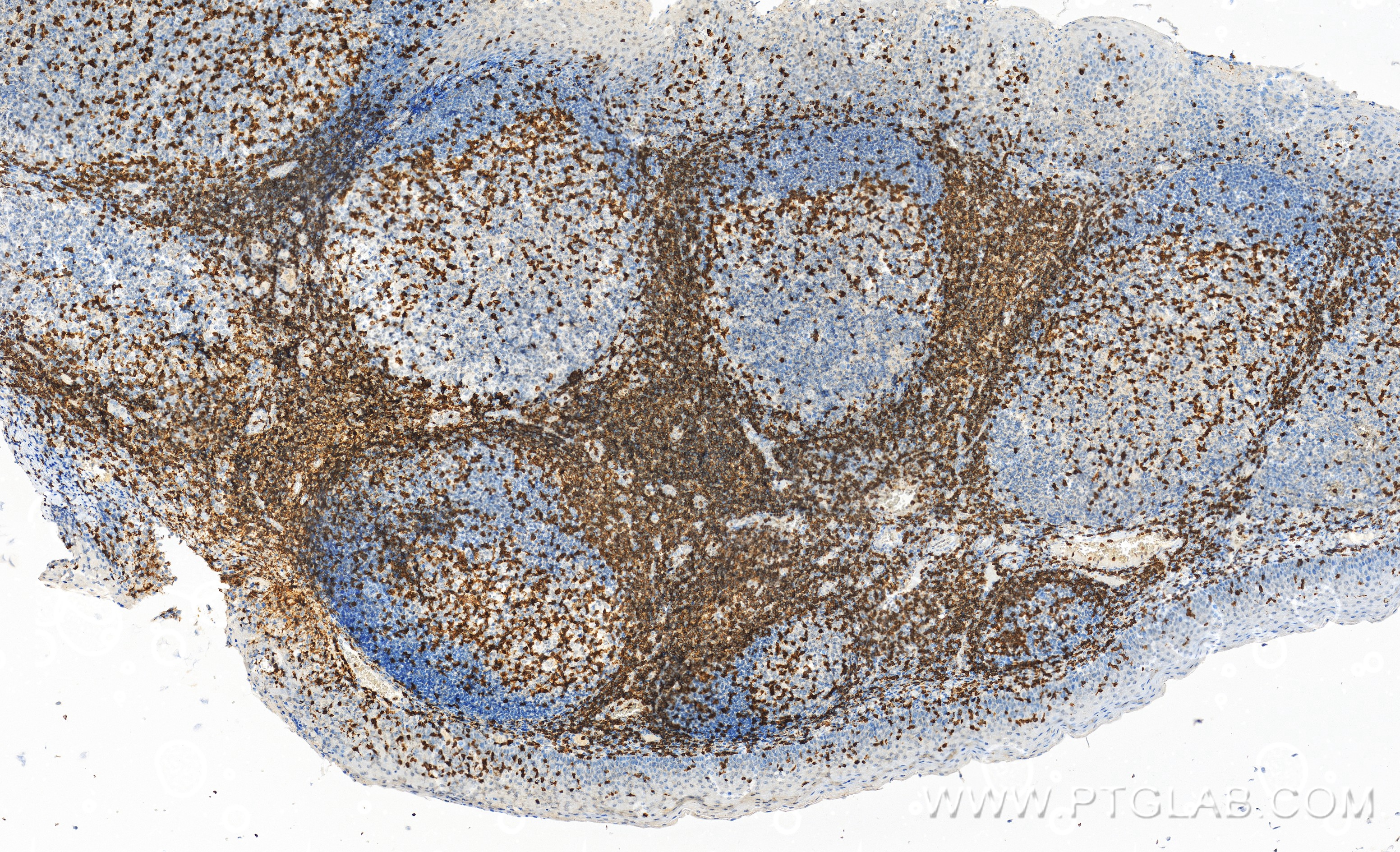
 at dilution of 1:10000 (under 10x lens). Heat mediated antigen retrieval with Tris-EDTA buffer (pH 9.0).")
 at dilution of 1:10000 (under 40x lens). Heat mediated antigen retrieval with Tris-EDTA buffer (pH 9.0).")
 at dilution of 1:10000 (under 40x lens). Heat mediated antigen retrieval with Tris-EDTA buffer (pH 9.0).")
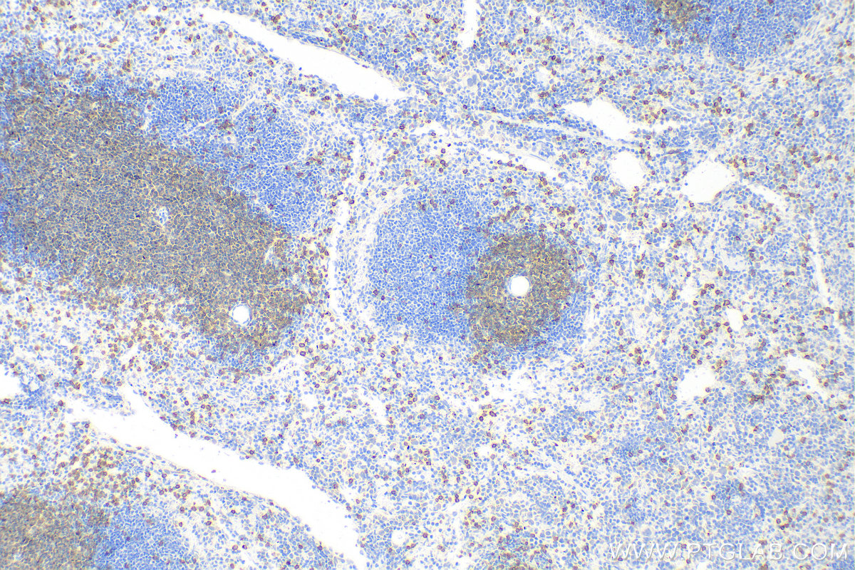
 at dilution of 1:10000 (under 10x lens). Heat mediated antigen retrieval with Tris-EDTA buffer (pH 9.0).")
 at dilution of 1:10000 (under 40x lens). Heat mediated antigen retrieval with Tris-EDTA buffer (pH 9.0).")
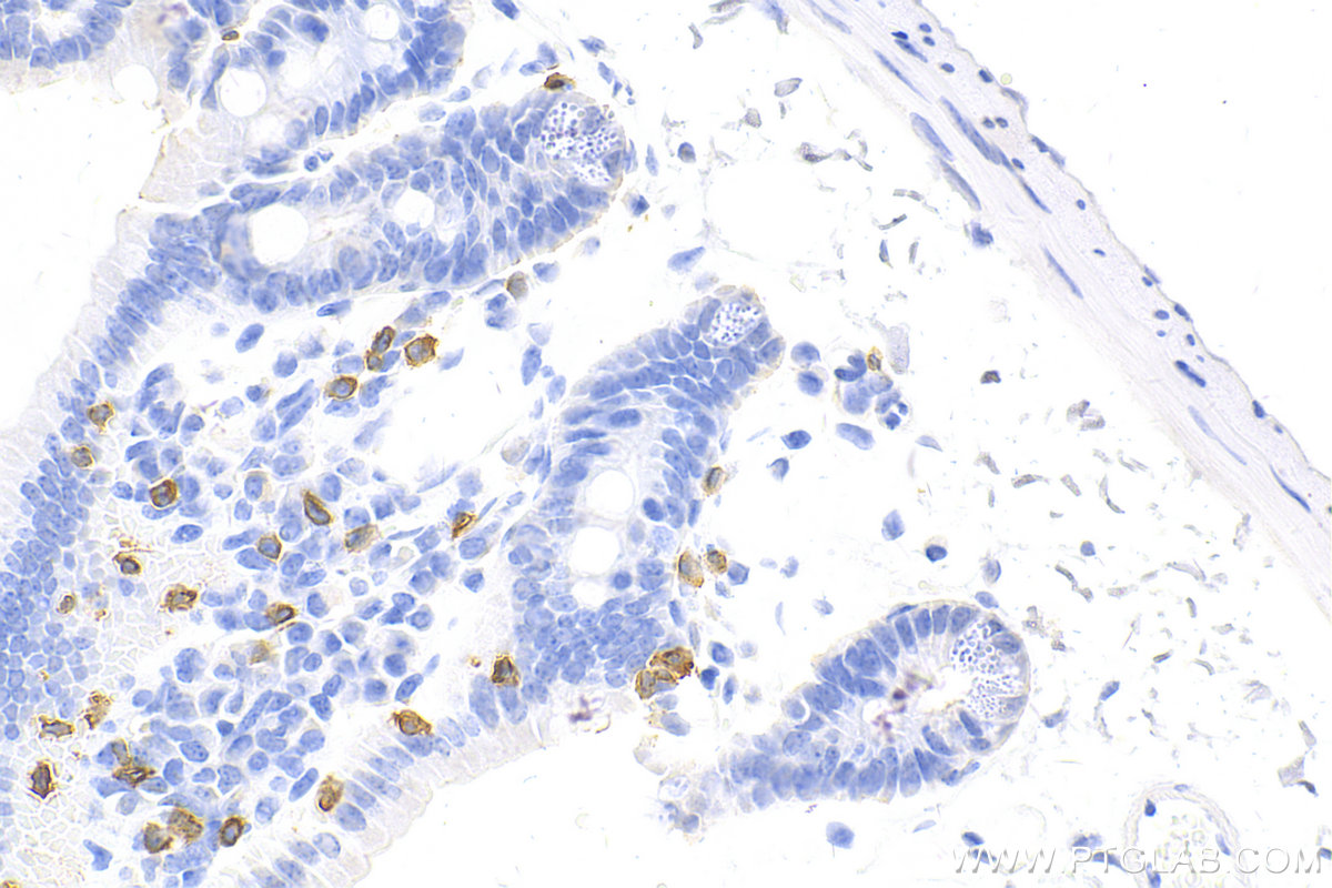
 at dilution of 1:5000 (under 10x lens). Heat mediated antigen retrieval with Tris-EDTA buffer (pH 9.0).")
 at dilution of 1:5000 (under 40x lens). Heat mediated antigen retrieval with Tris-EDTA buffer (pH 9.0).")
 at dilution of 1:10000 (under 40x lens). Heat mediated antigen retrieval with Tris-EDTA buffer (pH 9.0).")
 at dilution of 1:10000 (under 10x lens). Heat mediated antigen retrieval with Tris-EDTA buffer (pH 9.0).")
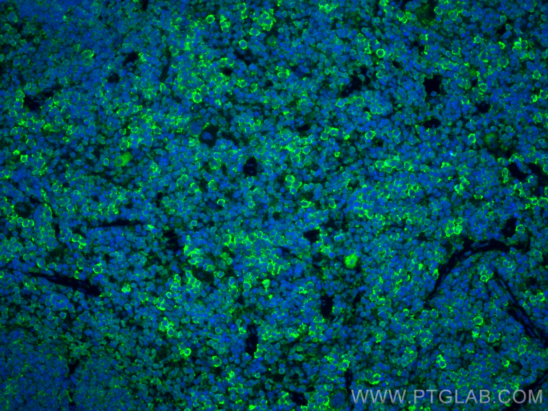
 fixed paraffin-embedded mouse spleen tissue using CD3 antibody (81324-1-RR, Clone: 4I19 ) at dilution of 1:200 and CoraLite®488-Conjugated Goat Anti-Rabbit IgG(H+L) (SA00013-2). Heat mediated antigen retrieval with Tris-EDTA buffer (pH 9.0).")
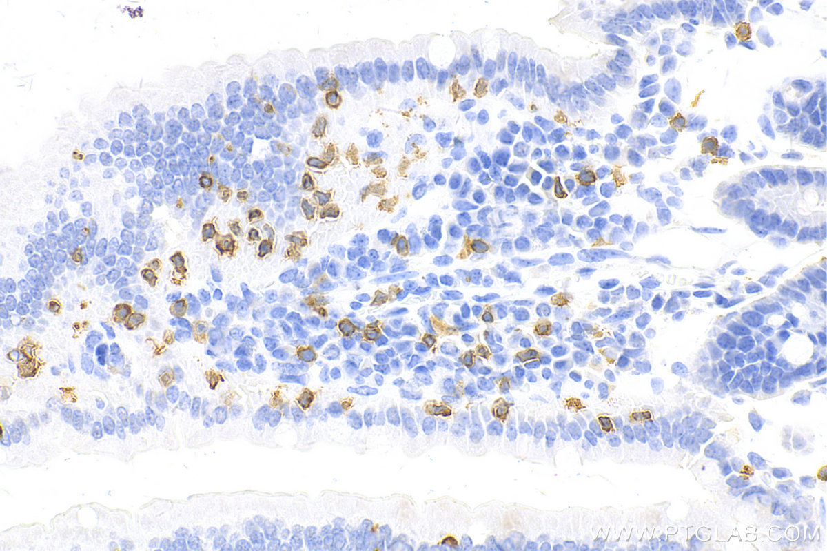
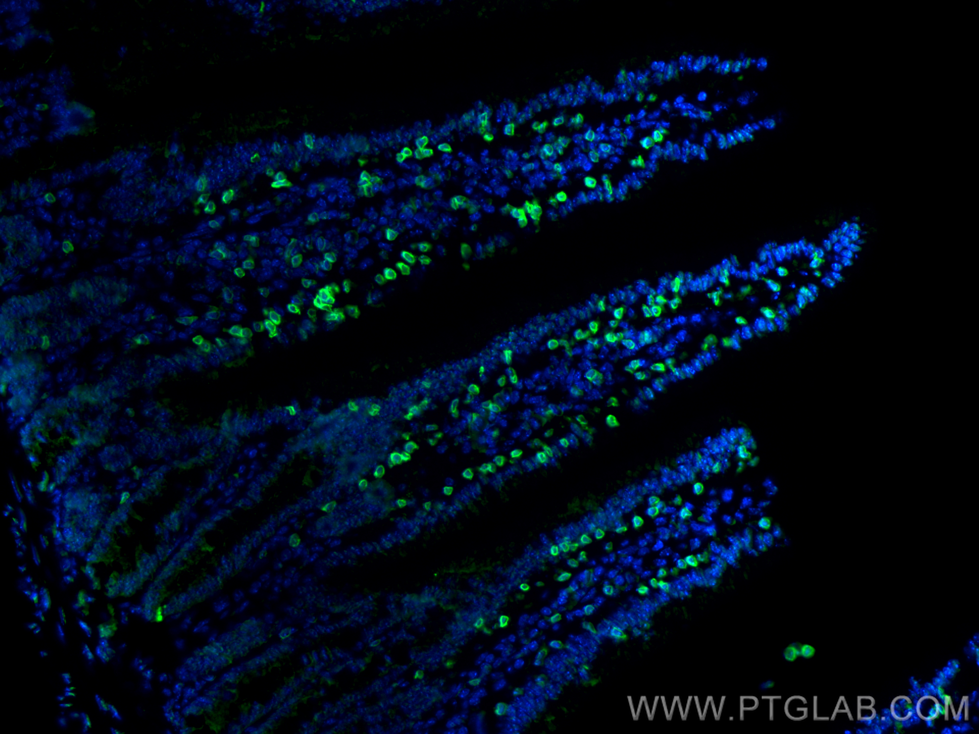
 fixed paraffin-embedded mouse small intestine tissue using CD3 antibody (81324-1-RR, Clone: 4I19 ) at dilution of 1:1000 and CoraLite®488-Conjugated Goat Anti-Rabbit IgG(H+L) (SA00013-2). Heat mediated antigen retrieval with Tris-EDTA buffer (pH 9.0).")
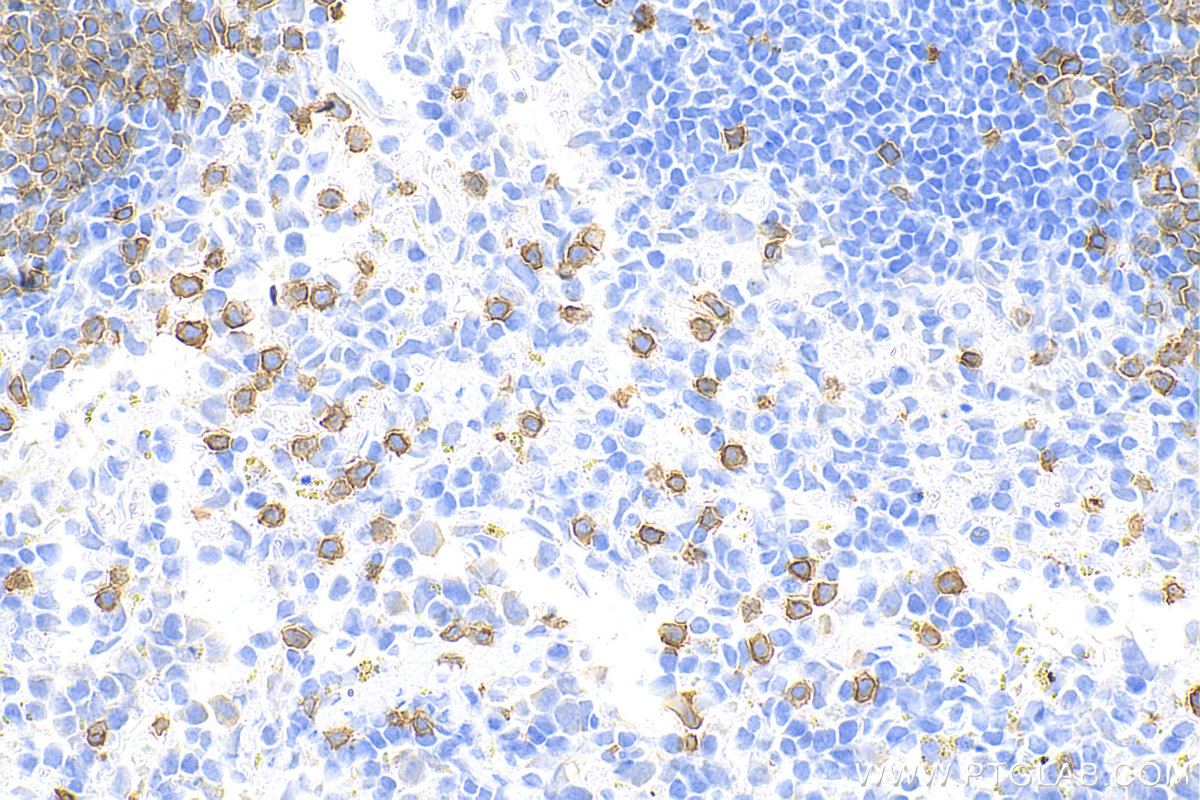
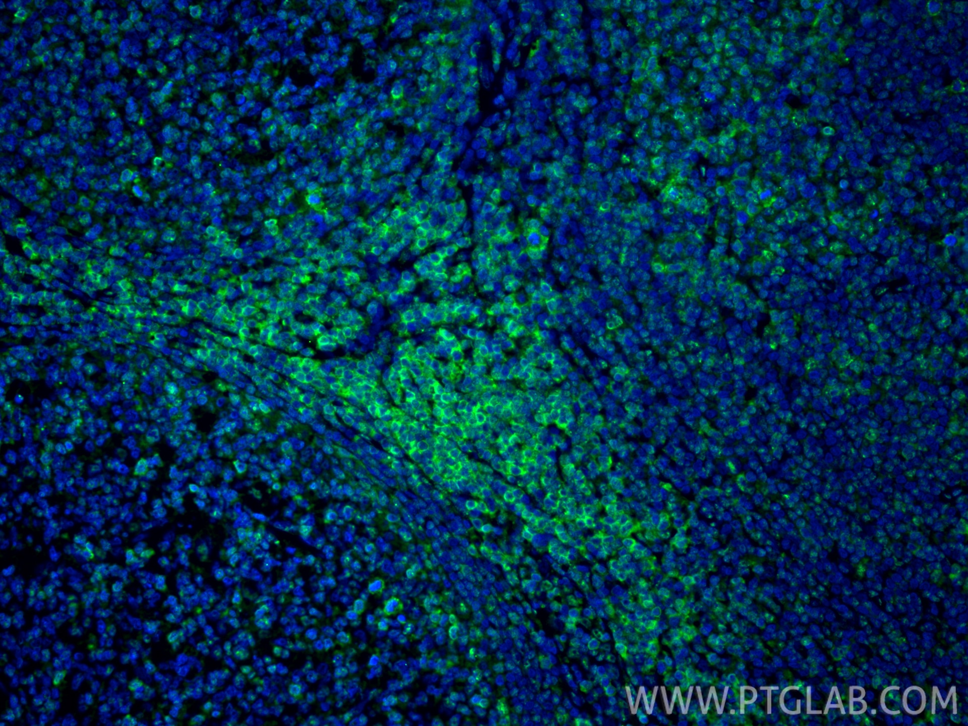
 fixed paraffin-embedded human tonsillitis tissue using CD3 antibody (81324-1-RR, Clone: 4I19 ) at dilution of 1:200 and CoraLite®488-Conjugated Goat Anti-Rabbit IgG(H+L) (SA00013-2). Heat mediated antigen retrieval with Tris-EDTA buffer (pH 9.0).")
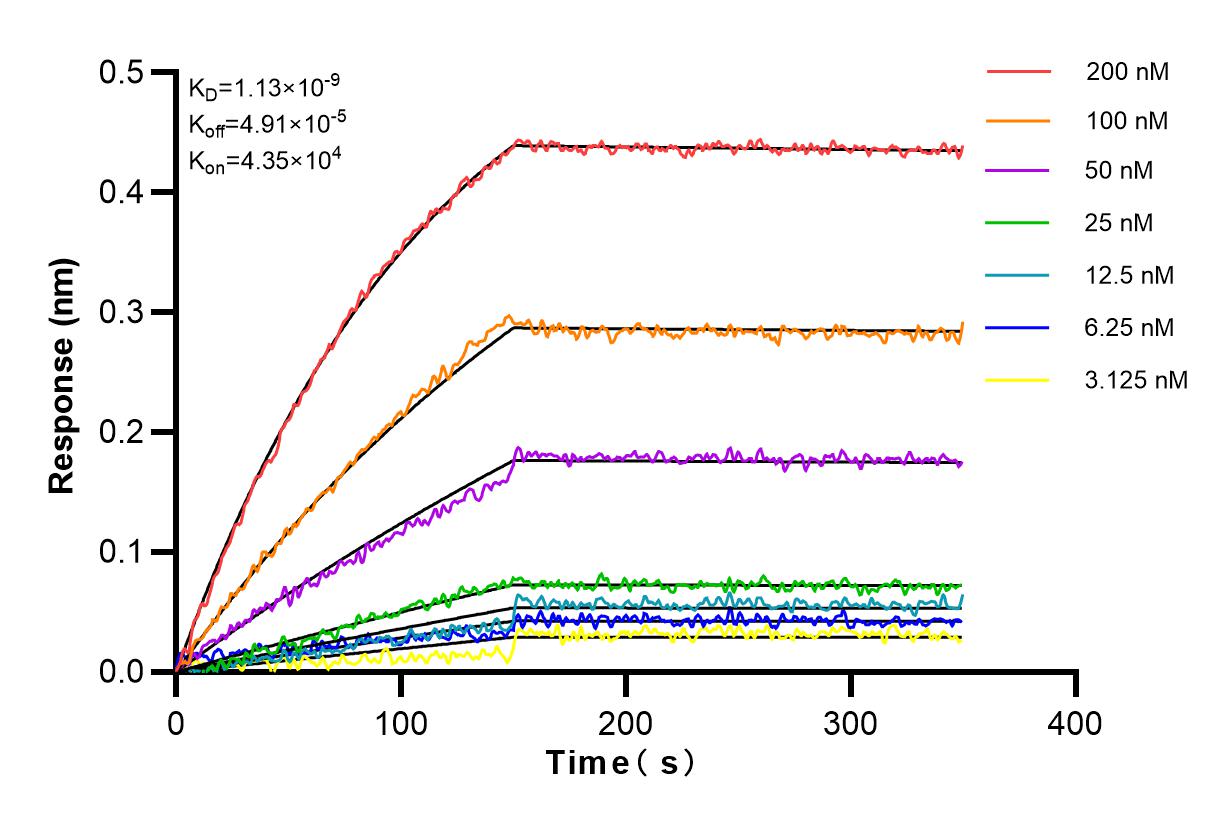
 kinetic assays of 81324-1-RR against Human CD3 were performed. The affinity constant is 1.13 nM.")
Tested Applications
| Positive WB detected in | Jurkat cells, MOLT-4 cells, pig thymus tissue, rat thymus tissue, mouse thymus tissue |
| Positive IHC detected in | human colon tissue, human tonsillitis tissue, mouse small intestine tissue, mouse spleen tissue Note: suggested antigen retrieval with TE buffer pH 9.0; (*) Alternatively, antigen retrieval may be performed with citrate buffer pH 6.0 |
| Positive IF-P detected in | mouse spleen tissue, mouse small intestine tissue, human tonsillitis tissue |
Recommended dilution
| Application | Dilution |
|---|---|
| Western Blot (WB) | WB : 1:5000-1:50000 |
| Immunohistochemistry (IHC) | IHC : 1:5000-1:20000 |
| Immunofluorescence (IF)-P | IF-P : 1:50-1:500 |
| It is recommended that this reagent should be titrated in each testing system to obtain optimal results. | |
| Sample-dependent, Check data in validation data gallery. | |
Published Applications
| IHC | See 1 publications below |
| IF | See 1 publications below |
Product Information
81324-1-RR targets CD3 in WB, IHC, IF-P, ELISA applications and shows reactivity with human, mouse, rat, pig samples.
| Tested Reactivity | human, mouse, rat, pig |
| Cited Reactivity | human |
| Host / Isotype | Rabbit / IgG |
| Class | Recombinant |
| Type | Antibody |
| Immunogen |
CatNo: Ag11797 Product name: Recombinant human CD3E protein Source: e coli.-derived, PET28a Tag: 6*His Domain: 1-207 aa of BC049847 Sequence: MQSGTHWRVLGLCLLSVGVWGQDGNEEMGGITQTPYKVSISGTTVILTCPQYPGSEILWQHNDKNIGGDEDDKNIGSDEDHLSLKEFSELEQSGYYVCYPRGSKPEDANFYLYLRARVCENCMEMDVMSVATIVIVDICITGGLLLLVYYWSKNRKAKAKPVTRGAGAGGRQRGQNKERPPPVPNPDYEPIRKGQRDLYSGLNQRRI Predict reactive species |
| Full Name | CD3e molecule, epsilon (CD3-TCR complex) |
| Calculated Molecular Weight | 207 aa, 23 kDa |
| Observed Molecular Weight | 23-25 kDa |
| GenBank Accession Number | BC049847 |
| Gene Symbol | CD3 |
| Gene ID (NCBI) | 916 |
| ENSEMBL Gene ID | ENSG00000198851 |
| RRID | AB_2935548 |
| Conjugate | Unconjugated |
| Form | Liquid |
| Purification Method | Protein A purification |
| UNIPROT ID | P07766 |
| Storage Buffer | PBS with 0.02% sodium azide and 50% glycerol, pH 7.3. |
| Storage Conditions | Store at -20°C. Stable for one year after shipment. Aliquoting is unnecessary for -20oC storage. 20ul sizes contain 0.1% BSA. |
Background Information
CD3 is a complex of proteins that directly associates with the T cell receptor (TCR). The TCR/CD3 complex of T-lymphocytes consists of either a TCR alpha/beta or TCR gamma/delta heterodimer coexpressed at the cell surface with the invariant subunits of CD3 labeled gamma, delta, epsilon, zeta, and eta. The TCR recognizes antigens bound to major histocompatibility complex (MHC) molecules. TCR-mediated peptide-MHC recognition is transmitted to the CD3 complex, leading to the intracellular signal transduction. CD3 is considered to be a pan-T cell marker for detection of normal and neoplastic T cells. This antibody is directed against the epsilon chain of human CD3 molecule.
Protocols
| Product Specific Protocols | |
|---|---|
| IF protocol for CD3 antibody 81324-1-RR | Download protocol |
| IHC protocol for CD3 antibody 81324-1-RR | Download protocol |
| WB protocol for CD3 antibody 81324-1-RR | Download protocol |
| Standard Protocols | |
|---|---|
| Click here to view our Standard Protocols |
Publications
| Species | Application | Title |
|---|---|---|
Front Immunol Efficacy and safety evaluation of cross-reactive Fibroblast activation protein scFv-based CAR-T cells | ||
Cancer Immunol Immunother A high proportion of CD38 (high) CD16 (low) NK cells in colorectal cancer can interrupt immune surveillance and favor tumor growth | ||
J Transl Med Prognostic implications and characterization of tumor-associated tertiary lymphoid structures genes in pancreatic cancer | ||
Viruses Early Highly Pathogenic Porcine Reproductive and Respiratory Syndrome Virus Infection Induces Necroptosis in Immune Cells of Peripheral Lymphoid Organs |