Tested Applications
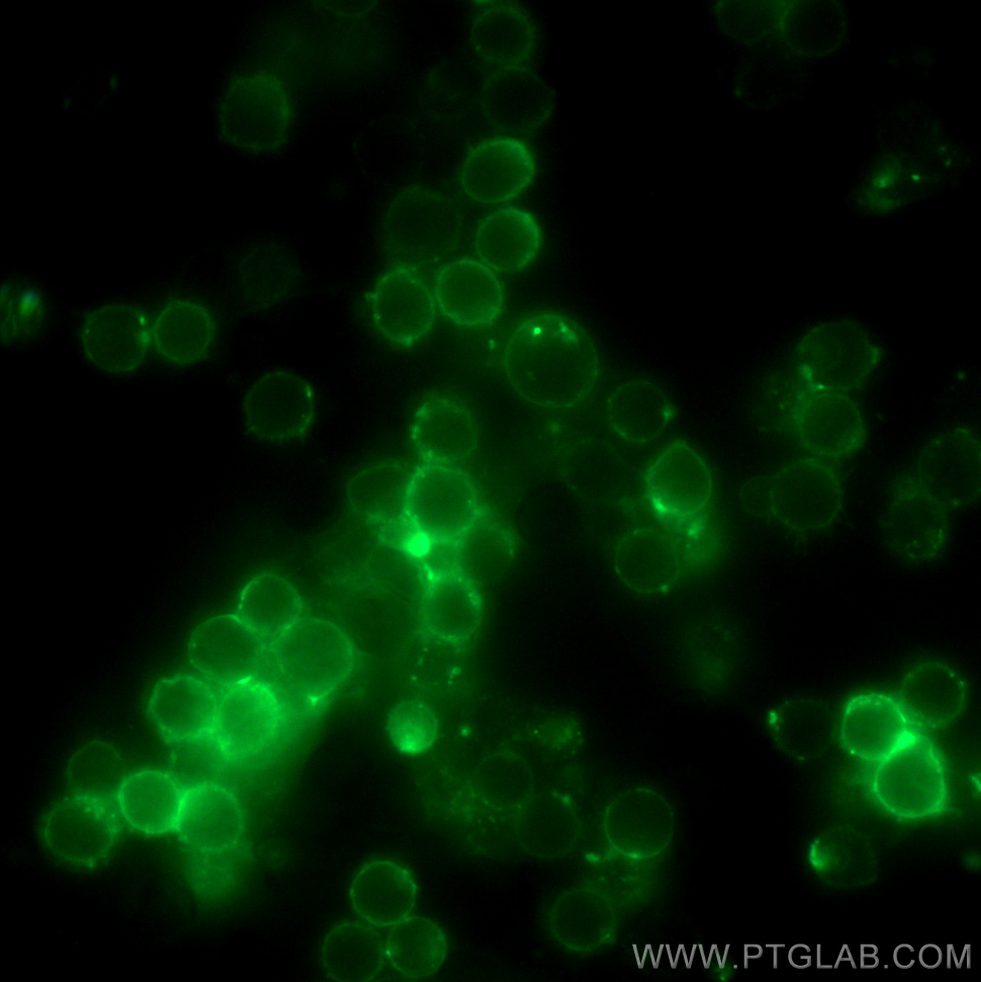
| Positive IF/ICC detected in | mouse splenocytes |
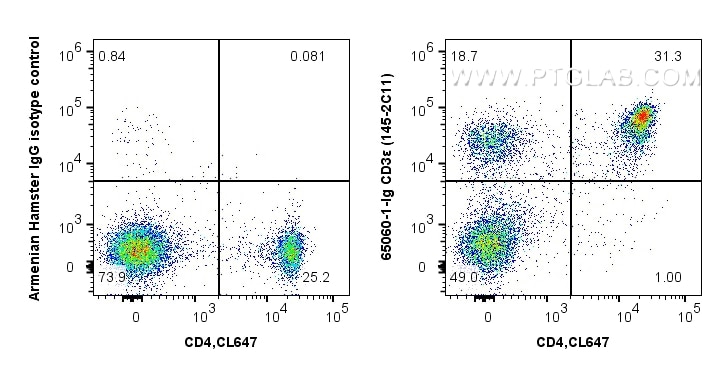
| Positive FC detected in | C57BL/6 mouse splenocytes |
Recommended dilution
| Application | Dilution |
|---|---|
| Immunofluorescence (IF)/ICC | IF/ICC : 1:250-1:1000 |
| Flow Cytometry (FC) | FC : 0.5 ug per 10^6 cells in 100 μl suspension |
| This reagent has been tested for flow cytometric analysis. It is recommended that this reagent should be titrated in each testing system to obtain optimal results. | |
| Sample-dependent, Check data in validation data gallery. | |
Product Information
65060-1-Ig targets CD3e in IF/ICC, FC applications and shows reactivity with mouse samples.
| Tested Reactivity | mouse |
| Host / Isotype | Armenian Hamster / IgG |
| Class | Monoclonal |
| Type | Antibody |
| Immunogen |
H-2Kb-specific murine cytotoxic T-lymphocyte (CTL) clone BM10-37 Predict reactive species |
| Full Name | CD3 antigen, epsilon polypeptide |
| GenBank Accession Number | BC098236 |
| Gene Symbol | CD3ε |
| Gene ID (NCBI) | 12501 |
| RRID | AB_2918368 |
| Conjugate | Unconjugated |
| Form | Liquid |
| Purification Method | Affinity purification |
| UNIPROT ID | P22646 |
| Storage Buffer | PBS with 0.09% sodium azide, pH 7.3. |
| Storage Conditions | Store at 2-8°C. Stable for one year after shipment. |
Background Information
CD3 is a multimeric protein associated with the T-cell receptor (TCR) to form a complex involved in antigen recognition and signal transduction (PMID: 15885124). CD3 is composed of CD3γ, δ, ε, and ζ chains (PMID: 1826255). It is expressed by thymocytes in a developmentally regulated manner, T cells, and some NK cells (PMID: 3289580). The TCR recognizes antigens bound to major histocompatibility complex (MHC) molecules. TCR-mediated peptide-MHC recognition is transmitted to the CD3 complex, leading to the intracellular signal transduction (PMID: 11985657).
Protocols
| Product Specific Protocols | |
|---|---|
| FC protocol for CD3e antibody 65060-1-Ig | Download protocol |
| IF protocol for CD3e antibody 65060-1-Ig | Download protocol |
| Standard Protocols | |
|---|---|
| Click here to view our Standard Protocols |
Reviews
The reviews below have been submitted by verified Proteintech customers who received an incentive for providing their feedback.
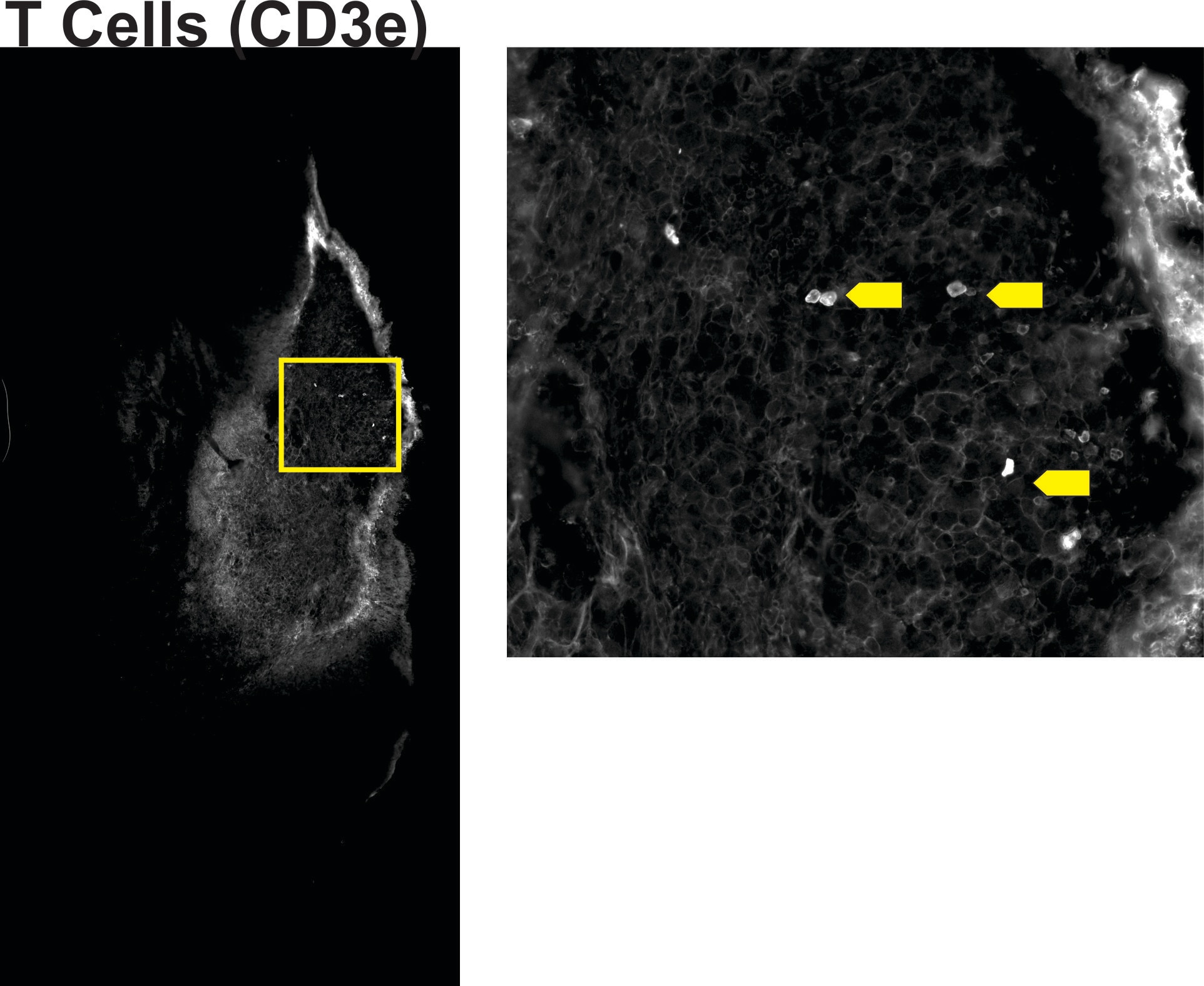
FH Helena (Verified Customer) (07-22-2022) | Not many t cells to detect but they were observable in this tissue section.
 |


