human spleen tissue were subjected to SDS PAGE followed by western blot with 66953-1-Ig (BLNK antibody) at dilution of 1:5000 incubated at room temperature for 1.5 hours.
human spleen tissue were subjected to SDS PAGE followed by western blot with 66953-1-Ig (BLNK antibody) at dilution of 1:5000 incubated at room temperature for 1.5 hours.
WB analysis of Ramos using 66953-1-Ig
Ramos cells were subjected to SDS PAGE followed by western blot with 66953-1-Ig (BLNK antibody) at dilution of 1:3000 incubated at room temperature for 1.5 hours.
Ramos cells were subjected to SDS PAGE followed by western blot with 66953-1-Ig (BLNK antibody) at dilution of 1:3000 incubated at room temperature for 1.5 hours.
WB analysis of pig spleen using 66953-1-Ig
pig spleen tissue were subjected to SDS PAGE followed by western blot with 66953-1-Ig (BLNK antibody) at dilution of 1:3000 incubated at room temperature for 1.5 hours.
pig spleen tissue were subjected to SDS PAGE followed by western blot with 66953-1-Ig (BLNK antibody) at dilution of 1:3000 incubated at room temperature for 1.5 hours.
WB analysis of Raji using 66953-1-Ig
Raji cells were subjected to SDS PAGE followed by western blot with 66953-1-Ig (BLNK antibody) at dilution of 1:5000 incubated at room temperature for 1.5 hours.
Raji cells were subjected to SDS PAGE followed by western blot with 66953-1-Ig (BLNK antibody) at dilution of 1:5000 incubated at room temperature for 1.5 hours.
WB analysis of Daudi using 66953-1-Ig
Daudi cells were subjected to SDS PAGE followed by western blot with 66953-1-Ig (BLNK antibody) at dilution of 1:5000 incubated at room temperature for 1.5 hours.
Daudi cells were subjected to SDS PAGE followed by western blot with 66953-1-Ig (BLNK antibody) at dilution of 1:5000 incubated at room temperature for 1.5 hours.
IHC staining of human appendicitis using 66953-1-Ig
Immunohistochemical analysis of paraffin-embedded human appendicitis tissue slide using 66953-1-Ig (BLNK antibody) at dilution of 1:500 (under 10x lens). Heat mediated antigen retrieval with Tris-EDTA buffer (pH 9.0).
Immunohistochemical analysis of paraffin-embedded human appendicitis tissue slide using 66953-1-Ig (BLNK antibody) at dilution of 1:500 (under 10x lens). Heat mediated antigen retrieval with Tris-EDTA buffer (pH 9.0).
IHC staining of human appendicitis using 66953-1-Ig
Immunohistochemical analysis of paraffin-embedded human appendicitis tissue slide using 66953-1-Ig (BLNK antibody) at dilution of 1:500 (under 40x lens). Heat mediated antigen retrieval with Tris-EDTA buffer (pH 9.0).
Immunohistochemical analysis of paraffin-embedded human appendicitis tissue slide using 66953-1-Ig (BLNK antibody) at dilution of 1:500 (under 40x lens). Heat mediated antigen retrieval with Tris-EDTA buffer (pH 9.0).
IHC staining of mouse thymus using 66953-1-Ig
Immunohistochemical analysis of paraffin-embedded mouse thymus tissue slide using 66953-1-Ig (BLNK antibody) at dilution of 1:500 (under 10x lens). Heat mediated antigen retrieval with Tris-EDTA buffer (pH 9.0).
Immunohistochemical analysis of paraffin-embedded mouse thymus tissue slide using 66953-1-Ig (BLNK antibody) at dilution of 1:500 (under 40x lens). Heat mediated antigen retrieval with Tris-EDTA buffer (pH 9.0).
IHC staining of rat spleen using 66953-1-Ig
Immunohistochemical analysis of paraffin-embedded rat spleen tissue slide using 66953-1-Ig (BLNK antibody) at dilution of 1:500 (under 10x lens). Heat mediated antigen retrieval with Tris-EDTA buffer (pH 9.0).
Immunohistochemical analysis of paraffin-embedded rat spleen tissue slide using 66953-1-Ig (BLNK antibody) at dilution of 1:500 (under 10x lens). Heat mediated antigen retrieval with Tris-EDTA buffer (pH 9.0).
IHC staining of rat spleen using 66953-1-Ig
Immunohistochemical analysis of paraffin-embedded rat spleen tissue slide using 66953-1-Ig (BLNK antibody) at dilution of 1:500 (under 40x lens). Heat mediated antigen retrieval with Tris-EDTA buffer (pH 9.0).
Immunohistochemical analysis of paraffin-embedded rat spleen tissue slide using 66953-1-Ig (BLNK antibody) at dilution of 1:500 (under 40x lens). Heat mediated antigen retrieval with Tris-EDTA buffer (pH 9.0).
IF Staining of Raji using 66953-1-Ig
Immunofluorescent analysis of (4% PFA) fixed Raji cells using BLNK antibody (66953-1-Ig, Clone: 1A1A1 ) at dilution of 1:400 and CoraLite®488-Conjugated Goat Anti-Mouse IgG(H+L).
Immunofluorescent analysis of (4% PFA) fixed Raji cells using BLNK antibody (66953-1-Ig, Clone: 1A1A1 ) at dilution of 1:400 and CoraLite®488-Conjugated Goat Anti-Mouse IgG(H+L).
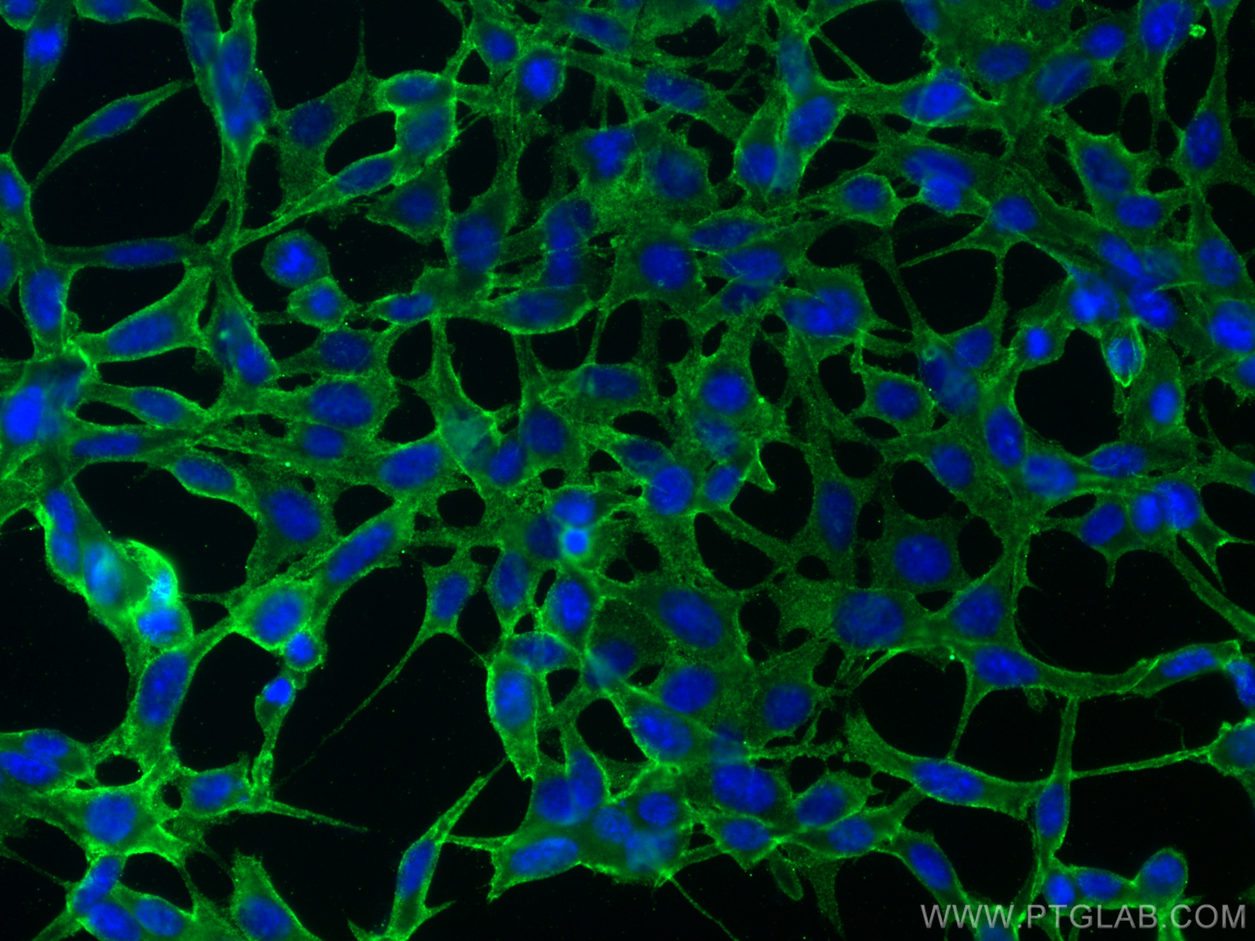
IF Staining of NIH/3T3 using 66953-1-Ig
Immunofluorescent analysis of (-20°C Ethanol) fixed NIH/3T3 cells using BLNK antibody (66953-1-Ig, Clone: 1A1A1 ) at dilution of 1:800 and CoraLite®488-Conjugated Goat Anti-Mouse IgG(H+L) (SA00013-1).
Immunofluorescent analysis of (-20°C Ethanol) fixed NIH/3T3 cells using BLNK antibody (66953-1-Ig, Clone: 1A1A1 ) at dilution of 1:800 and CoraLite®488-Conjugated Goat Anti-Mouse IgG(H+L) (SA00013-1).
The Proteintech guarantee covers Proteintech antibodies in any species and any application, including those not listed on the datasheet. If the antibody doesn’t perform, you can receive a hassle-free refund or credit note.
human appendicitis tissue, mouse thymus tissue, rat spleen tissue Note: suggested antigen retrieval with TE buffer pH 9.0; (*) Alternatively, antigen retrieval may be performed with citrate buffer pH 6.0
Positive IF/ICC detected in
Raji cells, NIH/3T3 cells
Recommended dilution
Application
Dilution
Western Blot (WB)
WB : 1:3000-1:8000
Immunohistochemistry (IHC)
IHC : 1:250-1:1000
Immunofluorescence (IF)/ICC
IF/ICC : 1:200-1:800
It is recommended that this reagent should be titrated in each testing system to obtain optimal results.
Sample-dependent, Check data in validation data gallery.
Product Information
66953-1-Ig targets BLNK in WB, IHC, IF/ICC, ELISA applications and shows reactivity with Human, pig, mouse, rat samples.
PBS with 0.02% sodium azide and 50% glycerol , pH 7.3
Storage Conditions
Store at -20°C. Stable for one year after shipment. Aliquoting is unnecessary for -20oC storage. 20ul sizes contain 0.1% BSA.
Background Information
BLNK (also known as SLP-65 or BASH) is an important adaptor protein selectively expressed in B-lineage cells. BLNK bridges B cell receptor-associated kinase activation with downstream signaling pathways, thereby affecting various biological functions. BLNK has been shown to be necessary for BCR-mediated Ca2+ mobilization, for the activation of mitogen-activated protein kinases such as ERK, JNK, and p38 in a chicken B cell line DT40, and for activation of transcription factors such as NF-AT and NF-κB in human or mouse B cells. BLNK plays a crucial role in pre-BCR-dependent progression of B cell development, BCR-mediated B cell survival, activation, proliferation, and T-independent immune responses.
human spleen tissue were subjected to SDS PAGE followed by western blot with 66953-1-Ig (BLNK antibody) at dilution of 1:5000 incubated at room temperature for 1.5 hours.
WB analysis of Ramos using 66953-1-Ig
Ramos cells were subjected to SDS PAGE followed by western blot with 66953-1-Ig (BLNK antibody) at dilution of 1:3000 incubated at room temperature for 1.5 hours.
WB analysis of pig spleen using 66953-1-Ig
pig spleen tissue were subjected to SDS PAGE followed by western blot with 66953-1-Ig (BLNK antibody) at dilution of 1:3000 incubated at room temperature for 1.5 hours.
WB analysis of Raji using 66953-1-Ig
Raji cells were subjected to SDS PAGE followed by western blot with 66953-1-Ig (BLNK antibody) at dilution of 1:5000 incubated at room temperature for 1.5 hours.
WB analysis of Daudi using 66953-1-Ig
Daudi cells were subjected to SDS PAGE followed by western blot with 66953-1-Ig (BLNK antibody) at dilution of 1:5000 incubated at room temperature for 1.5 hours.
IHC Figures
IHC staining of human appendicitis using 66953-1-Ig
Immunohistochemical analysis of paraffin-embedded human appendicitis tissue slide using 66953-1-Ig (BLNK antibody) at dilution of 1:500 (under 10x lens). Heat mediated antigen retrieval with Tris-EDTA buffer (pH 9.0).
IHC staining of human appendicitis using 66953-1-Ig
Immunohistochemical analysis of paraffin-embedded human appendicitis tissue slide using 66953-1-Ig (BLNK antibody) at dilution of 1:500 (under 40x lens). Heat mediated antigen retrieval with Tris-EDTA buffer (pH 9.0).
IHC staining of mouse thymus using 66953-1-Ig
Immunohistochemical analysis of paraffin-embedded mouse thymus tissue slide using 66953-1-Ig (BLNK antibody) at dilution of 1:500 (under 10x lens). Heat mediated antigen retrieval with Tris-EDTA buffer (pH 9.0).
IHC staining of mouse thymus using 66953-1-Ig
Immunohistochemical analysis of paraffin-embedded mouse thymus tissue slide using 66953-1-Ig (BLNK antibody) at dilution of 1:500 (under 40x lens). Heat mediated antigen retrieval with Tris-EDTA buffer (pH 9.0).
IHC staining of rat spleen using 66953-1-Ig
Immunohistochemical analysis of paraffin-embedded rat spleen tissue slide using 66953-1-Ig (BLNK antibody) at dilution of 1:500 (under 10x lens). Heat mediated antigen retrieval with Tris-EDTA buffer (pH 9.0).
IHC staining of rat spleen using 66953-1-Ig
Immunohistochemical analysis of paraffin-embedded rat spleen tissue slide using 66953-1-Ig (BLNK antibody) at dilution of 1:500 (under 40x lens). Heat mediated antigen retrieval with Tris-EDTA buffer (pH 9.0).
IF/ICC Figures
IF Staining of Raji using 66953-1-Ig
Immunofluorescent analysis of (4% PFA) fixed Raji cells using BLNK antibody (66953-1-Ig, Clone: 1A1A1 ) at dilution of 1:400 and CoraLite®488-Conjugated Goat Anti-Mouse IgG(H+L).
IF Staining of NIH/3T3 using 66953-1-Ig
Immunofluorescent analysis of (-20°C Ethanol) fixed NIH/3T3 cells using BLNK antibody (66953-1-Ig, Clone: 1A1A1 ) at dilution of 1:800 and CoraLite®488-Conjugated Goat Anti-Mouse IgG(H+L) (SA00013-1).
The species listed in Tested Reactivity are in-house verified and applicable species. For unlisted species, please refer to the homology analysis of the immunogen sequence and related species. For rabbit polyclonal antibodies, homology >70% is recommended. For mouse monoclonal antibodies and rabbit recombinant antibodies, homology >90% is recommended. Generally, the higher the homology, the greater the applicability. However, there will be certain differences in protein expression in different species, tissues or cells. Therefore, the homology analysis results are for reference only and do not serve as a guarantee.
At Proteintech, we pride ourselves on our antibody quality, customer service and transparency. As such, we are comparing our antibodies with other vendors, enabling easy identification and comparisons of key data to help you choose the suitable antibody for your needs.
We have selected the top cited antibodies from these vendors for you to compare.
 at dilution of 1:5000 incubated at room temperature for 1.5 hours.")
 at dilution of 1:3000 incubated at room temperature for 1.5 hours.")
 at dilution of 1:3000 incubated at room temperature for 1.5 hours.")
 at dilution of 1:5000 incubated at room temperature for 1.5 hours.")
 at dilution of 1:5000 incubated at room temperature for 1.5 hours.")
 at dilution of 1:500 (under 10x lens). Heat mediated antigen retrieval with Tris-EDTA buffer (pH 9.0).")
 at dilution of 1:500 (under 40x lens). Heat mediated antigen retrieval with Tris-EDTA buffer (pH 9.0).")
 at dilution of 1:500 (under 10x lens). Heat mediated antigen retrieval with Tris-EDTA buffer (pH 9.0).")
 at dilution of 1:500 (under 40x lens). Heat mediated antigen retrieval with Tris-EDTA buffer (pH 9.0).")
 at dilution of 1:500 (under 10x lens). Heat mediated antigen retrieval with Tris-EDTA buffer (pH 9.0).")
 at dilution of 1:500 (under 40x lens). Heat mediated antigen retrieval with Tris-EDTA buffer (pH 9.0).")
 fixed Raji cells using BLNK antibody (66953-1-Ig, Clone: 1A1A1 ) at dilution of 1:400 and CoraLite®488-Conjugated Goat Anti-Mouse IgG(H+L).")
 fixed NIH/3T3 cells using BLNK antibody (66953-1-Ig, Clone: 1A1A1 ) at dilution of 1:800 and CoraLite®488-Conjugated Goat Anti-Mouse IgG(H+L) (SA00013-1).")