at dilution of 1:4000 incubated at room temperature for 1.5 hours.")
 at dilution of 1:800 incubated at room temperature for 1.5 hours.")
 with RAW 264.7 cells lysate 4000ug.")
 at dilution of 1:200 (under 10x lens). Heat mediated antigen retrieval with Tris-EDTA buffer (pH 9.0).")
 at dilution of 1:200 (under 40x lens). Heat mediated antigen retrieval with Tris-EDTA buffer (pH 9.0).")
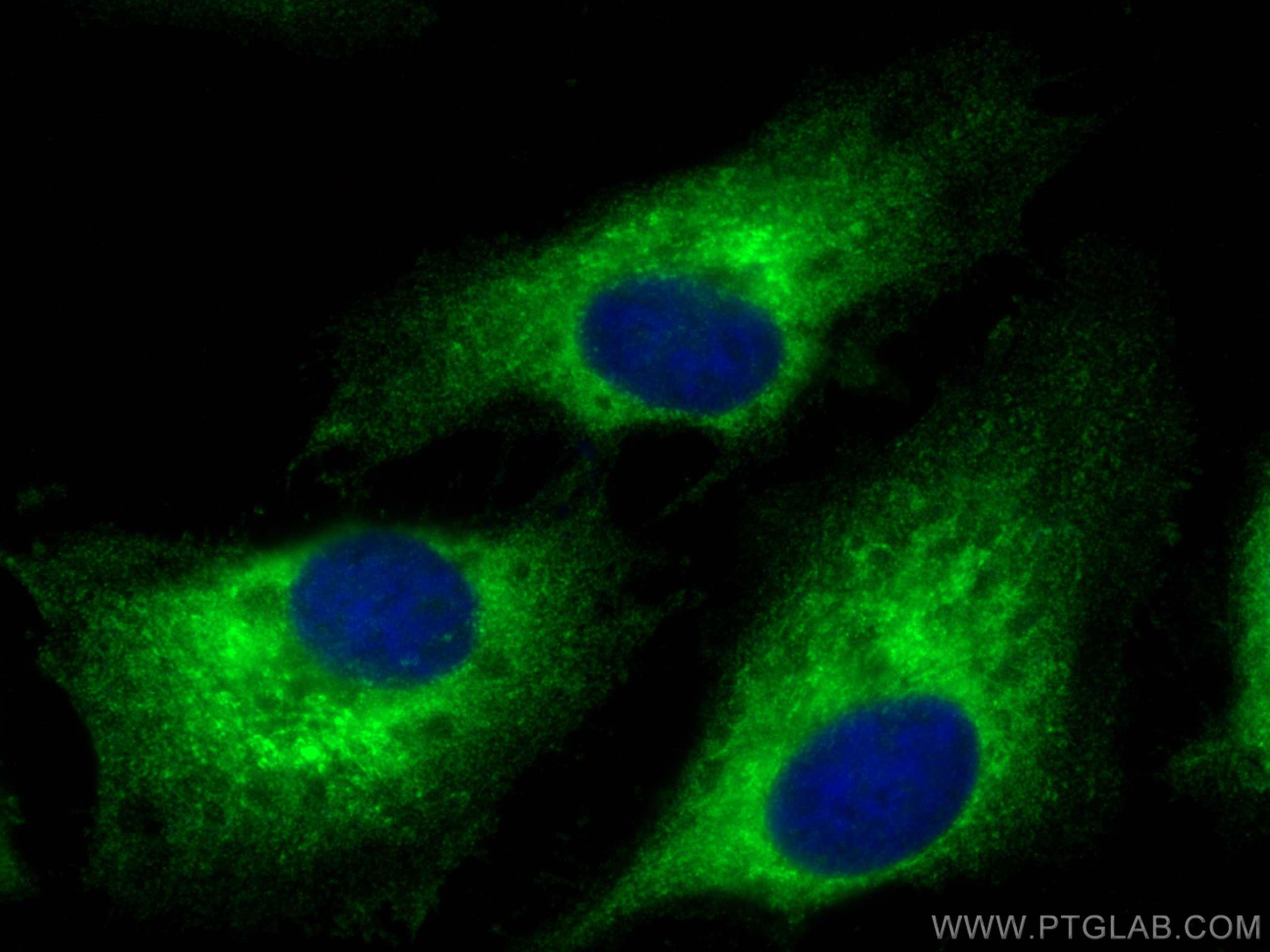
 fixed HeLa cells using p130Cas / BCAR1 antibody (16815-1-AP) at dilution of 1:200 and CoraLite®488-Conjugated AffiniPure Goat Anti-Rabbit IgG(H+L).")
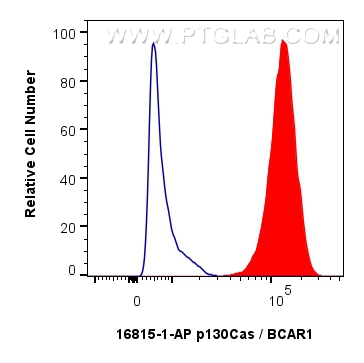
 and CoraLite®488-Conjugated Goat Anti-Rabbit IgG(H+L) (SA00013-2)(red), or 0.25 ug rabbit IgG isotype control (blue). Cells were fixed with 4% PFA and permeabilized with Flow Cytometry Perm Buffer.")
Tested Applications
| Positive WB detected in | HeLa cells, A431 cells, HepG2 cells, T-47D cells |
| Positive IP detected in | RAW 264.7 cells |
| Positive IHC detected in | human colon cancer tissue Note: suggested antigen retrieval with TE buffer pH 9.0; (*) Alternatively, antigen retrieval may be performed with citrate buffer pH 6.0 |
| Positive IF/ICC detected in | HeLa cells |
| Positive FC (Intra) detected in | A431 cells |
Recommended dilution
| Application | Dilution |
|---|---|
| Western Blot (WB) | WB : 1:1000-1:8000 |
| Immunoprecipitation (IP) | IP : 0.5-4.0 ug for 1.0-3.0 mg of total protein lysate |
| Immunohistochemistry (IHC) | IHC : 1:50-1:500 |
| Immunofluorescence (IF)/ICC | IF/ICC : 1:50-1:500 |
| Flow Cytometry (FC) (INTRA) | FC (INTRA) : 0.25 ug per 10^6 cells in a 100 µl suspension |
| It is recommended that this reagent should be titrated in each testing system to obtain optimal results. | |
| Sample-dependent, Check data in validation data gallery. | |
Product Information
16815-1-AP targets p130Cas / BCAR1 in WB, IHC, IF/ICC, FC (Intra), IP, ELISA applications and shows reactivity with human, mouse, rat samples.
| Tested Reactivity | human, mouse, rat |
| Host / Isotype | Rabbit / IgG |
| Class | Polyclonal |
| Type | Antibody |
| Immunogen |
CatNo: Ag10354 Product name: Recombinant human BCAR1 protein Source: e coli.-derived, PGEX-4T Tag: GST Domain: 1-420 aa of BC062556 Sequence: MNHLNVLAKALYDNVAESPDELSFRKGDIMTVLEQDTQGLDGWWLCSLHGRQGIVPGNRLKILVGMYDKKPAGPGSGPPATPAQPQPGLHAPAPPASQYTPMLPNTYQPQPDSVYLVPTPSKAQQGLYQVPGPSPQFQSPPAKQTSTFSKQTPHHPFPNPATDLYQVPPGPGGPAQDIYQVPPSAGMGHDIYQVPPSMDTRSWEGTKPPAKVVVPTRVGQGYVYEAAQPEQDEYDIPRHLLAPGPQDIYDVPPVRGLLPSQYGQEVYDTPPMAVKGPNGRDPLLEVYDVPPSVEKGLPPSNHHAVYDVPPSVSKDVPDGPLLREETYDVPPAFAKAKPFDPARTPLVLAAPPPDSPPAEDVYDVPPPAPDLYDVPPGLRRPGPGTLYDVPRERVLPPEVADGGVVDSGVYAVPPPAEREA Predict reactive species |
| Full Name | breast cancer anti-estrogen resistance 1 |
| Calculated Molecular Weight | 870 aa, 93 kDa |
| Observed Molecular Weight | 116 kDa |
| GenBank Accession Number | BC062556 |
| Gene Symbol | BCAR1 |
| Gene ID (NCBI) | 9564 |
| RRID | AB_2065460 |
| Conjugate | Unconjugated |
| Form | Liquid |
| Purification Method | Antigen affinity purification |
| UNIPROT ID | P56945 |
| Storage Buffer | PBS with 0.02% sodium azide and 50% glycerol, pH 7.3. |
| Storage Conditions | Store at -20°C. Stable for one year after shipment. Aliquoting is unnecessary for -20oC storage. 20ul sizes contain 0.1% BSA. |
Protocols
| Product Specific Protocols | |
|---|---|
| FC protocol for p130Cas / BCAR1 antibody 16815-1-AP | Download protocol |
| IF protocol for p130Cas / BCAR1 antibody 16815-1-AP | Download protocol |
| IHC protocol for p130Cas / BCAR1 antibody 16815-1-AP | Download protocol |
| IP protocol for p130Cas / BCAR1 antibody 16815-1-AP | Download protocol |
| WB protocol for p130Cas / BCAR1 antibody 16815-1-AP | Download protocol |
| Standard Protocols | |
|---|---|
| Click here to view our Standard Protocols |