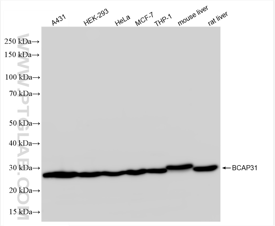
Various lysates were subjected to SDS PAGE followed by western blot with 84584-5-RR (BAP31 antibody) at dilution of 1:10000 incubated at room temperature for 1.5 hours.
Various lysates were subjected to SDS PAGE followed by western blot with 84584-5-RR (BAP31 antibody) at dilution of 1:10000 incubated at room temperature for 1.5 hours.
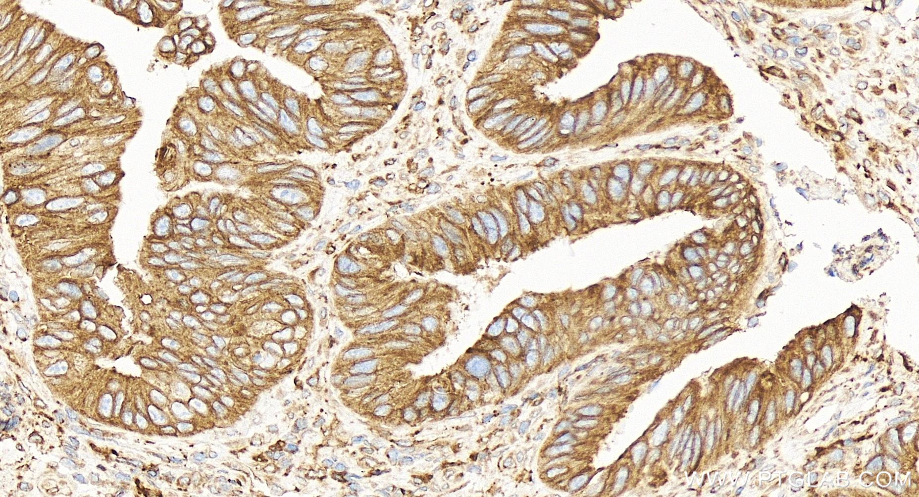
IHC staining of human colon cancer using 84584-5-RR
Immunohistochemical analysis of paraffin-embedded human colon cancer tissue slide using 84584-5-RR (BAP31 antibody) at dilution of 1:800 (under 40x lens). Heat mediated antigen retrieval with Tris-EDTA buffer (pH 9.0).
Immunohistochemical analysis of paraffin-embedded human colon cancer tissue slide using 84584-5-RR (BAP31 antibody) at dilution of 1:800 (under 40x lens). Heat mediated antigen retrieval with Tris-EDTA buffer (pH 9.0).
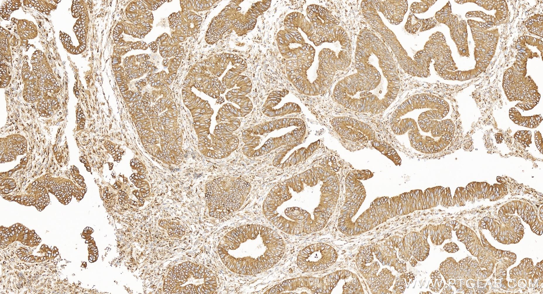
IHC staining of human colon cancer using 84584-5-RR
Immunohistochemical analysis of paraffin-embedded human colon cancer tissue slide using 84584-5-RR (BAP31 antibody) at dilution of 1:800 (under 10x lens). Heat mediated antigen retrieval with Tris-EDTA buffer (pH 9.0).
Immunohistochemical analysis of paraffin-embedded human colon cancer tissue slide using 84584-5-RR (BAP31 antibody) at dilution of 1:800 (under 10x lens). Heat mediated antigen retrieval with Tris-EDTA buffer (pH 9.0).
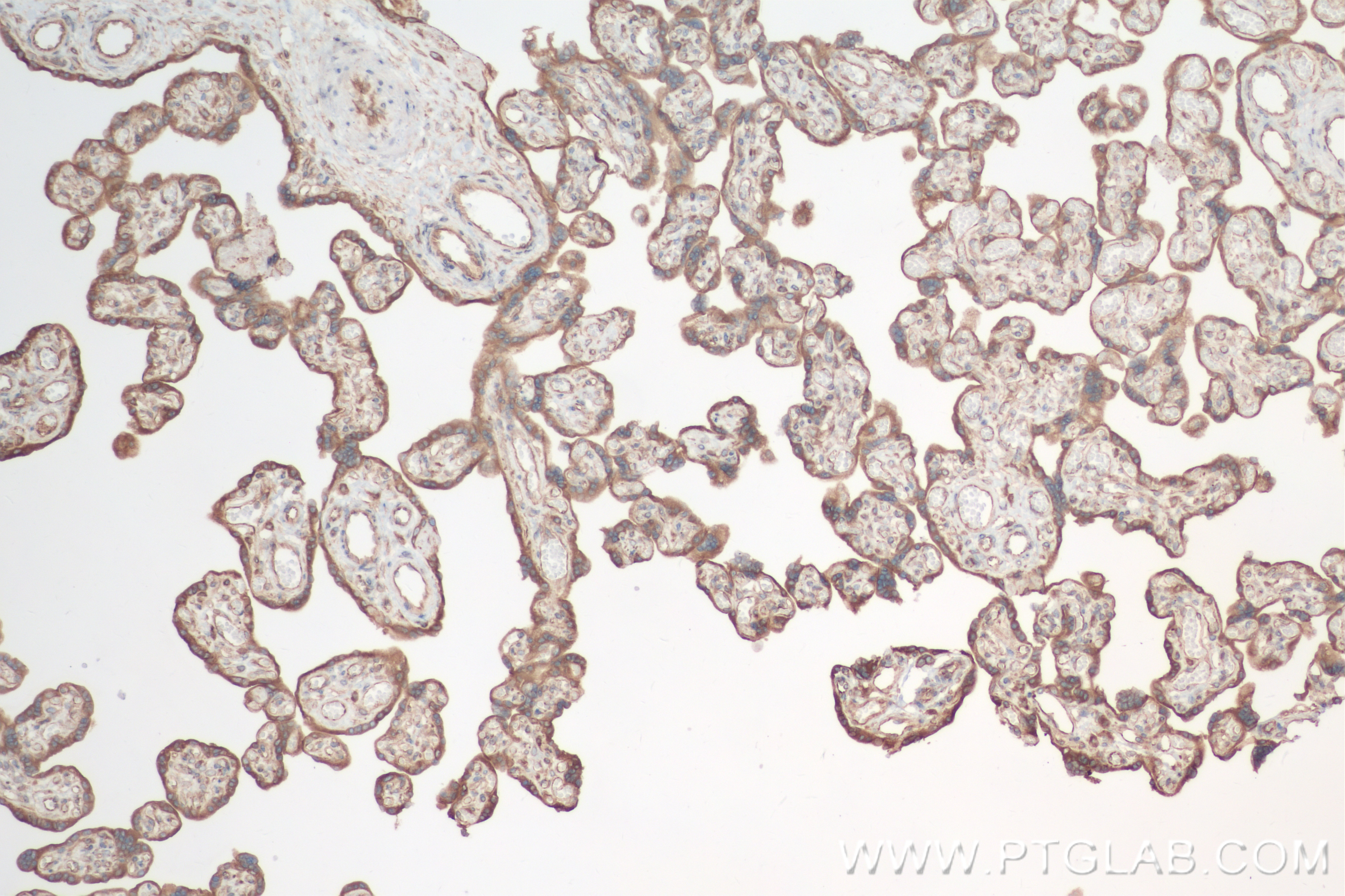
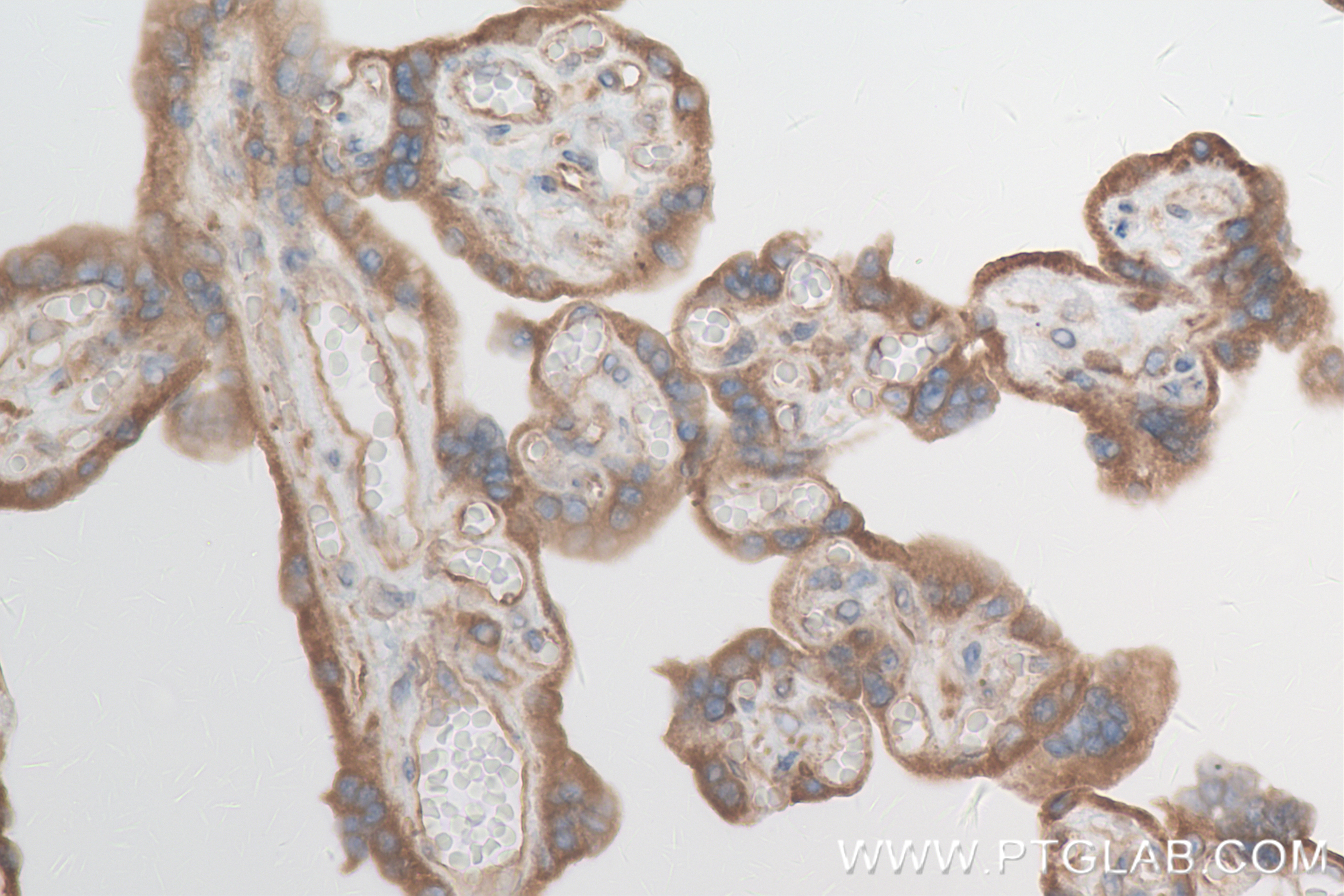
IHC staining of human placenta using 84584-5-RR
Immunohistochemical analysis of paraffin-embedded human placenta tissue slide using 84584-5-RR (BAP31 antibody) at dilution of 1:800 (under 10x lens). Heat mediated antigen retrieval with Tris-EDTA buffer (pH 9.0).
Immunohistochemical analysis of paraffin-embedded human placenta tissue slide using 84584-5-RR (BAP31 antibody) at dilution of 1:800 (under 10x lens). Heat mediated antigen retrieval with Tris-EDTA buffer (pH 9.0).
IHC staining of human placenta using 84584-5-RR
Immunohistochemical analysis of paraffin-embedded human placenta tissue slide using 84584-5-RR (BAP31 antibody) at dilution of 1:800 (under 40x lens). Heat mediated antigen retrieval with Tris-EDTA buffer (pH 9.0).
Immunohistochemical analysis of paraffin-embedded human placenta tissue slide using 84584-5-RR (BAP31 antibody) at dilution of 1:800 (under 40x lens). Heat mediated antigen retrieval with Tris-EDTA buffer (pH 9.0).
IF Staining of HeLa using 84584-5-RR
Immunofluorescent analysis of (4% PFA) fixed HeLa cells using BAP31 antibody (84584-5-RR, Clone: 241978D10 ) at dilution of 1:400 and CoraLite®488-Conjugated Goat Anti-Rabbit IgG(H+L) (SA00013-2).
Immunofluorescent analysis of (4% PFA) fixed HeLa cells using BAP31 antibody (84584-5-RR, Clone: 241978D10 ) at dilution of 1:400 and CoraLite®488-Conjugated Goat Anti-Rabbit IgG(H+L) (SA00013-2).
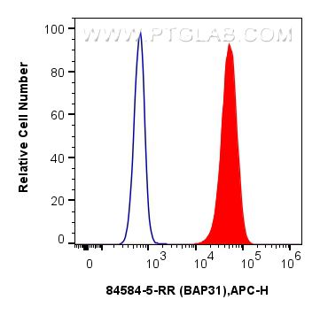
FC experiment of HepG2 using 84584-5-RR
1x10^6 HepG2 cells were intracellularly stained with 0.25 ug BAP31 Recombinant antibody (84584-5-RR, Clone:241978D10) and APC-Conjugated Goat Anti-Rabbit IgG(H+L)(red), or 0.25 ug Rabbit IgG Isotype Control Recombinant Antibody (98136-1-RR, Clone: 240953C9) (blue). Cells were fixed with 4% PFA and permeabilized with Flow Cytometry Perm Buffer (PF00011-C).
1x10^6 HepG2 cells were intracellularly stained with 0.25 ug BAP31 Recombinant antibody (84584-5-RR, Clone:241978D10) and APC-Conjugated Goat Anti-Rabbit IgG(H+L)(red), or 0.25 ug Rabbit IgG Isotype Control Recombinant Antibody (98136-1-RR, Clone: 240953C9) (blue). Cells were fixed with 4% PFA and permeabilized with Flow Cytometry Perm Buffer (PF00011-C).
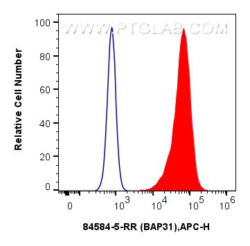
FC experiment of PC-3 using 84584-5-RR
1x10^6 PC-3 cells were intracellularly stained with 0.25 ug BAP31 Recombinant antibody (84584-5-RR, Clone:241978D10) and APC-Conjugated Goat Anti-Rabbit IgG(H+L)(red), or 0.25 ug Rabbit IgG Isotype Control Recombinant Antibody (98136-1-RR, Clone: 240953C9) (blue). Cells were fixed with 4% PFA and permeabilized with Flow Cytometry Perm Buffer (PF00011-C).
1x10^6 PC-3 cells were intracellularly stained with 0.25 ug BAP31 Recombinant antibody (84584-5-RR, Clone:241978D10) and APC-Conjugated Goat Anti-Rabbit IgG(H+L)(red), or 0.25 ug Rabbit IgG Isotype Control Recombinant Antibody (98136-1-RR, Clone: 240953C9) (blue). Cells were fixed with 4% PFA and permeabilized with Flow Cytometry Perm Buffer (PF00011-C).
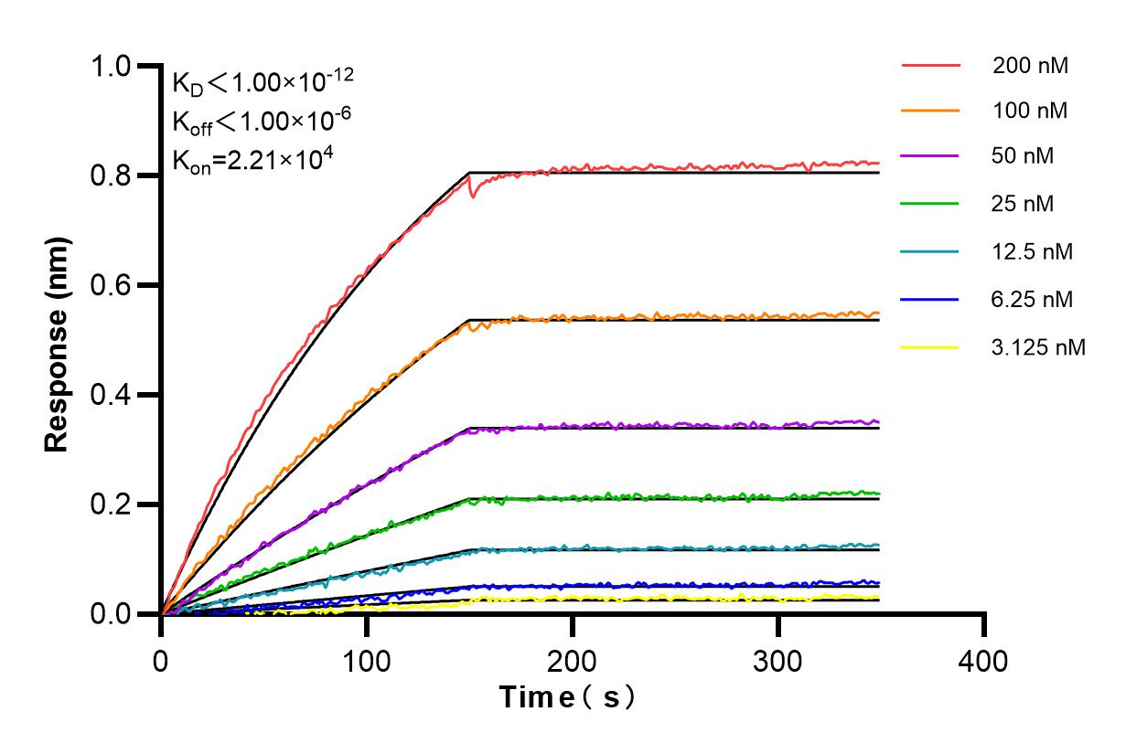
Affinity and Kinetic Characterization of 84584-5-RR
Biolayer interferometry (BLl) kinetic assays of 84584-5-RR against Human BAP31 were performed. The affinity constant is below 1 pM.
The Proteintech guarantee covers Proteintech antibodies in any species and any application, including those not listed on the datasheet. If the antibody doesn’t perform, you can receive a hassle-free refund or credit note.
A431 cells, HEK-293 cells, HeLa cells, MCF-7 cells, THP-1 cells, mouse liver tissue, rat liver tisssue
Positive IHC detected in
human colon cancer tissue, human placenta tissue Note: suggested antigen retrieval with TE buffer pH 9.0; (*) Alternatively, antigen retrieval may be performed with citrate buffer pH 6.0
Positive IF/ICC detected in
HeLa cells
Positive FC (Intra) detected in
HepG2 cells, PC-3 cells
Recommended dilution
Application
Dilution
Western Blot (WB)
WB : 1:5000-1:50000
Immunohistochemistry (IHC)
IHC : 1:400-1:1600
Immunofluorescence (IF)/ICC
IF/ICC : 1:200-1:800
Flow Cytometry (FC) (INTRA)
FC (INTRA) : 0.25 ug per 10^6 cells in a 100 µl suspension
It is recommended that this reagent should be titrated in each testing system to obtain optimal results.
Sample-dependent, Check data in validation data gallery.
Product Information
84584-5-RR targets BAP31 in WB, IHC, IF/ICC, FC (Intra), ELISA applications and shows reactivity with human, mouse, rat samples.
PBS with 0.02% sodium azide and 50% glycerol, pH 7.3.
Storage Conditions
Store at -20°C. Stable for one year after shipment. Aliquoting is unnecessary for -20oC storage. 20ul sizes contain 0.1% BSA.
Background Information
BAP31, also known as BCAP31, is a chaperone protein abundant in the endoplasmic reticulum (ER). BAP31 plays a role in the export of secreted proteins in the ER, the recognition of abnormally folded proteins, and their targeting to the ER-associated-degradation (ERAD). CDIP1 and BAP31 interact upon ER stress to regulate the mitochondrial apoptosis pathway(PMID: 24139803). It also serves as a cargo receptor for the export of transmembrane proteins. BAP31 may be involved in apoptosis.
Various lysates were subjected to SDS PAGE followed by western blot with 84584-5-RR (BAP31 antibody) at dilution of 1:10000 incubated at room temperature for 1.5 hours.
IHC Figures
IHC staining of human colon cancer using 84584-5-RR
Immunohistochemical analysis of paraffin-embedded human colon cancer tissue slide using 84584-5-RR (BAP31 antibody) at dilution of 1:800 (under 40x lens). Heat mediated antigen retrieval with Tris-EDTA buffer (pH 9.0).
IHC staining of human colon cancer using 84584-5-RR
Immunohistochemical analysis of paraffin-embedded human colon cancer tissue slide using 84584-5-RR (BAP31 antibody) at dilution of 1:800 (under 10x lens). Heat mediated antigen retrieval with Tris-EDTA buffer (pH 9.0).
IHC staining of human placenta using 84584-5-RR
Immunohistochemical analysis of paraffin-embedded human placenta tissue slide using 84584-5-RR (BAP31 antibody) at dilution of 1:800 (under 10x lens). Heat mediated antigen retrieval with Tris-EDTA buffer (pH 9.0).
IHC staining of human placenta using 84584-5-RR
Immunohistochemical analysis of paraffin-embedded human placenta tissue slide using 84584-5-RR (BAP31 antibody) at dilution of 1:800 (under 40x lens). Heat mediated antigen retrieval with Tris-EDTA buffer (pH 9.0).
IF/ICC Figures
IF Staining of HeLa using 84584-5-RR
Immunofluorescent analysis of (4% PFA) fixed HeLa cells using BAP31 antibody (84584-5-RR, Clone: 241978D10 ) at dilution of 1:400 and CoraLite®488-Conjugated Goat Anti-Rabbit IgG(H+L) (SA00013-2).
FC (INTRA) Figures
FC experiment of HepG2 using 84584-5-RR
1x10^6 HepG2 cells were intracellularly stained with 0.25 ug BAP31 Recombinant antibody (84584-5-RR, Clone:241978D10) and APC-Conjugated Goat Anti-Rabbit IgG(H+L)(red), or 0.25 ug Rabbit IgG Isotype Control Recombinant Antibody (98136-1-RR, Clone: 240953C9) (blue). Cells were fixed with 4% PFA and permeabilized with Flow Cytometry Perm Buffer (PF00011-C).
FC experiment of PC-3 using 84584-5-RR
1x10^6 PC-3 cells were intracellularly stained with 0.25 ug BAP31 Recombinant antibody (84584-5-RR, Clone:241978D10) and APC-Conjugated Goat Anti-Rabbit IgG(H+L)(red), or 0.25 ug Rabbit IgG Isotype Control Recombinant Antibody (98136-1-RR, Clone: 240953C9) (blue). Cells were fixed with 4% PFA and permeabilized with Flow Cytometry Perm Buffer (PF00011-C).
AFFINITY Figures
Affinity and Kinetic Characterization of 84584-5-RR
Biolayer interferometry (BLl) kinetic assays of 84584-5-RR against Human BAP31 were performed. The affinity constant is below 1 pM.
The species listed in Tested Reactivity are in-house verified and applicable species. For unlisted species, please refer to the homology analysis of the immunogen sequence and related species. For rabbit polyclonal antibodies, homology >70% is recommended. For mouse monoclonal antibodies and rabbit recombinant antibodies, homology >90% is recommended. Generally, the higher the homology, the greater the applicability. However, there will be certain differences in protein expression in different species, tissues or cells. Therefore, the homology analysis results are for reference only and do not serve as a guarantee.
At Proteintech, we pride ourselves on our antibody quality, customer service and transparency. As such, we are comparing our antibodies with other vendors, enabling easy identification and comparisons of key data to help you choose the suitable antibody for your needs.
We have selected the top cited antibodies from these vendors for you to compare.
Proteintech
BAP31 Recombinant antibody
Catalog Number
84584-5-RR
Citations
-
Dilutions
WB : 1:5000-1:50000 IHC : 1:400-1:1600 IF/ICC : 1:200-1:800 FC (INTRA) : 0.25 ug per 10^6 cells in a 100 µl suspension
Applications
WB, IHC, IF/ICC, FC (Intra), ELISA
Reactivity
human, mouse, rat
Product Guarantee
Covers any species including not listed on datasheet
Covers any applications including not listed on datasheet
 at dilution of 1:10000 incubated at room temperature for 1.5 hours.")
 at dilution of 1:800 (under 40x lens). Heat mediated antigen retrieval with Tris-EDTA buffer (pH 9.0).")
 at dilution of 1:800 (under 10x lens). Heat mediated antigen retrieval with Tris-EDTA buffer (pH 9.0).")
 at dilution of 1:800 (under 10x lens). Heat mediated antigen retrieval with Tris-EDTA buffer (pH 9.0).")
 at dilution of 1:800 (under 40x lens). Heat mediated antigen retrieval with Tris-EDTA buffer (pH 9.0).")
 fixed HeLa cells using BAP31 antibody (84584-5-RR, Clone: 241978D10 ) at dilution of 1:400 and CoraLite®488-Conjugated Goat Anti-Rabbit IgG(H+L) (SA00013-2).")
 and APC-Conjugated Goat Anti-Rabbit IgG(H+L)(red), or 0.25 ug Rabbit IgG Isotype Control Recombinant Antibody (98136-1-RR, Clone: 240953C9) (blue). Cells were fixed with 4% PFA and permeabilized with Flow Cytometry Perm Buffer (PF00011-C).")
 and APC-Conjugated Goat Anti-Rabbit IgG(H+L)(red), or 0.25 ug Rabbit IgG Isotype Control Recombinant Antibody (98136-1-RR, Clone: 240953C9) (blue). Cells were fixed with 4% PFA and permeabilized with Flow Cytometry Perm Buffer (PF00011-C).")
 kinetic assays of 84584-5-RR against Human BAP31 were performed. The affinity constant is below 1 pM.")