MCF-7 cells were subjected to SDS PAGE followed by western blot with 67057-1-Ig (ATX3,ATXN3 antibody) at dilution of 1:10000 incubated at room temperature for 1.5 hours.
MCF-7 cells were subjected to SDS PAGE followed by western blot with 67057-1-Ig (ATX3,ATXN3 antibody) at dilution of 1:10000 incubated at room temperature for 1.5 hours.
WB analysis of Jurkat using 67057-1-Ig
Jurkat cells were subjected to SDS PAGE followed by western blot with 67057-1-Ig (ATX3,ATXN3 antibody) at dilution of 1:10000 incubated at room temperature for 1.5 hours.
Jurkat cells were subjected to SDS PAGE followed by western blot with 67057-1-Ig (ATX3,ATXN3 antibody) at dilution of 1:10000 incubated at room temperature for 1.5 hours.
WB analysis of pig brain using 67057-1-Ig
pig brain tissue were subjected to SDS PAGE followed by western blot with 67057-1-Ig (ATX3,ATXN3 antibody) at dilution of 1:10000 incubated at room temperature for 1.5 hours.
pig brain tissue were subjected to SDS PAGE followed by western blot with 67057-1-Ig (ATX3,ATXN3 antibody) at dilution of 1:10000 incubated at room temperature for 1.5 hours.
WB analysis of mouse brain using 67057-1-Ig
mouse brain tissue were subjected to SDS PAGE followed by western blot with 67057-1-Ig (ATX3,ATXN3 antibody) at dilution of 1:10000 incubated at room temperature for 1.5 hours.
mouse brain tissue were subjected to SDS PAGE followed by western blot with 67057-1-Ig (ATX3,ATXN3 antibody) at dilution of 1:10000 incubated at room temperature for 1.5 hours.
WB analysis of HeLa using 67057-1-Ig
HeLa cells were subjected to SDS PAGE followed by western blot with 67057-1-Ig (ATX3,ATXN3 antibody) at dilution of 1:10000 incubated at room temperature for 1.5 hours.
HeLa cells were subjected to SDS PAGE followed by western blot with 67057-1-Ig (ATX3,ATXN3 antibody) at dilution of 1:10000 incubated at room temperature for 1.5 hours.
WB analysis of HEK-293 using 67057-1-Ig
HEK-293 cells were subjected to SDS PAGE followed by western blot with 67057-1-Ig (ATX3,ATXN3 antibody) at dilution of 1:10000 incubated at room temperature for 1.5 hours.
HEK-293 cells were subjected to SDS PAGE followed by western blot with 67057-1-Ig (ATX3,ATXN3 antibody) at dilution of 1:10000 incubated at room temperature for 1.5 hours.
IHC staining of human pancreas cancer using 67057-1-Ig
Immunohistochemical analysis of paraffin-embedded human pancreas cancer tissue slide using 67057-1-Ig (ATX3,ATXN3 antibody) at dilution of 1:1000 (under 10x lens). Heat mediated antigen retrieval with Tris-EDTA buffer (pH 9.0).
Immunohistochemical analysis of paraffin-embedded human pancreas cancer tissue slide using 67057-1-Ig (ATX3,ATXN3 antibody) at dilution of 1:1000 (under 10x lens). Heat mediated antigen retrieval with Tris-EDTA buffer (pH 9.0).
IHC staining of human pancreas cancer using 67057-1-Ig
Immunohistochemical analysis of paraffin-embedded human pancreas cancer tissue slide using 67057-1-Ig (ATX3,ATXN3 antibody) at dilution of 1:1000 (under 40x lens). Heat mediated antigen retrieval with Tris-EDTA buffer (pH 9.0).
Immunohistochemical analysis of paraffin-embedded human pancreas cancer tissue slide using 67057-1-Ig (ATX3,ATXN3 antibody) at dilution of 1:1000 (under 40x lens). Heat mediated antigen retrieval with Tris-EDTA buffer (pH 9.0).
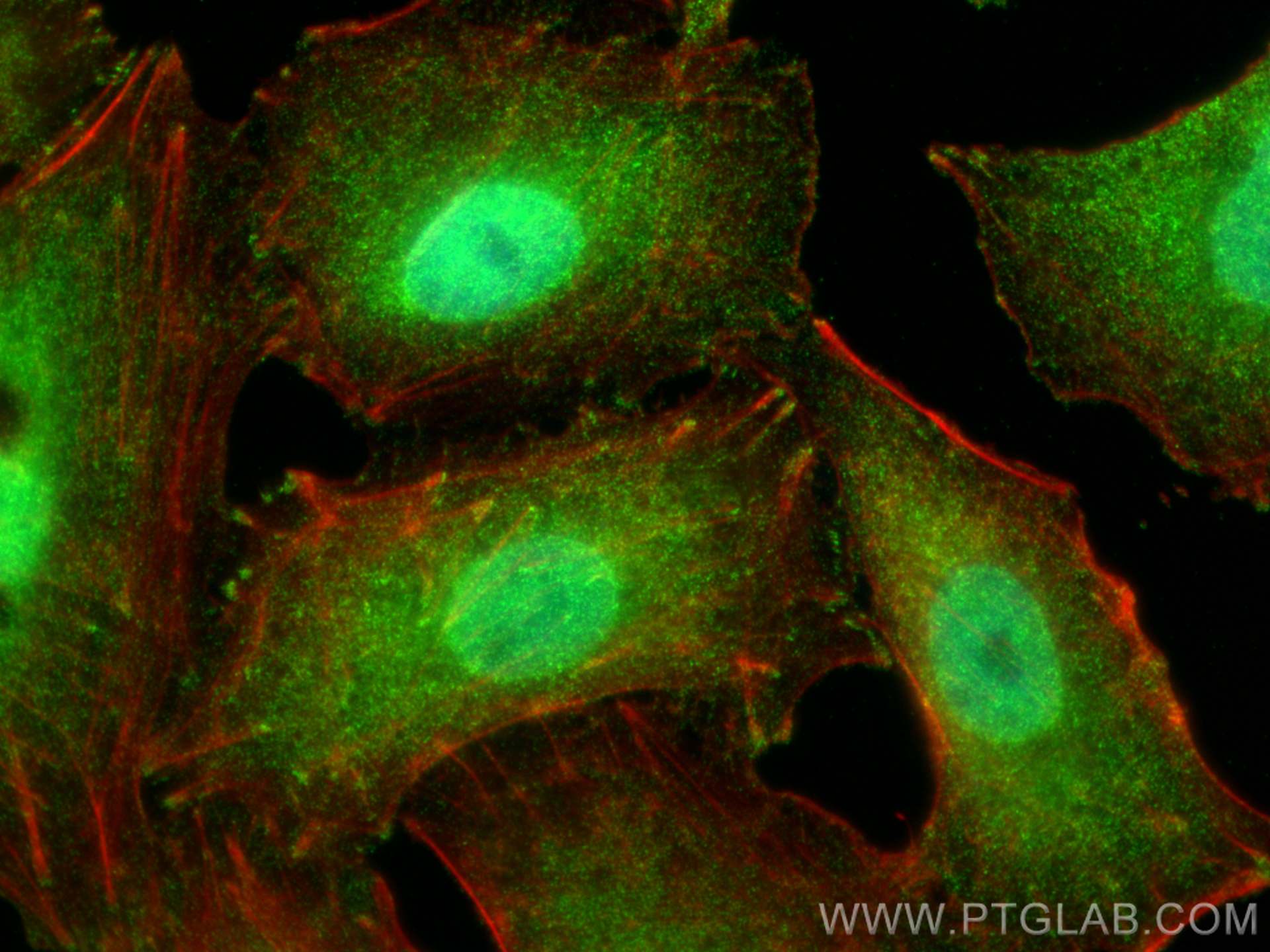
IF Staining of A549 using 67057-1-Ig
Immunofluorescent analysis of (4% PFA) fixed A549 cells using ATX3,ATXN3 antibody (67057-1-Ig, Clone: 1F7E10 ) at dilution of 1:800 and CoraLite®488-Conjugated Goat Anti-Mouse IgG(H+L), CL594-phalloidin (red).
The Proteintech guarantee covers Proteintech antibodies in any species and any application, including those not listed on the datasheet. If the antibody doesn’t perform, you can receive a hassle-free refund or credit note.
human pancreas cancer tissue Note: suggested antigen retrieval with TE buffer pH 9.0; (*) Alternatively, antigen retrieval may be performed with citrate buffer pH 6.0
Positive IF/ICC detected in
A549 cells
Recommended dilution
Application
Dilution
Western Blot (WB)
WB : 1:5000-1:50000
Immunohistochemistry (IHC)
IHC : 1:500-1:2000
Immunofluorescence (IF)/ICC
IF/ICC : 1:400-1:1600
It is recommended that this reagent should be titrated in each testing system to obtain optimal results.
Sample-dependent, Check data in validation data gallery.
PBS with 0.02% sodium azide and 50% glycerol, pH 7.3.
Storage Conditions
Store at -20°C. Aliquoting is unnecessary for -20oC storage. 20ul sizes contain 0.1% BSA.
Background Information
ATXN3, which has deubiquitinase activity and act as a component of the ubiquitin proteasome system, plays a role in transcriptional regulation and neuroprotection. ATXN3 interacts with RAD23, HHR23A and HHR23B, involves in the pathology of MJD. ATXN3 is a mixed-linkage, chain-editing enzyme and that the UIM region of ATXN3 regulates its substrate specificity. Contains an N-terminal deubiquitinating domain, called the Josephin domain, followed by 2 ubiquitin-interacting motifs (UIMs) and a polyQ tract near the C terminus. ATXN3 can be phosphorylated in a protein casein kinase-2-dependent manner, thus the MW would be larger than the predicted one.
MCF-7 cells were subjected to SDS PAGE followed by western blot with 67057-1-Ig (ATX3,ATXN3 antibody) at dilution of 1:10000 incubated at room temperature for 1.5 hours.
WB analysis of Jurkat using 67057-1-Ig
Jurkat cells were subjected to SDS PAGE followed by western blot with 67057-1-Ig (ATX3,ATXN3 antibody) at dilution of 1:10000 incubated at room temperature for 1.5 hours.
WB analysis of pig brain using 67057-1-Ig
pig brain tissue were subjected to SDS PAGE followed by western blot with 67057-1-Ig (ATX3,ATXN3 antibody) at dilution of 1:10000 incubated at room temperature for 1.5 hours.
WB analysis of mouse brain using 67057-1-Ig
mouse brain tissue were subjected to SDS PAGE followed by western blot with 67057-1-Ig (ATX3,ATXN3 antibody) at dilution of 1:10000 incubated at room temperature for 1.5 hours.
WB analysis of HeLa using 67057-1-Ig
HeLa cells were subjected to SDS PAGE followed by western blot with 67057-1-Ig (ATX3,ATXN3 antibody) at dilution of 1:10000 incubated at room temperature for 1.5 hours.
WB analysis of HEK-293 using 67057-1-Ig
HEK-293 cells were subjected to SDS PAGE followed by western blot with 67057-1-Ig (ATX3,ATXN3 antibody) at dilution of 1:10000 incubated at room temperature for 1.5 hours.
IHC Figures
IHC staining of human pancreas cancer using 67057-1-Ig
Immunohistochemical analysis of paraffin-embedded human pancreas cancer tissue slide using 67057-1-Ig (ATX3,ATXN3 antibody) at dilution of 1:1000 (under 10x lens). Heat mediated antigen retrieval with Tris-EDTA buffer (pH 9.0).
IHC staining of human pancreas cancer using 67057-1-Ig
Immunohistochemical analysis of paraffin-embedded human pancreas cancer tissue slide using 67057-1-Ig (ATX3,ATXN3 antibody) at dilution of 1:1000 (under 40x lens). Heat mediated antigen retrieval with Tris-EDTA buffer (pH 9.0).
IF/ICC Figures
IF Staining of A549 using 67057-1-Ig
Immunofluorescent analysis of (4% PFA) fixed A549 cells using ATX3,ATXN3 antibody (67057-1-Ig, Clone: 1F7E10 ) at dilution of 1:800 and CoraLite®488-Conjugated Goat Anti-Mouse IgG(H+L), CL594-phalloidin (red).
The species listed in Tested Reactivity are in-house verified and applicable species. For unlisted species, please refer to the homology analysis of the immunogen sequence and related species. For rabbit polyclonal antibodies, homology >70% is recommended. For mouse monoclonal antibodies and rabbit recombinant antibodies, homology >90% is recommended. Generally, the higher the homology, the greater the applicability. However, there will be certain differences in protein expression in different species, tissues or cells. Therefore, the homology analysis results are for reference only and do not serve as a guarantee.
At Proteintech, we pride ourselves on our antibody quality, customer service and transparency. As such, we are comparing our antibodies with other vendors, enabling easy identification and comparisons of key data to help you choose the suitable antibody for your needs.
We have selected the top cited antibodies from these vendors for you to compare.
 at dilution of 1:10000 incubated at room temperature for 1.5 hours.")
 at dilution of 1:10000 incubated at room temperature for 1.5 hours.")
 at dilution of 1:10000 incubated at room temperature for 1.5 hours.")
 at dilution of 1:10000 incubated at room temperature for 1.5 hours.")
 at dilution of 1:10000 incubated at room temperature for 1.5 hours.")
 at dilution of 1:10000 incubated at room temperature for 1.5 hours.")
 at dilution of 1:1000 (under 10x lens). Heat mediated antigen retrieval with Tris-EDTA buffer (pH 9.0).")
 at dilution of 1:1000 (under 40x lens). Heat mediated antigen retrieval with Tris-EDTA buffer (pH 9.0).")
 fixed A549 cells using ATX3,ATXN3 antibody (67057-1-Ig, Clone: 1F7E10 ) at dilution of 1:800 and CoraLite®488-Conjugated Goat Anti-Mouse IgG(H+L), CL594-phalloidin (red).")