Various lysates were subjected to SDS PAGE followed by western blot with 68204-1-Ig (ARF4 antibody) at dilution of 1:20000 incubated at room temperature for 1.5 hours.
Various lysates were subjected to SDS PAGE followed by western blot with 68204-1-Ig (ARF4 antibody) at dilution of 1:20000 incubated at room temperature for 1.5 hours.
WB analysis of A549 using 68204-1-Ig
A549 cells were subjected to SDS PAGE followed by western blot with 68204-1-Ig (ARF4 antibody) at dilution of 1:20000 incubated at room temperature for 1.5 hours.
A549 cells were subjected to SDS PAGE followed by western blot with 68204-1-Ig (ARF4 antibody) at dilution of 1:20000 incubated at room temperature for 1.5 hours.
WB analysis using 68204-1-Ig
Various lysates were subjected to SDS PAGE followed by western blot with 68204-1-Ig (ARF4 antibody) at dilution of 1:20000 incubated at room temperature for 1.5 hours.
Various lysates were subjected to SDS PAGE followed by western blot with 68204-1-Ig (ARF4 antibody) at dilution of 1:20000 incubated at room temperature for 1.5 hours.
WB analysis of HeLa using 68204-1-Ig
WB result of ARF4 antibody (68204-1-Ig; 1:10000; incubated at room temperature for 1.5 hours) with sh-Control and sh-ARF4 transfected HeLa cells.
WB result of ARF4 antibody (68204-1-Ig; 1:10000; incubated at room temperature for 1.5 hours) with sh-Control and sh-ARF4 transfected HeLa cells.
WB analysis using 68204-1-Ig
Various lysates were subjected to SDS PAGE followed by western blot with 68204-1-Ig (ARF4 antibody) at dilution of 1:20000 incubated at room temperature for 1.5 hours.
Various lysates were subjected to SDS PAGE followed by western blot with 68204-1-Ig (ARF4 antibody) at dilution of 1:20000 incubated at room temperature for 1.5 hours.
WB analysis of HEK-293 using 68204-1-Ig
HEK-293 cells were subjected to SDS PAGE followed by western blot with 68204-1-Ig (ARF4 antibody) at dilution of 1:20000 incubated at room temperature for 1.5 hours.
HEK-293 cells were subjected to SDS PAGE followed by western blot with 68204-1-Ig (ARF4 antibody) at dilution of 1:20000 incubated at room temperature for 1.5 hours.
WB analysis of mouse brain using 68204-1-Ig
mouse brain tissue were subjected to SDS PAGE followed by western blot with 68204-1-Ig (ARF4 antibody) at dilution of 1:20000 incubated at room temperature for 1.5 hours.
mouse brain tissue were subjected to SDS PAGE followed by western blot with 68204-1-Ig (ARF4 antibody) at dilution of 1:20000 incubated at room temperature for 1.5 hours.
IHC staining of human stomach cancer using 68204-1-Ig
Immunohistochemical analysis of paraffin-embedded human stomach cancer tissue slide using 68204-1-Ig (ARF4 antibody) at dilution of 1:2000 (under 10x lens). Heat mediated antigen retrieval with Tris-EDTA buffer (pH 9.0).
Immunohistochemical analysis of paraffin-embedded human stomach cancer tissue slide using 68204-1-Ig (ARF4 antibody) at dilution of 1:2000 (under 10x lens). Heat mediated antigen retrieval with Tris-EDTA buffer (pH 9.0).
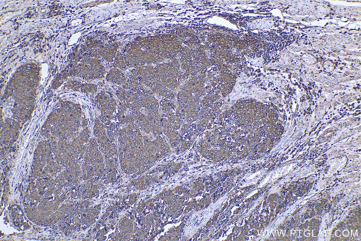
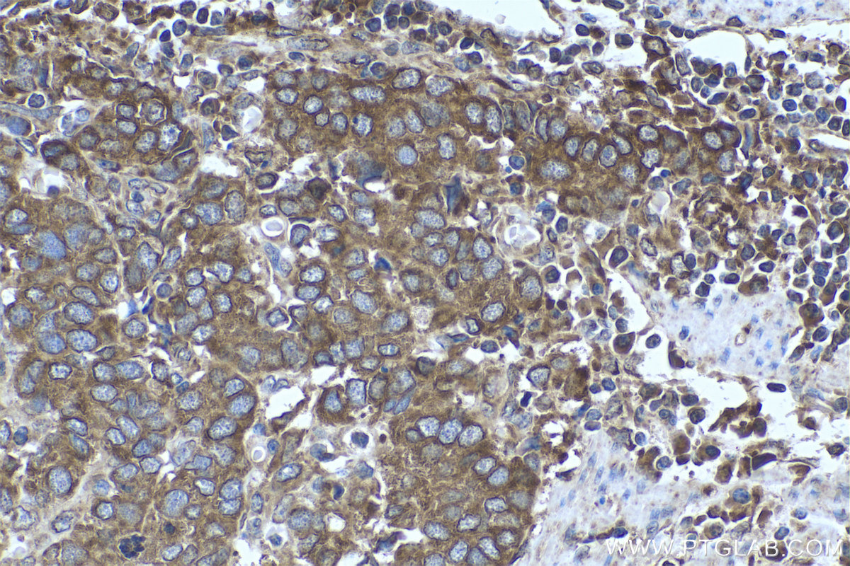
IHC staining of human stomach cancer using 68204-1-Ig
Immunohistochemical analysis of paraffin-embedded human stomach cancer tissue slide using 68204-1-Ig (ARF4 antibody) at dilution of 1:2000 (under 40x lens). Heat mediated antigen retrieval with Tris-EDTA buffer (pH 9.0).
Immunohistochemical analysis of paraffin-embedded human stomach cancer tissue slide using 68204-1-Ig (ARF4 antibody) at dilution of 1:2000 (under 40x lens). Heat mediated antigen retrieval with Tris-EDTA buffer (pH 9.0).
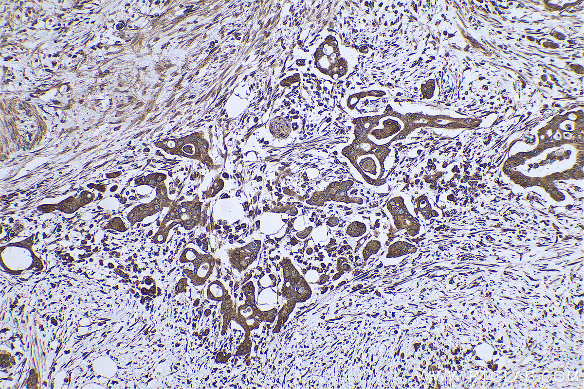
IHC staining of human pancreas cancer using 68204-1-Ig
Immunohistochemical analysis of paraffin-embedded human pancreas cancer tissue slide using 68204-1-Ig (ARF4 antibody) at dilution of 1:2000 (under 10x lens). Heat mediated antigen retrieval with Tris-EDTA buffer (pH 9.0).
Immunohistochemical analysis of paraffin-embedded human pancreas cancer tissue slide using 68204-1-Ig (ARF4 antibody) at dilution of 1:2000 (under 10x lens). Heat mediated antigen retrieval with Tris-EDTA buffer (pH 9.0).
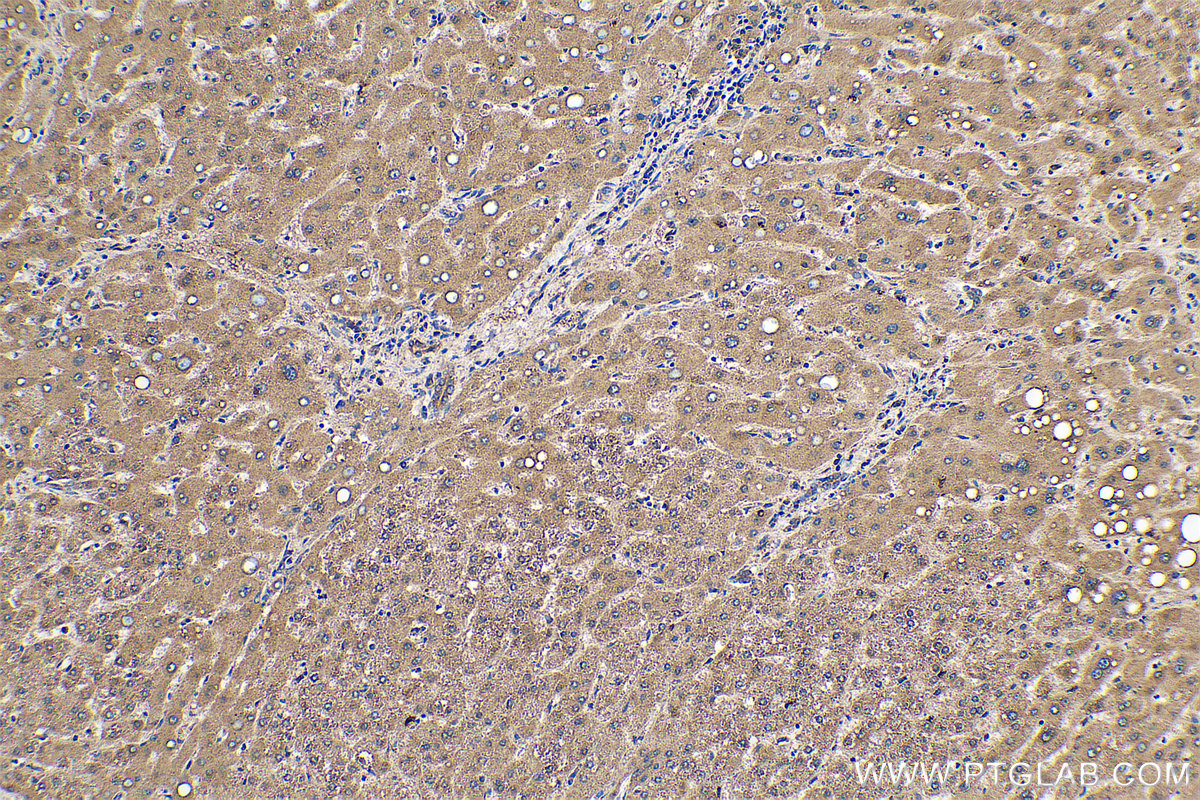
IHC staining of human liver using 68204-1-Ig
Immunohistochemical analysis of paraffin-embedded human liver tissue slide using 68204-1-Ig (ARF4 antibody) at dilution of 1:8000 (under 10x lens). Heat mediated antigen retrieval with Tris-EDTA buffer (pH 9.0).
Immunohistochemical analysis of paraffin-embedded human liver tissue slide using 68204-1-Ig (ARF4 antibody) at dilution of 1:8000 (under 10x lens). Heat mediated antigen retrieval with Tris-EDTA buffer (pH 9.0).
IF Staining of HepG2 using 68204-1-Ig
Immunofluorescent analysis of (4% PFA) fixed HepG2 cells using ARF4 antibody (68204-1-Ig, Clone: 2A4B8 ) at dilution of 1:800 and CoraLite®488-Conjugated AffiniPure Goat Anti-Mouse IgG(H+L).
Immunofluorescent analysis of (4% PFA) fixed HepG2 cells using ARF4 antibody (68204-1-Ig, Clone: 2A4B8 ) at dilution of 1:800 and CoraLite®488-Conjugated AffiniPure Goat Anti-Mouse IgG(H+L).
FC experiment of K-562 using 68204-1-Ig
1X10^6 K-562 cells were intracellularly stained with 0.4 ug Anti-Human ARF4 (68204-1-Ig, Clone:2A4B8) and CoraLite®488-Conjugated AffiniPure Goat Anti-Mouse IgG(H+L) at dilution 1:1000 (red), or 0.4 ug Mouse IgG2b Isotype Control (66360-3-Ig, Clone: K11B8C4B5) (blue). Cells were fixed with 4% PFA and permeabilized with Flow Cytometry Perm Buffer (PF00011-C).
1X10^6 K-562 cells were intracellularly stained with 0.4 ug Anti-Human ARF4 (68204-1-Ig, Clone:2A4B8) and CoraLite®488-Conjugated AffiniPure Goat Anti-Mouse IgG(H+L) at dilution 1:1000 (red), or 0.4 ug Mouse IgG2b Isotype Control (66360-3-Ig, Clone: K11B8C4B5) (blue). Cells were fixed with 4% PFA and permeabilized with Flow Cytometry Perm Buffer (PF00011-C).
The Proteintech guarantee covers Proteintech antibodies in any species and any application, including those not listed on the datasheet. If the antibody doesn’t perform, you can receive a hassle-free refund or credit note.
LNCaP cells, HEK-293 cells, rat brain tissue, A549 cells, mouse brain tissue, HeLa cells, HepG2 cells, Jurkat cells
Positive IHC detected in
human stomach cancer tissue, human liver tissue, human pancreas cancer tissue Note: suggested antigen retrieval with TE buffer pH 9.0; (*) Alternatively, antigen retrieval may be performed with citrate buffer pH 6.0
Positive IF/ICC detected in
HepG2 cells
Positive FC (Intra) detected in
K-562 cells
Recommended dilution
Application
Dilution
Western Blot (WB)
WB : 1:5000-1:50000
Immunohistochemistry (IHC)
IHC : 1:1000-1:4000
Immunofluorescence (IF)/ICC
IF/ICC : 1:400-1:1600
Flow Cytometry (FC) (INTRA)
FC (INTRA) : 0.40 ug per 10^6 cells in a 100 µl suspension
It is recommended that this reagent should be titrated in each testing system to obtain optimal results.
Sample-dependent, Check data in validation data gallery.
Product Information
68204-1-Ig targets ARF4 in WB, IHC, IF/ICC, FC (Intra), ELISA applications and shows reactivity with human, mouse, rat samples.
PBS with 0.02% sodium azide and 50% glycerol , pH 7.3
Storage Conditions
Store at -20°C. Stable for one year after shipment. Aliquoting is unnecessary for -20oC storage. 20ul sizes contain 0.1% BSA.
Background Information
ARF4, also named as ARF2, belongs to the small GTPase superfamily and Arf family. ADP-ribosylation factors (ARFs) are members of the ARF family of GTP-binding proteins of the Ras superfamily, with 20 kDa protein size. ARFs bind and regulate GTP/GDP cycle by alternating between the active GTP-bound and inactive GDP-bound conformations. ARF family proteins are essential and ubiquitous in eukaryotes. Six highly conserved members of the family have been identified in mammalian cells. They function in vesicular traffic and actin remodelling and other bioprocesses in cells. ARF4 and ARF5 are class II ARFs. Their function are not fully described. (PMID: 7759471, PMID: 16042562). This antibody can bind ARFs for the close sequences.
Various lysates were subjected to SDS PAGE followed by western blot with 68204-1-Ig (ARF4 antibody) at dilution of 1:20000 incubated at room temperature for 1.5 hours.
WB analysis of A549 using 68204-1-Ig
A549 cells were subjected to SDS PAGE followed by western blot with 68204-1-Ig (ARF4 antibody) at dilution of 1:20000 incubated at room temperature for 1.5 hours.
WB analysis using 68204-1-Ig
Various lysates were subjected to SDS PAGE followed by western blot with 68204-1-Ig (ARF4 antibody) at dilution of 1:20000 incubated at room temperature for 1.5 hours.
WB analysis of HeLa using 68204-1-Ig
WB result of ARF4 antibody (68204-1-Ig; 1:10000; incubated at room temperature for 1.5 hours) with sh-Control and sh-ARF4 transfected HeLa cells.
WB analysis using 68204-1-Ig
Various lysates were subjected to SDS PAGE followed by western blot with 68204-1-Ig (ARF4 antibody) at dilution of 1:20000 incubated at room temperature for 1.5 hours.
WB analysis using 68204-1-Ig
Various lysates were subjected to SDS PAGE followed by western blot with 68204-1-Ig (ARF4 antibody) at dilution of 1:20000 incubated at room temperature for 1.5 hours.
WB analysis of HEK-293 using 68204-1-Ig
HEK-293 cells were subjected to SDS PAGE followed by western blot with 68204-1-Ig (ARF4 antibody) at dilution of 1:20000 incubated at room temperature for 1.5 hours.
WB analysis of mouse brain using 68204-1-Ig
mouse brain tissue were subjected to SDS PAGE followed by western blot with 68204-1-Ig (ARF4 antibody) at dilution of 1:20000 incubated at room temperature for 1.5 hours.
IHC Figures
IHC staining of human stomach cancer using 68204-1-Ig
Immunohistochemical analysis of paraffin-embedded human stomach cancer tissue slide using 68204-1-Ig (ARF4 antibody) at dilution of 1:2000 (under 10x lens). Heat mediated antigen retrieval with Tris-EDTA buffer (pH 9.0).
IHC staining of human stomach cancer using 68204-1-Ig
Immunohistochemical analysis of paraffin-embedded human stomach cancer tissue slide using 68204-1-Ig (ARF4 antibody) at dilution of 1:2000 (under 40x lens). Heat mediated antigen retrieval with Tris-EDTA buffer (pH 9.0).
IHC staining of human pancreas cancer using 68204-1-Ig
Immunohistochemical analysis of paraffin-embedded human pancreas cancer tissue slide using 68204-1-Ig (ARF4 antibody) at dilution of 1:2000 (under 10x lens). Heat mediated antigen retrieval with Tris-EDTA buffer (pH 9.0).
IHC staining of human liver using 68204-1-Ig
Immunohistochemical analysis of paraffin-embedded human liver tissue slide using 68204-1-Ig (ARF4 antibody) at dilution of 1:8000 (under 10x lens). Heat mediated antigen retrieval with Tris-EDTA buffer (pH 9.0).
IF/ICC Figures
IF Staining of HepG2 using 68204-1-Ig
Immunofluorescent analysis of (4% PFA) fixed HepG2 cells using ARF4 antibody (68204-1-Ig, Clone: 2A4B8 ) at dilution of 1:800 and CoraLite®488-Conjugated AffiniPure Goat Anti-Mouse IgG(H+L).
FC (INTRA) Figures
FC experiment of K-562 using 68204-1-Ig
1X10^6 K-562 cells were intracellularly stained with 0.4 ug Anti-Human ARF4 (68204-1-Ig, Clone:2A4B8) and CoraLite®488-Conjugated AffiniPure Goat Anti-Mouse IgG(H+L) at dilution 1:1000 (red), or 0.4 ug Mouse IgG2b Isotype Control (66360-3-Ig, Clone: K11B8C4B5) (blue). Cells were fixed with 4% PFA and permeabilized with Flow Cytometry Perm Buffer (PF00011-C).
The species listed in Tested Reactivity are in-house verified and applicable species. For unlisted species, please refer to the homology analysis of the immunogen sequence and related species. For rabbit polyclonal antibodies, homology >70% is recommended. For mouse monoclonal antibodies and rabbit recombinant antibodies, homology >90% is recommended. Generally, the higher the homology, the greater the applicability. However, there will be certain differences in protein expression in different species, tissues or cells. Therefore, the homology analysis results are for reference only and do not serve as a guarantee.
At Proteintech, we pride ourselves on our antibody quality, customer service and transparency. As such, we are comparing our antibodies with other vendors, enabling easy identification and comparisons of key data to help you choose the suitable antibody for your needs.
We have selected the top cited antibodies from these vendors for you to compare.
Proteintech
KD/KO VALIDATED
ARF4 Monoclonal antibody
$149 (20ul) $449 (150ul)
Catalog Number
68204-1-Ig
Citations
-
Dilutions
WB : 1:5000-1:50000 IHC : 1:1000-1:4000 IF/ICC : 1:400-1:1600 FC (INTRA) : 0.40 ug per 10^6 cells in a 100 µl suspension
Applications
WB, IHC, IF/ICC, FC (Intra), ELISA
Reactivity
human, mouse, rat
Product Guarantee
Covers any species including not listed on datasheet
Covers any applications including not listed on datasheet
 at dilution of 1:20000 incubated at room temperature for 1.5 hours.")
 at dilution of 1:20000 incubated at room temperature for 1.5 hours.")
 at dilution of 1:20000 incubated at room temperature for 1.5 hours.")
 with sh-Control and sh-ARF4 transfected HeLa cells.")
 at dilution of 1:20000 incubated at room temperature for 1.5 hours.")
 at dilution of 1:20000 incubated at room temperature for 1.5 hours.")
 at dilution of 1:20000 incubated at room temperature for 1.5 hours.")
 at dilution of 1:20000 incubated at room temperature for 1.5 hours.")
 at dilution of 1:2000 (under 10x lens). Heat mediated antigen retrieval with Tris-EDTA buffer (pH 9.0).")
 at dilution of 1:2000 (under 40x lens). Heat mediated antigen retrieval with Tris-EDTA buffer (pH 9.0).")
 at dilution of 1:2000 (under 10x lens). Heat mediated antigen retrieval with Tris-EDTA buffer (pH 9.0).")
 at dilution of 1:8000 (under 10x lens). Heat mediated antigen retrieval with Tris-EDTA buffer (pH 9.0).")
 fixed HepG2 cells using ARF4 antibody (68204-1-Ig, Clone: 2A4B8 ) at dilution of 1:800 and CoraLite®488-Conjugated AffiniPure Goat Anti-Mouse IgG(H+L).")
 and CoraLite®488-Conjugated AffiniPure Goat Anti-Mouse IgG(H+L) at dilution 1:1000 (red), or 0.4 ug Mouse IgG2b Isotype Control (66360-3-Ig, Clone: K11B8C4B5) (blue). Cells were fixed with 4% PFA and permeabilized with Flow Cytometry Perm Buffer (PF00011-C).")