Tested Applications
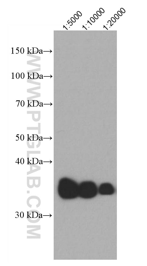
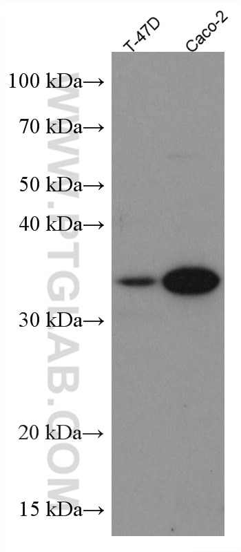
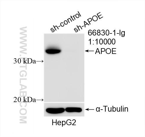
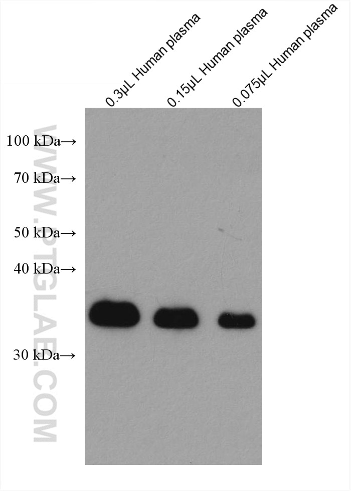
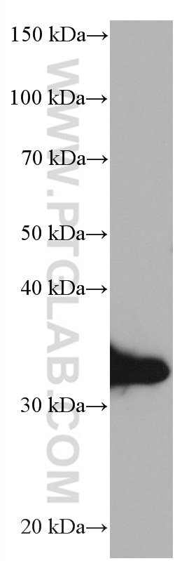
| Positive WB detected in | human plasma tissue, human blood, human plasma, T-47D cells, HepG2 cells, Caco-2 cells |
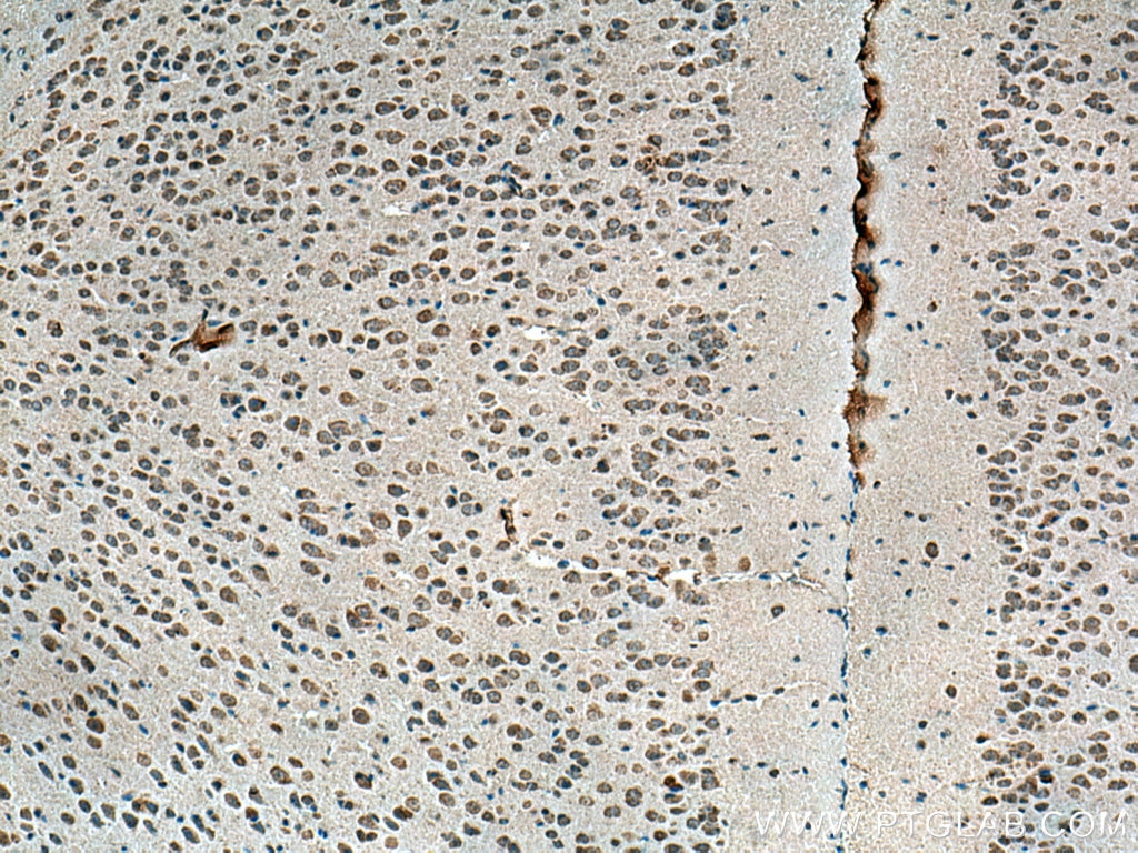
| Positive IHC detected in | mouse brain tissue Note: suggested antigen retrieval with TE buffer pH 9.0; (*) Alternatively, antigen retrieval may be performed with citrate buffer pH 6.0 |
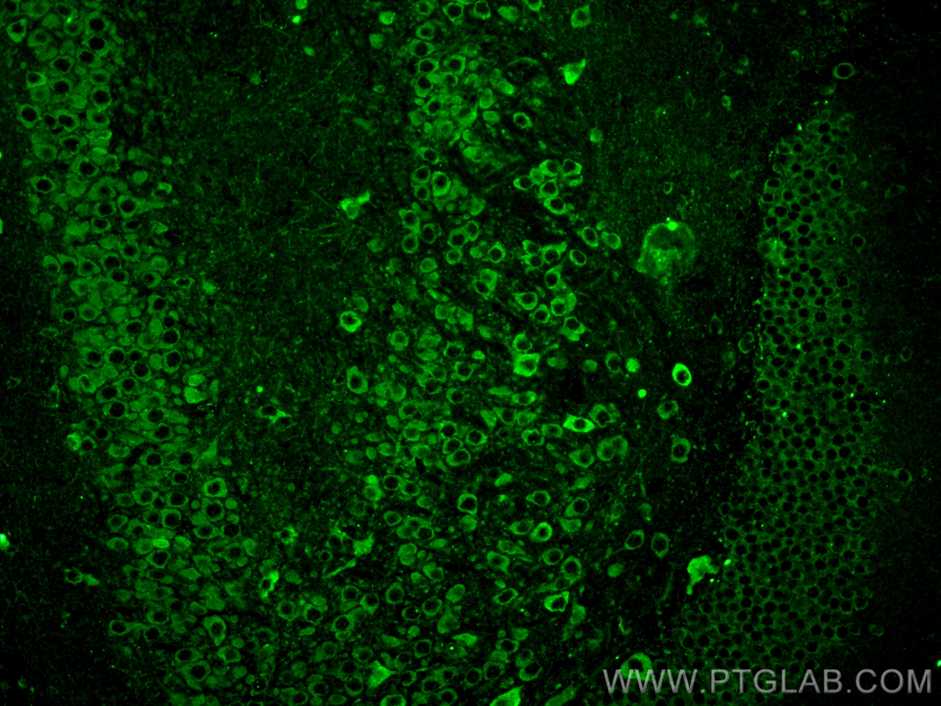
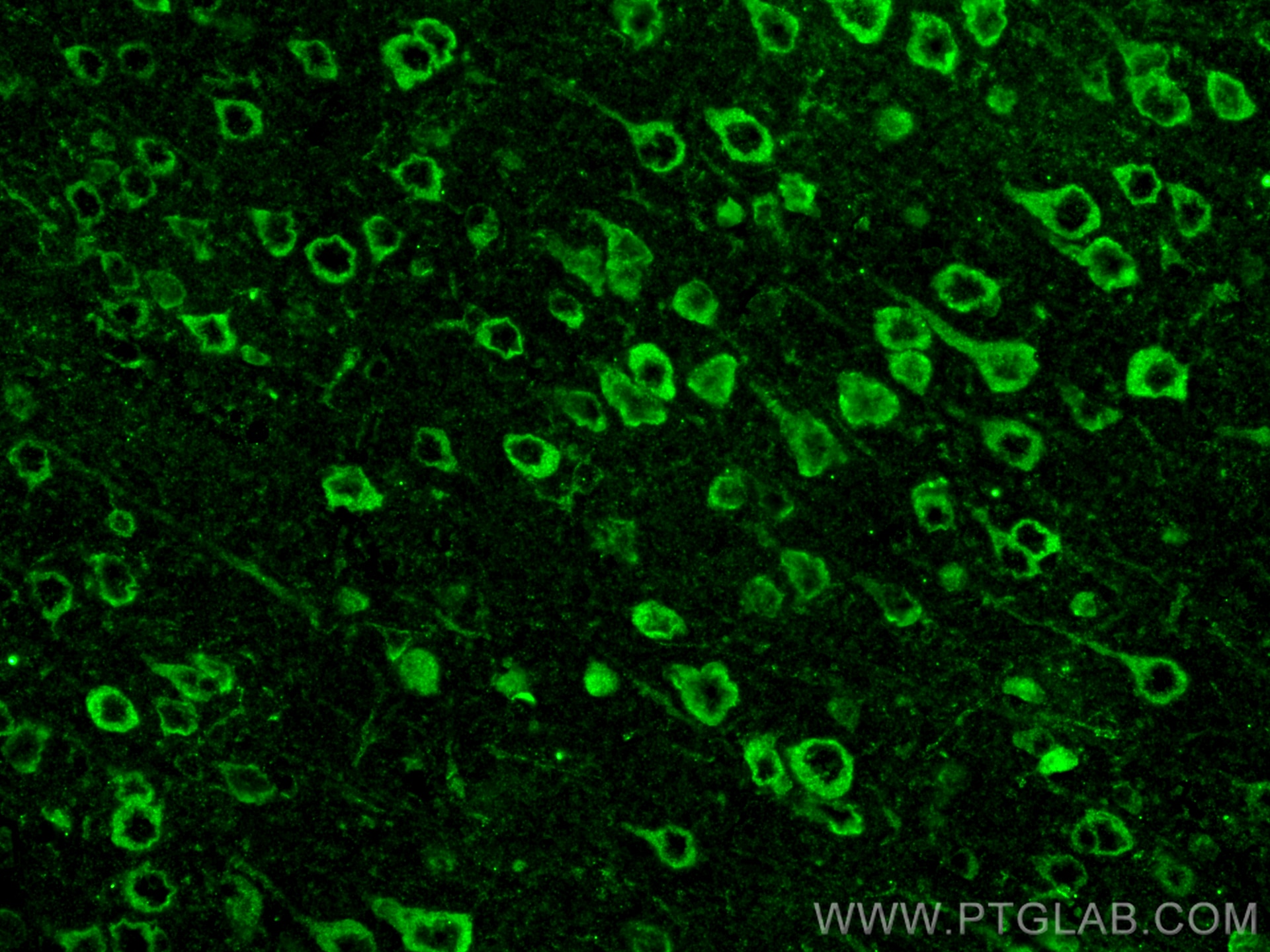
| Positive IF-P detected in | mouse brain tissue |
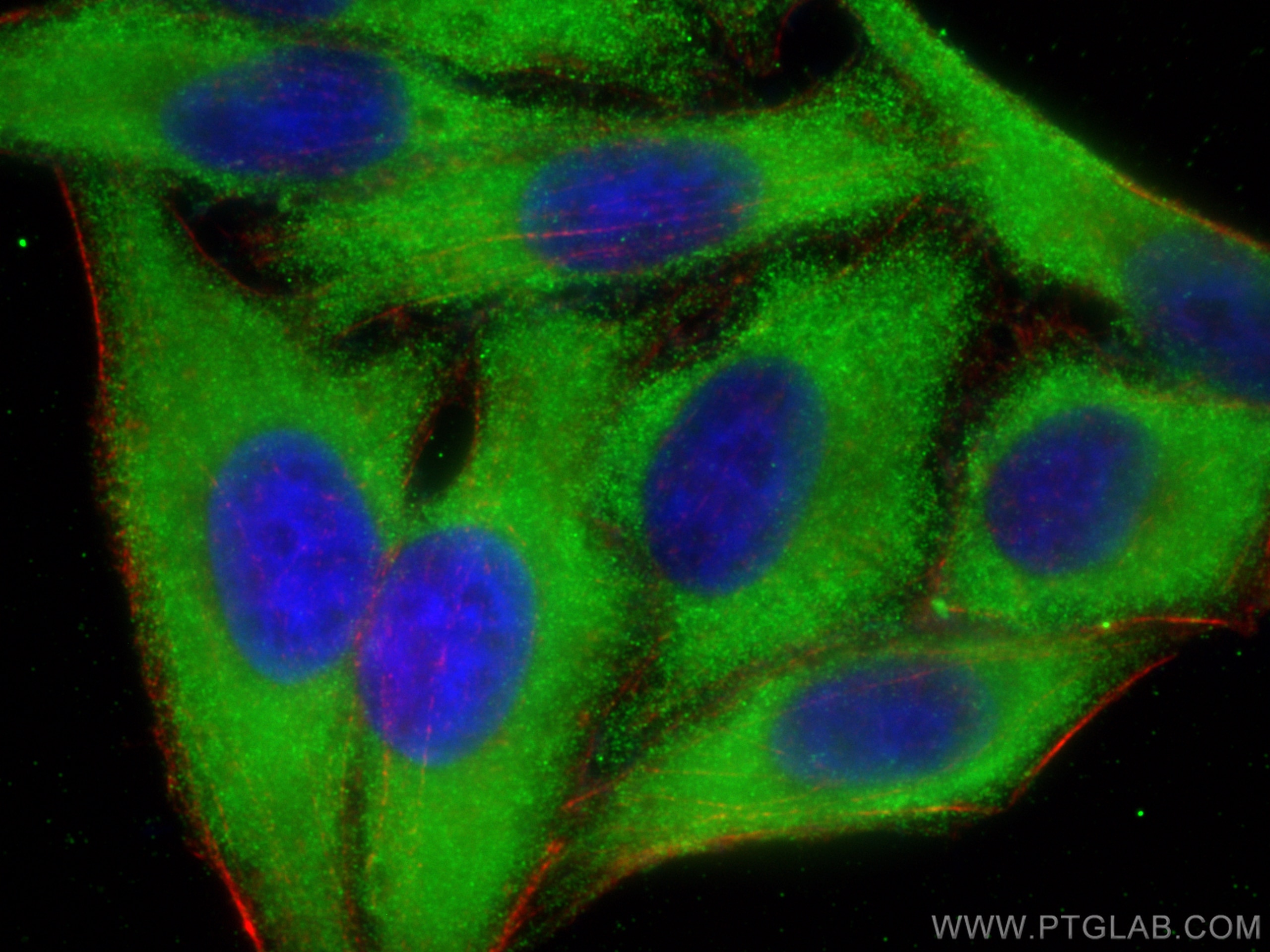
| Positive IF/ICC detected in | HepG2 cells |
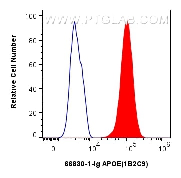
| Positive FC (Intra) detected in | HepG2 cells |
Recommended dilution
| Application | Dilution |
|---|---|
| Western Blot (WB) | WB : 1:3000-1:20000 |
| Immunohistochemistry (IHC) | IHC : 1:50-1:500 |
| Immunofluorescence (IF)-P | IF-P : 1:200-1:800 |
| Immunofluorescence (IF)/ICC | IF/ICC : 1:400-1:1600 |
| Flow Cytometry (FC) (INTRA) | FC (INTRA) : 0.80 ug per 10^6 cells in a 100 µl suspension |
| It is recommended that this reagent should be titrated in each testing system to obtain optimal results. | |
| Sample-dependent, Check data in validation data gallery. | |
Published Applications
| WB | See 8 publications below |
| IHC | See 2 publications below |
| IF | See 4 publications below |
Product Information
66830-1-Ig targets APOE in WB, IHC, IF/ICC, IF-P, FC (Intra), ELISA applications and shows reactivity with human, mouse samples.
| Tested Reactivity | human, mouse |
| Cited Reactivity | human, mouse |
| Host / Isotype | Mouse / IgG1 |
| Class | Monoclonal |
| Type | Antibody |
| Immunogen | APOE fusion protein Ag28186 Predict reactive species |
| Full Name | Apolipoprotein E |
| Calculated Molecular Weight | 36 kDa |
| Observed Molecular Weight | 34-36 kDa |
| GenBank Accession Number | BC003557 |
| Gene Symbol | APOE |
| Gene ID (NCBI) | 348 |
| RRID | AB_2882173 |
| Conjugate | Unconjugated |
| Form | Liquid |
| Purification Method | Protein G purification |
| UNIPROT ID | P02649 |
| Storage Buffer | PBS with 0.02% sodium azide and 50% glycerol , pH 7.3 |
| Storage Conditions | Store at -20°C. Stable for one year after shipment. Aliquoting is unnecessary for -20oC storage. 20ul sizes contain 0.1% BSA. |
Background Information
The apolipoprotein E (APOE) is a 299-amino acid polypeptide that mediates the binding, internalization, and catabolism of lipoprotein particles, and also serve as a ligand for the LDL (APO B/E) receptor and for the specific APOE receptor (chylomicron remnant) of hepatic tissues. The very strong association of the APOE ɛ4 allele with AD risk and its role in the accumulation of amyloid β in brains of people and animal models solidify the biological relevance of APOE isoforms but do not provide mechanistic insight.
Protocols
| Product Specific Protocols | |
|---|---|
| WB protocol for APOE antibody 66830-1-Ig | Download protocol |
| IHC protocol for APOE antibody 66830-1-Ig | Download protocol |
| IF protocol for APOE antibody 66830-1-Ig | Download protocol |
| Standard Protocols | |
|---|---|
| Click here to view our Standard Protocols |
Publications
| Species | Application | Title |
|---|---|---|
Brain Behav Immun Egln3 expression in microglia enhances the neuroinflammatory responses in Alzheimer's disease | ||
EMBO J Rab2A-mediated Golgi-lipid droplet interactions support very-low-density lipoprotein secretion in hepatocytes | ||
J Biol Chem Lack of ApoE inhibits ADan amyloidosis in a mouse model of Familial Danish Dementia | ||
Front Immunol Binding of Macrophage Receptor MARCO, LDL, and LDLR to Disease-Associated Crystalline Structures. | ||
mSphere Circulating extracellular vesicles from severe COVID-19 patients induce lung inflammation |
Reviews
The reviews below have been submitted by verified Proteintech customers who received an incentive for providing their feedback.
FH Silvia (Verified Customer) (12-03-2019) | Specific antibody for IF and WB on Human tissues
|
