Tested Applications
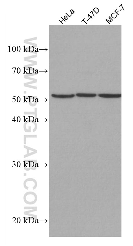
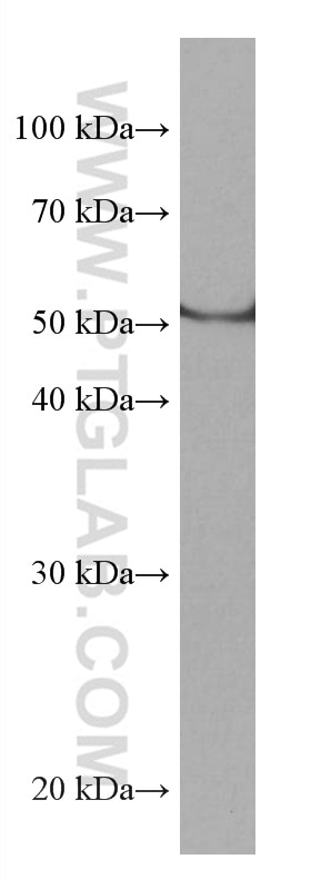
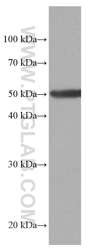
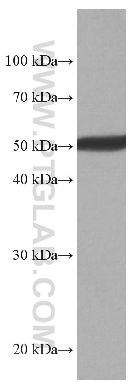
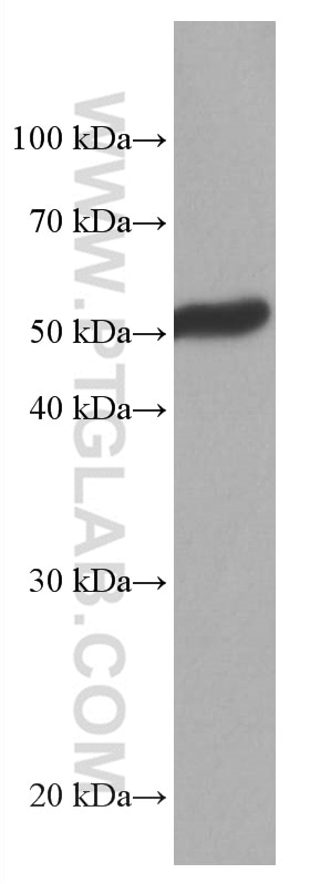
| Positive WB detected in | HeLa cells, PC-3 cells, rat brain tissue, mouse brain tissue, pig brain tissue, T-47D cells, MCF-7 cells |
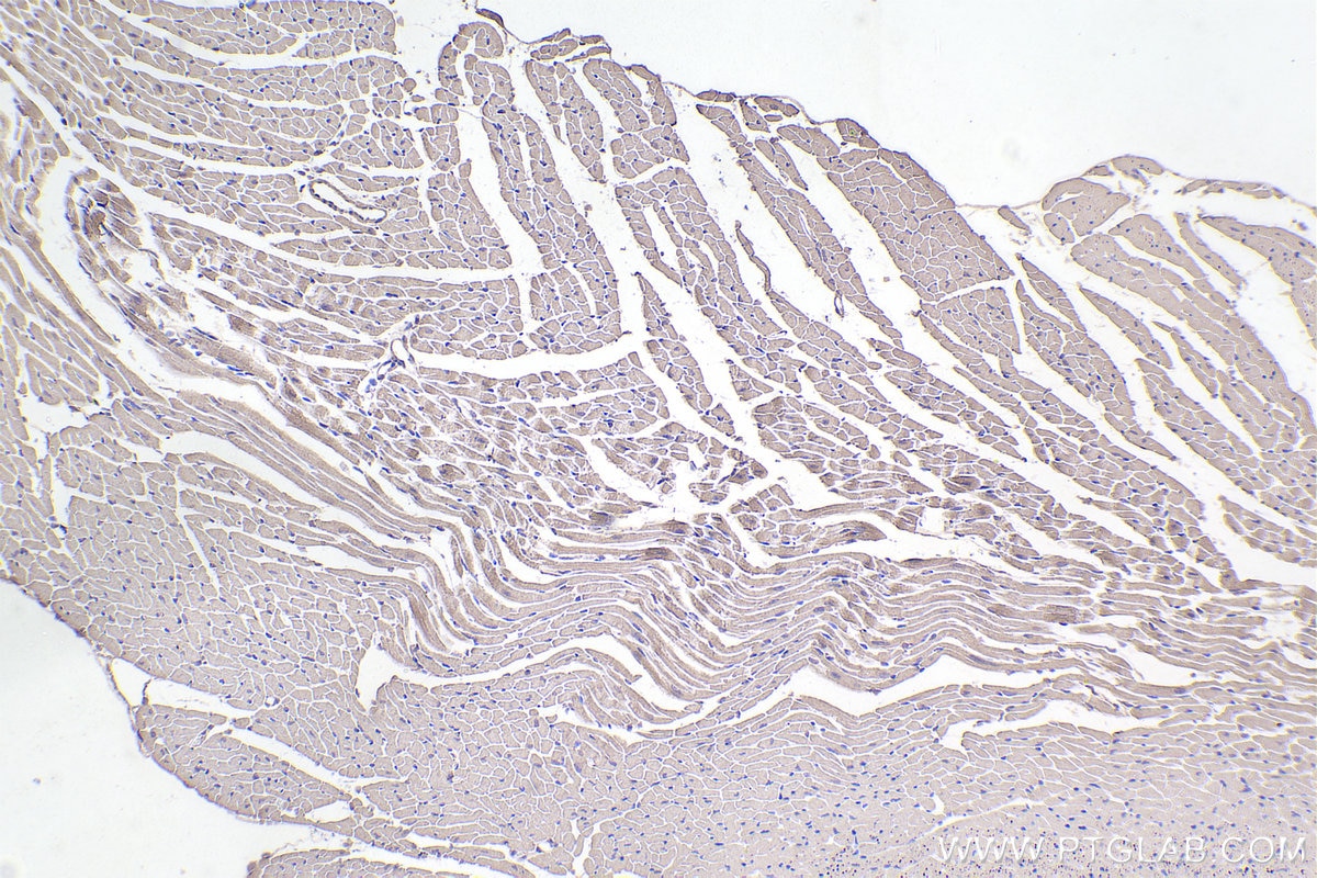
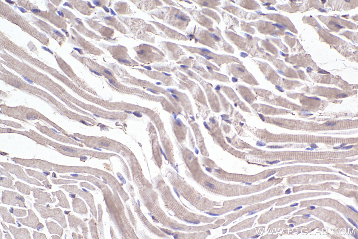
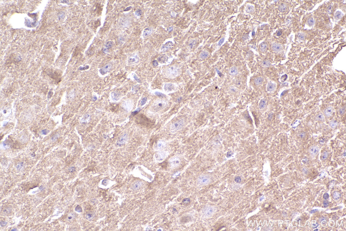
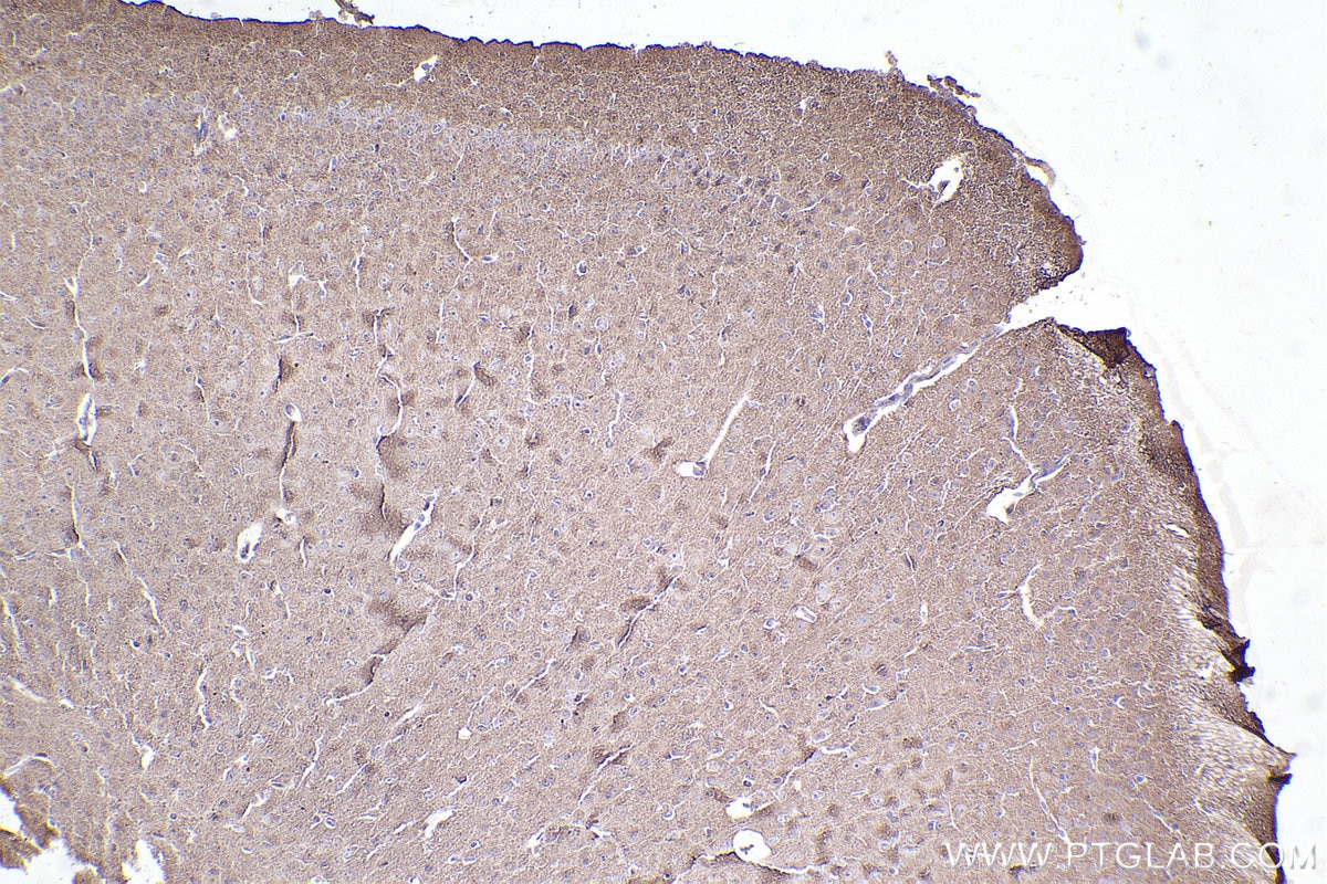
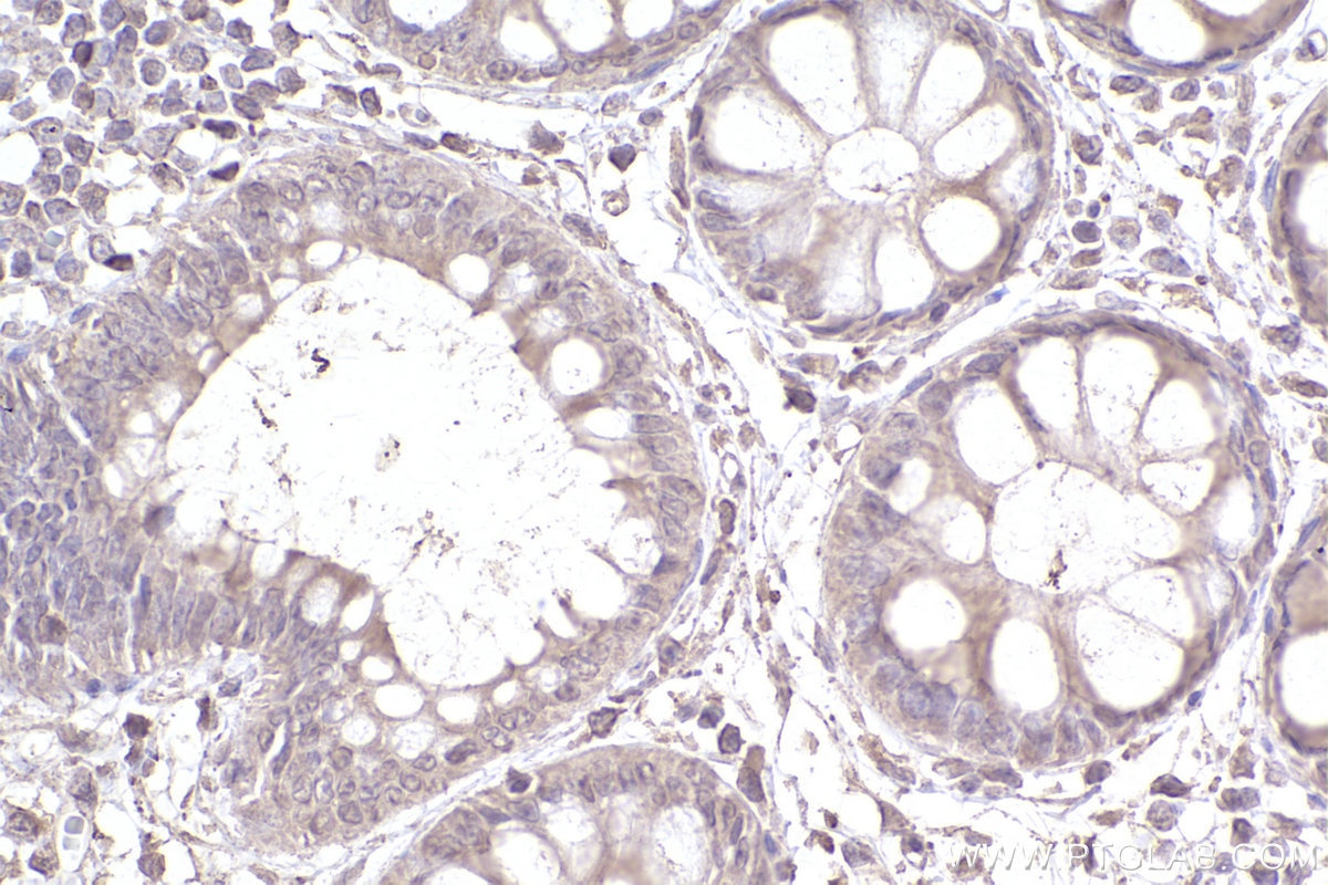
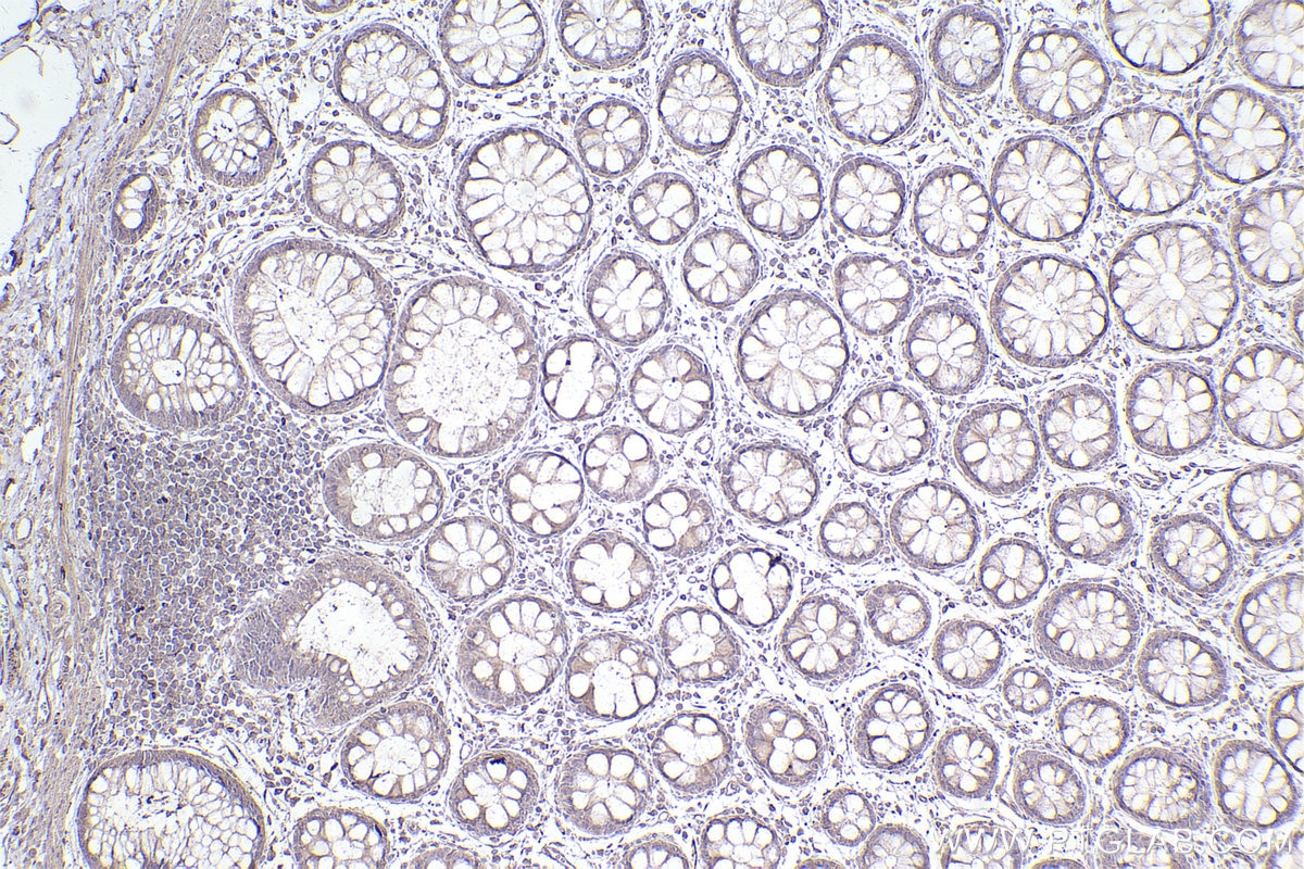
| Positive IHC detected in | mouse heart tissue, human rectal cancer tissue, mouse brain tissue Note: suggested antigen retrieval with TE buffer pH 9.0; (*) Alternatively, antigen retrieval may be performed with citrate buffer pH 6.0 |
Recommended dilution
| Application | Dilution |
|---|---|
| Western Blot (WB) | WB : 1:3000-1:10000 |
| Immunohistochemistry (IHC) | IHC : 1:500-1:2000 |
| It is recommended that this reagent should be titrated in each testing system to obtain optimal results. | |
| Sample-dependent, Check data in validation data gallery. | |
Published Applications
| WB | See 1 publications below |
Product Information
67033-1-Ig targets CYP1B1 in WB, IHC, ELISA applications and shows reactivity with human, mouse, rat, pig samples.
| Tested Reactivity | human, mouse, rat, pig |
| Cited Reactivity | human, mouse |
| Host / Isotype | Mouse / IgG1 |
| Class | Monoclonal |
| Type | Antibody |
| Immunogen | CYP1B1 fusion protein Ag13380 Predict reactive species |
| Full Name | cytochrome P450, family 1, subfamily B, polypeptide 1 |
| Calculated Molecular Weight | 61 kDa |
| Observed Molecular Weight | 52 kDa |
| GenBank Accession Number | BC012049 |
| Gene Symbol | CYP1B1 |
| Gene ID (NCBI) | 1545 |
| RRID | AB_2882348 |
| Conjugate | Unconjugated |
| Form | Liquid |
| Purification Method | Protein G purification |
| UNIPROT ID | Q16678 |
| Storage Buffer | PBS with 0.02% sodium azide and 50% glycerol, pH 7.3. |
| Storage Conditions | Store at -20°C. Stable for one year after shipment. Aliquoting is unnecessary for -20oC storage. 20ul sizes contain 0.1% BSA. |
Background Information
Cytochrome P450 CYP1B1 is a recently cloned dioxin-inducible form of the cytochrome P450 family of xenobiotic metabolizing enzymes. CYP1B1 was found to be expressed at a high frequency in a wide range of human cancers of different histogenetic types, including cancers of the breast, colon, lung, esophagus, skin, lymph node, brain, and testis. There was no detectable immunostaining for CYP1B1 in normal tissues.(PMID:9230218). CYP1B1 is a 60 kDa protein that it also can be detected a band of 51 kDa or 57 kDa through the western blot(PMID:9525272; PMID: 9525272).
Protocols
| Product Specific Protocols | |
|---|---|
| WB protocol for CYP1B1 antibody 67033-1-Ig | Download protocol |
| IHC protocol for CYP1B1 antibody 67033-1-Ig | Download protocol |
| Standard Protocols | |
|---|---|
| Click here to view our Standard Protocols |
