WB analysis of human skeletal muscle using 66205-1-Ig
human skeletal muscle tissue were subjected to SDS PAGE followed by western blot with 66205-1-Ig (Myoglobin antibody at dilution of 1:50000 incubated at room temperature for 1.5 hours.
human skeletal muscle tissue were subjected to SDS PAGE followed by western blot with 66205-1-Ig (Myoglobin antibody at dilution of 1:50000 incubated at room temperature for 1.5 hours.
WB analysis of pig heart using 66205-1-Ig
pig heart tissue were subjected to SDS PAGE followed by western blot with 66205-1-Ig (Myoglobin antibody) at dilution of 1:50000 incubated at room temperature for 1.5 hours.
pig heart tissue were subjected to SDS PAGE followed by western blot with 66205-1-Ig (Myoglobin antibody) at dilution of 1:50000 incubated at room temperature for 1.5 hours.
WB analysis of rat heart using 66205-1-Ig
rat heart tissue were subjected to SDS PAGE followed by western blot with 66205-1-Ig (Myoglobin antibody) at dilution of 1:50000 incubated at room temperature for 1.5 hours.
rat heart tissue were subjected to SDS PAGE followed by western blot with 66205-1-Ig (Myoglobin antibody) at dilution of 1:50000 incubated at room temperature for 1.5 hours.
WB analysis of mouse heart using 66205-1-Ig
mouse heart tissue were subjected to SDS PAGE followed by western blot with 66205-1-Ig (Myoglobin antibody) at dilution of 1:50000 incubated at room temperature for 1.5 hours.
mouse heart tissue were subjected to SDS PAGE followed by western blot with 66205-1-Ig (Myoglobin antibody) at dilution of 1:50000 incubated at room temperature for 1.5 hours.
WB analysis of rabbit heart using 66205-1-Ig
rabbit heart tissue were subjected to SDS PAGE followed by western blot with 66205-1-Ig (Myoglobin antibody) at dilution of 1:50000 incubated at room temperature for 1.5 hours.
rabbit heart tissue were subjected to SDS PAGE followed by western blot with 66205-1-Ig (Myoglobin antibody) at dilution of 1:50000 incubated at room temperature for 1.5 hours.
WB analysis of human heart using 66205-1-Ig
human heart tissue were subjected to SDS PAGE followed by western blot with 66205-1-Ig (Myoglobin antibody at dilution of 1:50000 incubated at room temperature for 1.5 hours.
human heart tissue were subjected to SDS PAGE followed by western blot with 66205-1-Ig (Myoglobin antibody at dilution of 1:50000 incubated at room temperature for 1.5 hours.
IHC staining of human heart using 66205-1-Ig
Immunohistochemical analysis of paraffin-embedded human heart tissue slide using 66205-1-Ig (Myoglobin antibody at dilution of 1:200 (under 10x lens).
Immunohistochemical analysis of paraffin-embedded human heart tissue slide using 66205-1-Ig (Myoglobin antibody at dilution of 1:200 (under 10x lens).
IHC staining of rat heart using 66205-1-Ig
Immunohistochemical analysis of paraffin-embedded rat heart tissue slide using 66205-1-Ig (Myoglobin antibody) at dilution of 1:8000 (under 10x lens). Heat mediated antigen retrieval with Tris-EDTA buffer (pH 9.0).
Immunohistochemical analysis of paraffin-embedded rat heart tissue slide using 66205-1-Ig (Myoglobin antibody) at dilution of 1:8000 (under 10x lens). Heat mediated antigen retrieval with Tris-EDTA buffer (pH 9.0).
IHC staining of mouse tongue using 66205-1-Ig
Immunohistochemical analysis of paraffin-embedded mouse tongue tissue slide using 66205-1-Ig (Myoglobin antibody) at dilution of 1:8000 (under 10x lens). Heat mediated antigen retrieval with Tris-EDTA buffer (pH 9.0).
Immunohistochemical analysis of paraffin-embedded mouse tongue tissue slide using 66205-1-Ig (Myoglobin antibody) at dilution of 1:8000 (under 10x lens). Heat mediated antigen retrieval with Tris-EDTA buffer (pH 9.0).
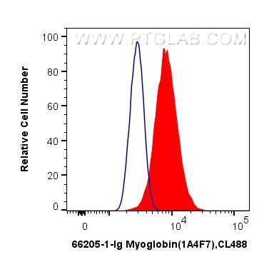
FC experiment of C2C12 using 66205-1-Ig
1X10^6 C2C12 cells were intracellularly stained with 0.4 ug Anti-Human Myoglobin (66205-1-Ig, Clone:1A4F7) and CoraLite®488-Conjugated AffiniPure Goat Anti-Mouse IgG(H+L) at dilution 1:1000 (red), or 0.4 ug Control Antibody. Cells were fixed with 4% PFA and permeabilized with Flow Cytometry Perm Buffer (PF00011-C).
1X10^6 C2C12 cells were intracellularly stained with 0.4 ug Anti-Human Myoglobin (66205-1-Ig, Clone:1A4F7) and CoraLite®488-Conjugated AffiniPure Goat Anti-Mouse IgG(H+L) at dilution 1:1000 (red), or 0.4 ug Control Antibody. Cells were fixed with 4% PFA and permeabilized with Flow Cytometry Perm Buffer (PF00011-C).
The Proteintech guarantee covers Proteintech antibodies in any species and any application, including those not listed on the datasheet. If the antibody doesn’t perform, you can receive a hassle-free refund or credit note.
human skeletal muscle tissue, pig heart tissue, rat heart tissue, mouse heart tissue, rabbit heart tissue, human heart tissue
Positive IHC detected in
human heart tissue, mouse tongue tissue, rat heart tissue Note: suggested antigen retrieval with TE buffer pH 9.0; (*) Alternatively, antigen retrieval may be performed with citrate buffer pH 6.0
Positive FC (Intra) detected in
C2C12 cells
Recommended dilution
Application
Dilution
Western Blot (WB)
WB : 1:5000-1:80000
Immunohistochemistry (IHC)
IHC : 1:50-1:500
Flow Cytometry (FC) (INTRA)
FC (INTRA) : 0.40 ug per 10^6 cells in a 100 µl suspension
It is recommended that this reagent should be titrated in each testing system to obtain optimal results.
Sample-dependent, Check data in validation data gallery.
PBS with 0.02% sodium azide and 50% glycerol, pH 7.3.
Storage Conditions
Store at -20°C. Stable for one year after shipment. Aliquoting is unnecessary for -20oC storage. 20ul sizes contain 0.1% BSA.
Background Information
Myoglobin is a cytoplasmic hemoprotein that is expressed primarily in cardiomyocytes and oxidative skeletal muscle fibers, functioning on facilitating oxygen transport and modulating nitric oxide homeostasis within cardiac and skeletal myocytes. Recent studies indicated that myoglobin was also expressed in non-muscle tissues. This antibody well recognized endogenous myoglobin in muscle lysates.
Time-restricted feeding relieves high temperature-induced impairment on meat quality by activating the Nrf2/HO-1 pathway, modification of muscle fiber composition, and enriching the polyunsaturated fatty acids in pigs
WB analysis of human skeletal muscle using 66205-1-Ig
human skeletal muscle tissue were subjected to SDS PAGE followed by western blot with 66205-1-Ig (Myoglobin antibody at dilution of 1:50000 incubated at room temperature for 1.5 hours.
WB analysis of pig heart using 66205-1-Ig
pig heart tissue were subjected to SDS PAGE followed by western blot with 66205-1-Ig (Myoglobin antibody) at dilution of 1:50000 incubated at room temperature for 1.5 hours.
WB analysis of rat heart using 66205-1-Ig
rat heart tissue were subjected to SDS PAGE followed by western blot with 66205-1-Ig (Myoglobin antibody) at dilution of 1:50000 incubated at room temperature for 1.5 hours.
WB analysis of mouse heart using 66205-1-Ig
mouse heart tissue were subjected to SDS PAGE followed by western blot with 66205-1-Ig (Myoglobin antibody) at dilution of 1:50000 incubated at room temperature for 1.5 hours.
WB analysis of rabbit heart using 66205-1-Ig
rabbit heart tissue were subjected to SDS PAGE followed by western blot with 66205-1-Ig (Myoglobin antibody) at dilution of 1:50000 incubated at room temperature for 1.5 hours.
WB analysis of human heart using 66205-1-Ig
human heart tissue were subjected to SDS PAGE followed by western blot with 66205-1-Ig (Myoglobin antibody at dilution of 1:50000 incubated at room temperature for 1.5 hours.
IHC Figures
IHC staining of human heart using 66205-1-Ig
Immunohistochemical analysis of paraffin-embedded human heart tissue slide using 66205-1-Ig (Myoglobin antibody at dilution of 1:200 (under 10x lens).
IHC staining of rat heart using 66205-1-Ig
Immunohistochemical analysis of paraffin-embedded rat heart tissue slide using 66205-1-Ig (Myoglobin antibody) at dilution of 1:8000 (under 10x lens). Heat mediated antigen retrieval with Tris-EDTA buffer (pH 9.0).
IHC staining of mouse tongue using 66205-1-Ig
Immunohistochemical analysis of paraffin-embedded mouse tongue tissue slide using 66205-1-Ig (Myoglobin antibody) at dilution of 1:8000 (under 10x lens). Heat mediated antigen retrieval with Tris-EDTA buffer (pH 9.0).
FC (INTRA) Figures
FC experiment of C2C12 using 66205-1-Ig
1X10^6 C2C12 cells were intracellularly stained with 0.4 ug Anti-Human Myoglobin (66205-1-Ig, Clone:1A4F7) and CoraLite®488-Conjugated AffiniPure Goat Anti-Mouse IgG(H+L) at dilution 1:1000 (red), or 0.4 ug Control Antibody. Cells were fixed with 4% PFA and permeabilized with Flow Cytometry Perm Buffer (PF00011-C).
The species listed in Tested Reactivity are in-house verified and applicable species. For unlisted species, please refer to the homology analysis of the immunogen sequence and related species. For rabbit polyclonal antibodies, homology >70% is recommended. For mouse monoclonal antibodies and rabbit recombinant antibodies, homology >90% is recommended. Generally, the higher the homology, the greater the applicability. However, there will be certain differences in protein expression in different species, tissues or cells. Therefore, the homology analysis results are for reference only and do not serve as a guarantee.
At Proteintech, we pride ourselves on our antibody quality, customer service and transparency. As such, we are comparing our antibodies with other vendors, enabling easy identification and comparisons of key data to help you choose the suitable antibody for your needs.
We have selected the top cited antibodies from these vendors for you to compare.
Proteintech
Myoglobin Monoclonal antibody
Catalog Number
66205-1-Ig
Citations
1
Dilutions
WB : 1:5000-1:80000 IHC : 1:50-1:500 FC (INTRA) : 0.40 ug per 10^6 cells in a 100 µl suspension
Applications
WB, IHC, FC (Intra), ELISA
Reactivity
human, mouse, rat, pig, rabbit
Product Guarantee
Covers any species including not listed on datasheet
Covers any applications including not listed on datasheet

 at dilution of 1:50000 incubated at room temperature for 1.5 hours.")
 at dilution of 1:50000 incubated at room temperature for 1.5 hours.")
 at dilution of 1:50000 incubated at room temperature for 1.5 hours.")
 at dilution of 1:50000 incubated at room temperature for 1.5 hours.")

.")
 at dilution of 1:8000 (under 10x lens). Heat mediated antigen retrieval with Tris-EDTA buffer (pH 9.0).")
 at dilution of 1:8000 (under 10x lens). Heat mediated antigen retrieval with Tris-EDTA buffer (pH 9.0).")
 and CoraLite®488-Conjugated AffiniPure Goat Anti-Mouse IgG(H+L) at dilution 1:1000 (red), or 0.4 ug Control Antibody. Cells were fixed with 4% PFA and permeabilized with Flow Cytometry Perm Buffer (PF00011-C).")