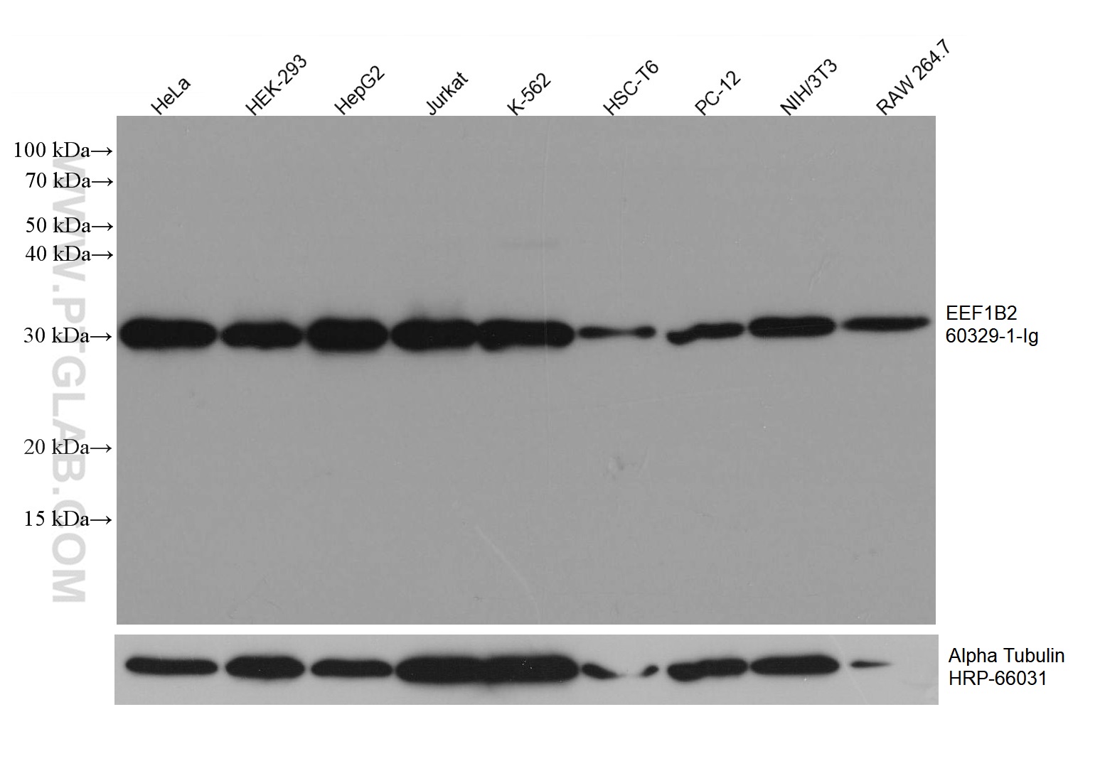
Various lysates were subjected to SDS PAGE followed by western blot with 60329-1-Ig (EEF1B2 antibody) at dilution of 1:2900 incubated at room temperature for 1.5 hours.
Various lysates were subjected to SDS PAGE followed by western blot with 60329-1-Ig (EEF1B2 antibody) at dilution of 1:2900 incubated at room temperature for 1.5 hours.
WB analysis of HeLa using 60329-1-Ig
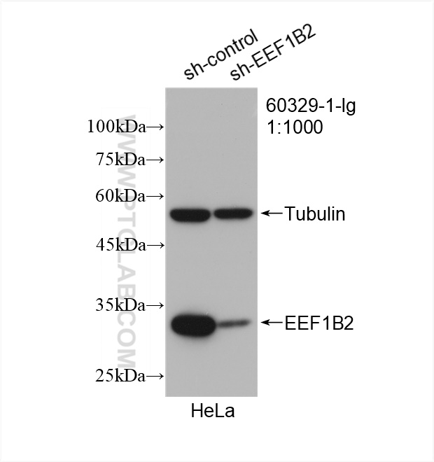
WB result of EEF1B2 antibody (60329-1-Ig; 1:1000; incubated at room temperature for 1.5 hours) with sh-Control and sh-EEF1B2 transfected HeLa cells.
WB result of EEF1B2 antibody (60329-1-Ig; 1:1000; incubated at room temperature for 1.5 hours) with sh-Control and sh-EEF1B2 transfected HeLa cells.
WB analysis using 60329-1-Ig
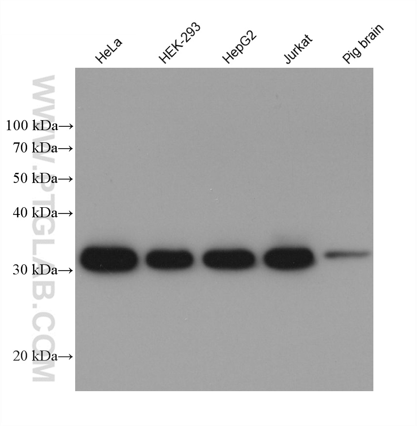
Various lysates were subjected to SDS PAGE followed by western blot with 60329-1-Ig (EEF1B2 antibody) at dilution of 1:10000 incubated at room temperature for 1.5 hours.
Various lysates were subjected to SDS PAGE followed by western blot with 60329-1-Ig (EEF1B2 antibody) at dilution of 1:10000 incubated at room temperature for 1.5 hours.
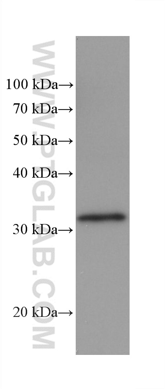
WB analysis of rabbit brain using 60329-1-Ig
rabbit brain tissue were subjected to SDS PAGE followed by western blot with 60329-1-Ig (EEF1B2 antibody) at dilution of 1:10000 incubated at room temperature for 1.5 hours.
rabbit brain tissue were subjected to SDS PAGE followed by western blot with 60329-1-Ig (EEF1B2 antibody) at dilution of 1:10000 incubated at room temperature for 1.5 hours.
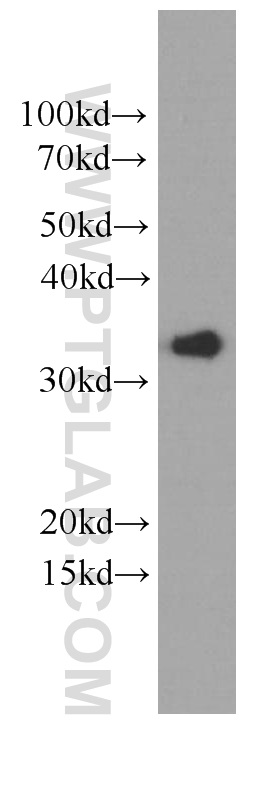
WB analysis of HeLa using 60329-1-Ig
HeLa cells were subjected to SDS PAGE followed by western blot with 60329-1-Ig (EEF1B2 Antibody) at dilution of 1:2000 incubated at room temperature for 1.5 hours.
HeLa cells were subjected to SDS PAGE followed by western blot with 60329-1-Ig (EEF1B2 Antibody) at dilution of 1:2000 incubated at room temperature for 1.5 hours.
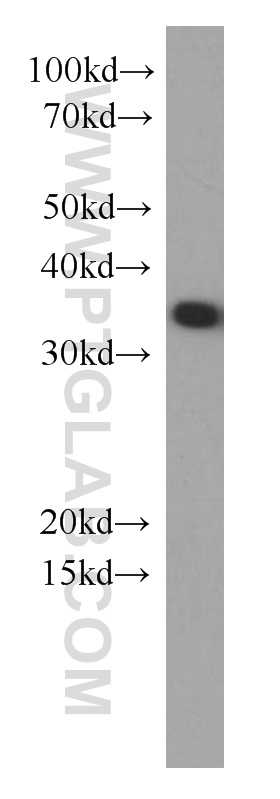
WB analysis of HEK-293 using 60329-1-Ig
HEK-293 cells were subjected to SDS PAGE followed by western blot with 60329-1-Ig (EEF1B2 Antibody) at dilution of 1:1000 incubated at room temperature for 1.5 hours.
HEK-293 cells were subjected to SDS PAGE followed by western blot with 60329-1-Ig (EEF1B2 Antibody) at dilution of 1:1000 incubated at room temperature for 1.5 hours.
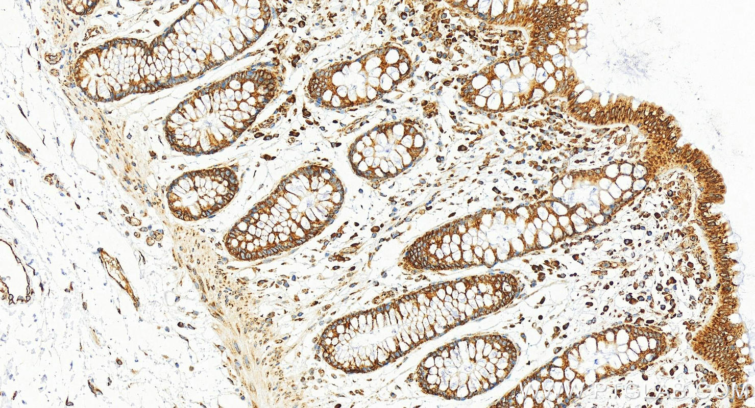
IHC staining of human colon using 60329-1-Ig
Immunohistochemical analysis of paraffin-embedded human colon tissue slide using 60329-1-Ig (EEF1B2 antibody) at dilution of 1:200 (under 20x lens). Heat mediated antigen retrieval with Tris-EDTA buffer (pH 9.0).
Immunohistochemical analysis of paraffin-embedded human colon tissue slide using 60329-1-Ig (EEF1B2 antibody) at dilution of 1:200 (under 20x lens). Heat mediated antigen retrieval with Tris-EDTA buffer (pH 9.0).
IF Staining of MCF-7 using 60329-1-Ig
Immunofluorescent analysis of (-20°C Methanol) fixed MCF-7 cells using EEF1B2 antibody (60329-1-Ig, Clone: 3E1C11 ) at dilution of 1:800 and CoraLite®488-Conjugated Goat Anti-Mouse IgG(H+L) (SA00013-1).
Immunofluorescent analysis of (-20°C Methanol) fixed MCF-7 cells using EEF1B2 antibody (60329-1-Ig, Clone: 3E1C11 ) at dilution of 1:800 and CoraLite®488-Conjugated Goat Anti-Mouse IgG(H+L) (SA00013-1).
The Proteintech guarantee covers Proteintech antibodies in any species and any application, including those not listed on the datasheet. If the antibody doesn’t perform, you can receive a hassle-free refund or credit note.
human colon tissue Note: suggested antigen retrieval with TE buffer pH 9.0; (*) Alternatively, antigen retrieval may be performed with citrate buffer pH 6.0
Positive IF/ICC detected in
MCF-7 cells
Recommended dilution
Application
Dilution
Western Blot (WB)
WB : 1:2000-1:10000
Immunohistochemistry (IHC)
IHC : 1:50-1:500
Immunofluorescence (IF)/ICC
IF/ICC : 1:400-1:1600
It is recommended that this reagent should be titrated in each testing system to obtain optimal results.
Sample-dependent, Check data in validation data gallery.
Product Information
60329-1-Ig targets EEF1B2 in WB, IHC, IF/ICC, ELISA applications and shows reactivity with human, mouse, rat, pig, rabbit samples.
PBS with 0.02% sodium azide and 50% glycerol, pH 7.3.
Storage Conditions
Store at -20°C. Stable for one year after shipment. Aliquoting is unnecessary for -20oC storage. 20ul sizes contain 0.1% BSA.
Background Information
In eukaryotes, the translation elongation factor eEF1A responsible for transporting amino-acylated tRNA to the ribosome forms a higher-order complex, eEF1H, with its guanine-nucleotide-exchange factor eEF1B. eEF1B consists of three subunits: eEF1B alpha, eEF1B beta and eEF1B gamma. The eEF1B2 possess the nucleotide-exchange activity. Although several models on the basis of in vitro experiments have been proposed for the macromolecular organization of the eEF1H complex, these models differ in various aspects. The human eukaryote elongation factor 1 beta 2 (eEF1B2) migrated as a 30-34 kDa protein in SDS-PAGE.
Various lysates were subjected to SDS PAGE followed by western blot with 60329-1-Ig (EEF1B2 antibody) at dilution of 1:2900 incubated at room temperature for 1.5 hours.
WB analysis of HeLa using 60329-1-Ig
WB result of EEF1B2 antibody (60329-1-Ig; 1:1000; incubated at room temperature for 1.5 hours) with sh-Control and sh-EEF1B2 transfected HeLa cells.
WB analysis using 60329-1-Ig
Various lysates were subjected to SDS PAGE followed by western blot with 60329-1-Ig (EEF1B2 antibody) at dilution of 1:10000 incubated at room temperature for 1.5 hours.
WB analysis of rabbit brain using 60329-1-Ig
rabbit brain tissue were subjected to SDS PAGE followed by western blot with 60329-1-Ig (EEF1B2 antibody) at dilution of 1:10000 incubated at room temperature for 1.5 hours.
WB analysis of HeLa using 60329-1-Ig
HeLa cells were subjected to SDS PAGE followed by western blot with 60329-1-Ig (EEF1B2 Antibody) at dilution of 1:2000 incubated at room temperature for 1.5 hours.
WB analysis of HEK-293 using 60329-1-Ig
HEK-293 cells were subjected to SDS PAGE followed by western blot with 60329-1-Ig (EEF1B2 Antibody) at dilution of 1:1000 incubated at room temperature for 1.5 hours.
IHC Figures
IHC staining of human colon using 60329-1-Ig
Immunohistochemical analysis of paraffin-embedded human colon tissue slide using 60329-1-Ig (EEF1B2 antibody) at dilution of 1:200 (under 20x lens). Heat mediated antigen retrieval with Tris-EDTA buffer (pH 9.0).
IF/ICC Figures
IF Staining of MCF-7 using 60329-1-Ig
Immunofluorescent analysis of (-20°C Methanol) fixed MCF-7 cells using EEF1B2 antibody (60329-1-Ig, Clone: 3E1C11 ) at dilution of 1:800 and CoraLite®488-Conjugated Goat Anti-Mouse IgG(H+L) (SA00013-1).
The species listed in Tested Reactivity are in-house verified and applicable species. For unlisted species, please refer to the homology analysis of the immunogen sequence and related species. For rabbit polyclonal antibodies, homology >70% is recommended. For mouse monoclonal antibodies and rabbit recombinant antibodies, homology >90% is recommended. Generally, the higher the homology, the greater the applicability. However, there will be certain differences in protein expression in different species, tissues or cells. Therefore, the homology analysis results are for reference only and do not serve as a guarantee.
At Proteintech, we pride ourselves on our antibody quality, customer service and transparency. As such, we are comparing our antibodies with other vendors, enabling easy identification and comparisons of key data to help you choose the suitable antibody for your needs.
We have selected the top cited antibodies from these vendors for you to compare.
 at dilution of 1:2900 incubated at room temperature for 1.5 hours.")
 with sh-Control and sh-EEF1B2 transfected HeLa cells.")
 at dilution of 1:10000 incubated at room temperature for 1.5 hours.")
 at dilution of 1:10000 incubated at room temperature for 1.5 hours.")
 at dilution of 1:2000 incubated at room temperature for 1.5 hours.")
 at dilution of 1:1000 incubated at room temperature for 1.5 hours.")
 at dilution of 1:200 (under 20x lens). Heat mediated antigen retrieval with Tris-EDTA buffer (pH 9.0).")
 fixed MCF-7 cells using EEF1B2 antibody (60329-1-Ig, Clone: 3E1C11 ) at dilution of 1:800 and CoraLite®488-Conjugated Goat Anti-Mouse IgG(H+L) (SA00013-1).")