Tested Applications
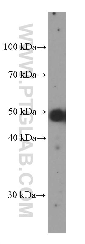
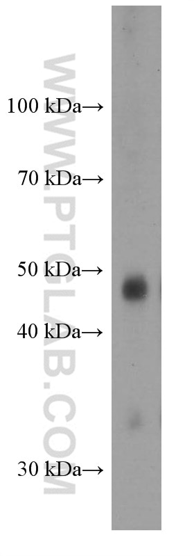
| Positive WB detected in | SKOV-3 cells, human thyroid gland tissue |
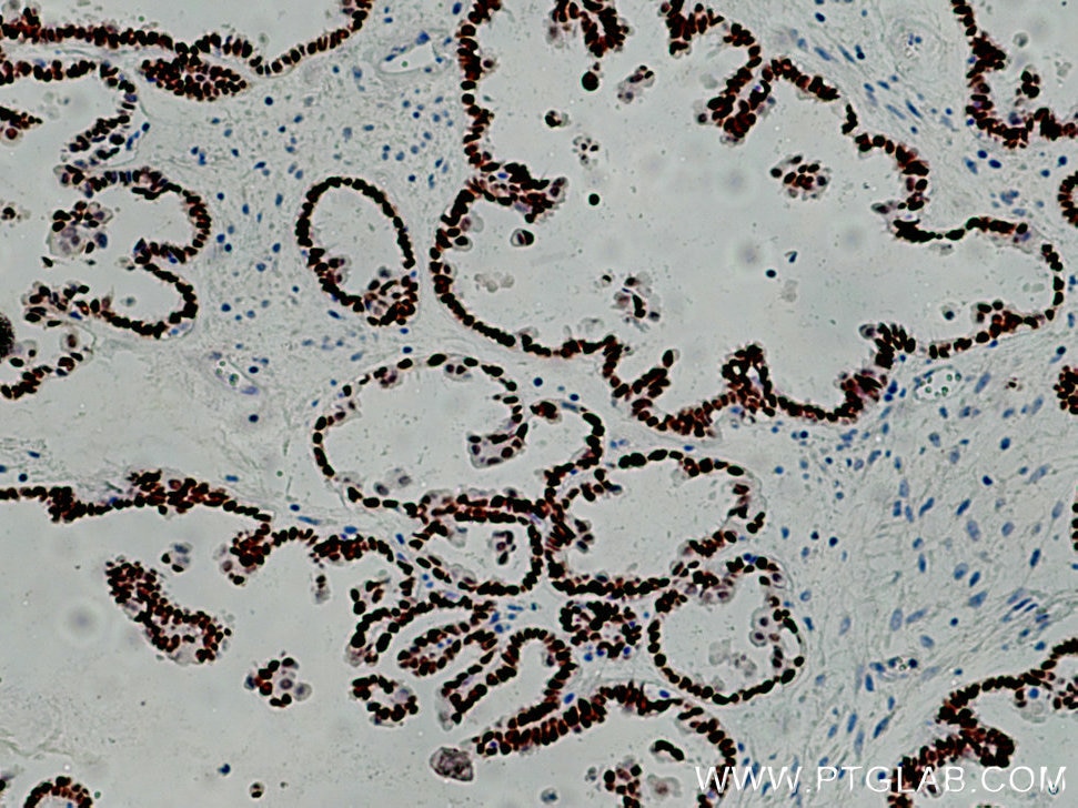
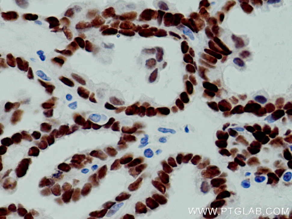
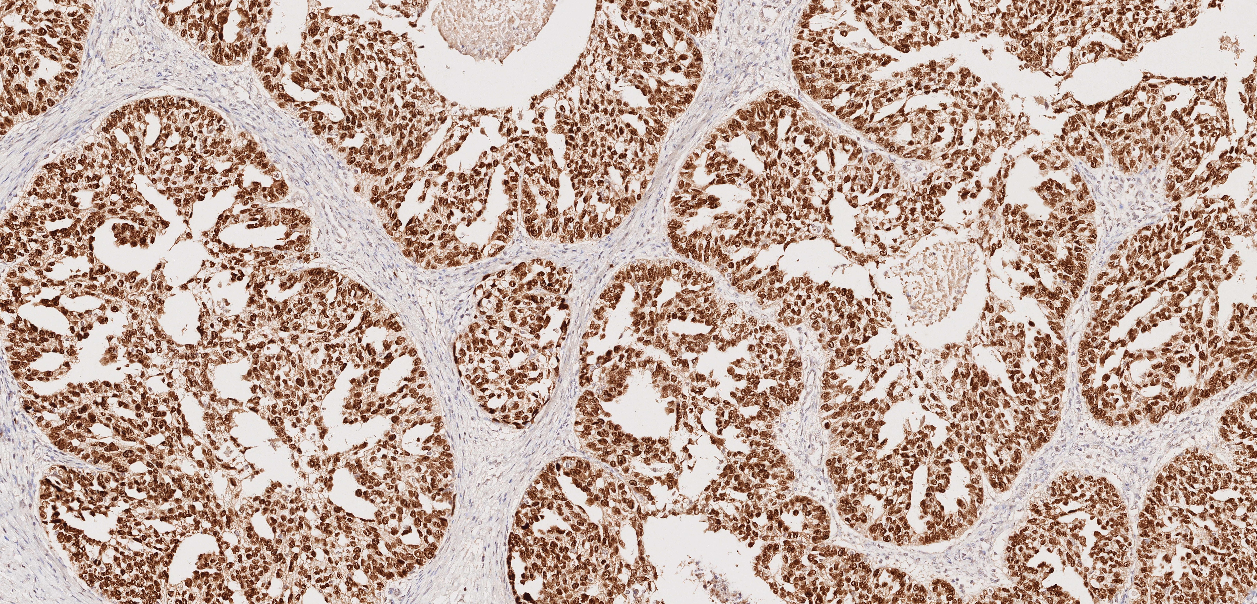
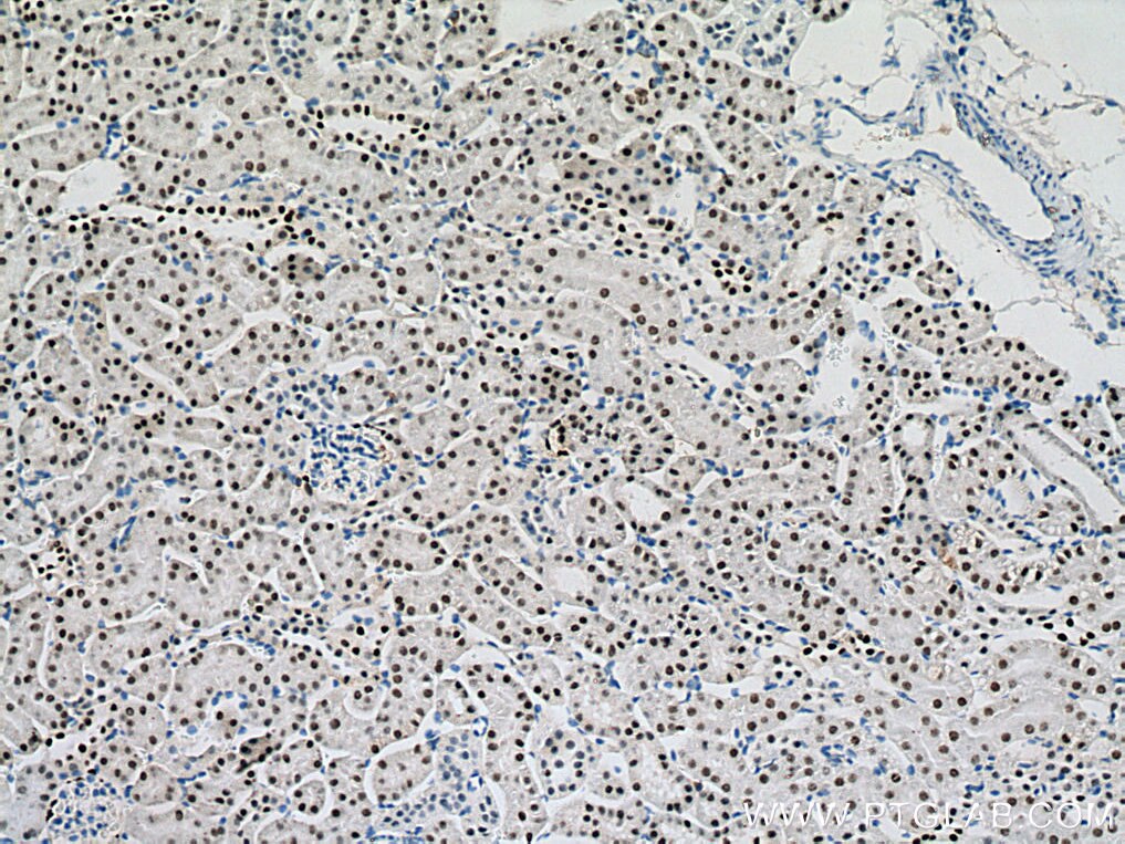
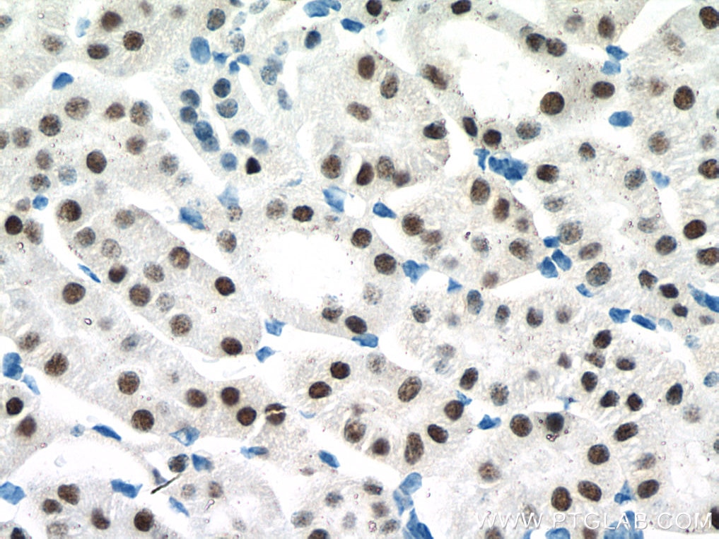
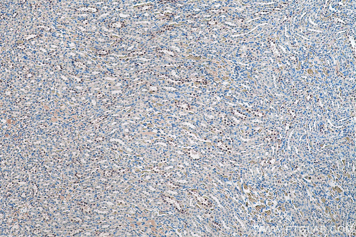
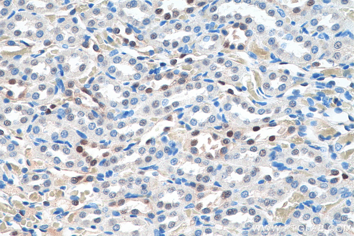
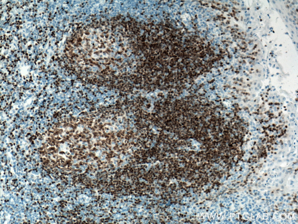
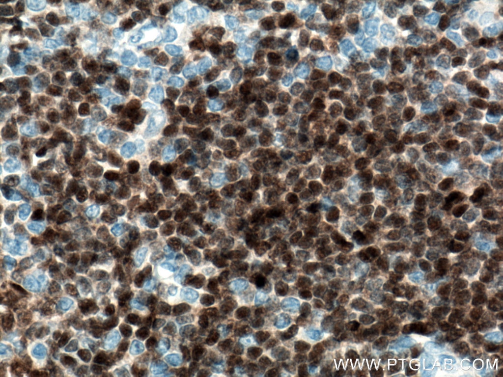
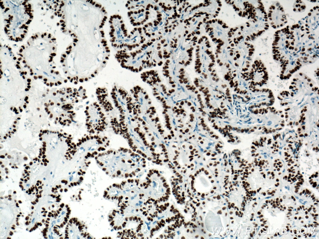
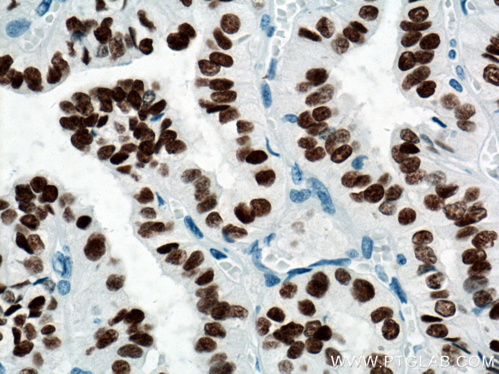
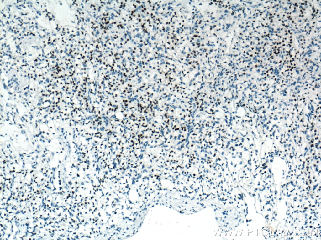
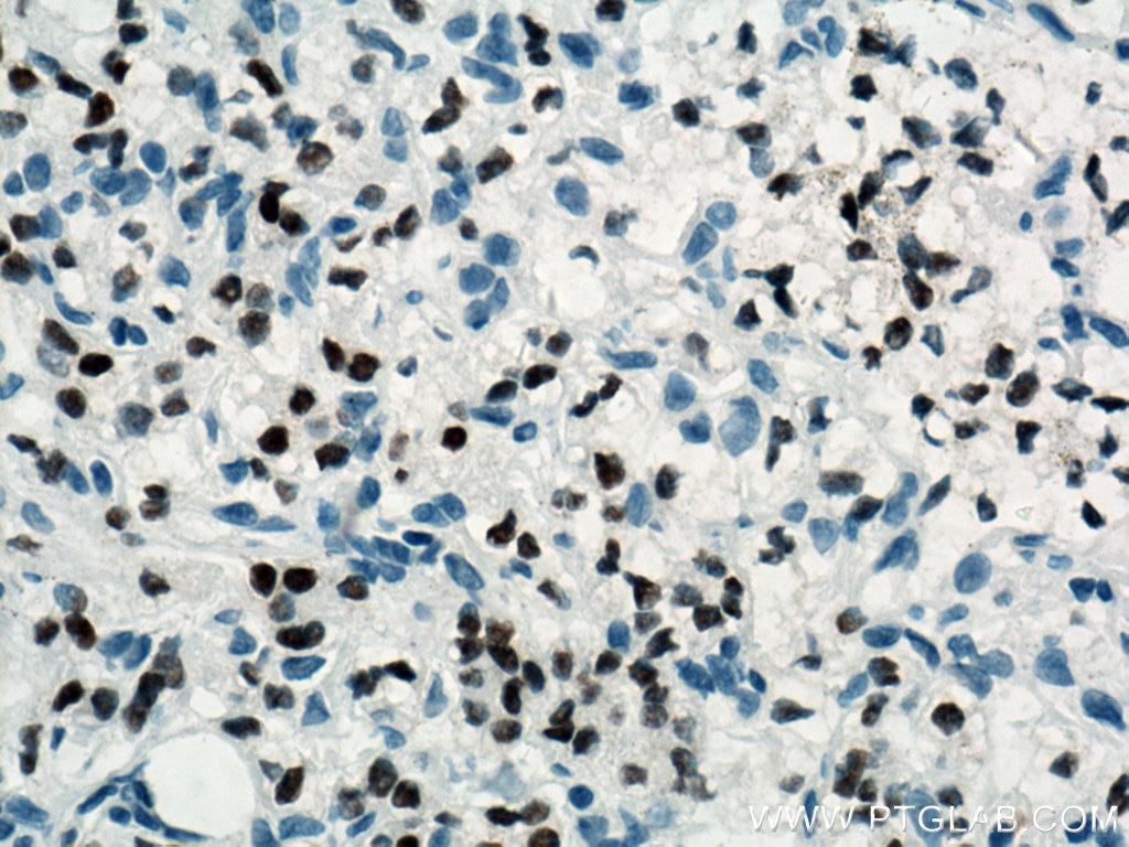
| Positive IHC detected in | human ovary tumor tissue, human ovary cancer tissue, human thyroid cancer tissue, human tonsillitis tissue, mouse kidney tissue, rat kidney tissue Note: suggested antigen retrieval with TE buffer pH 9.0; (*) Alternatively, antigen retrieval may be performed with citrate buffer pH 6.0 |
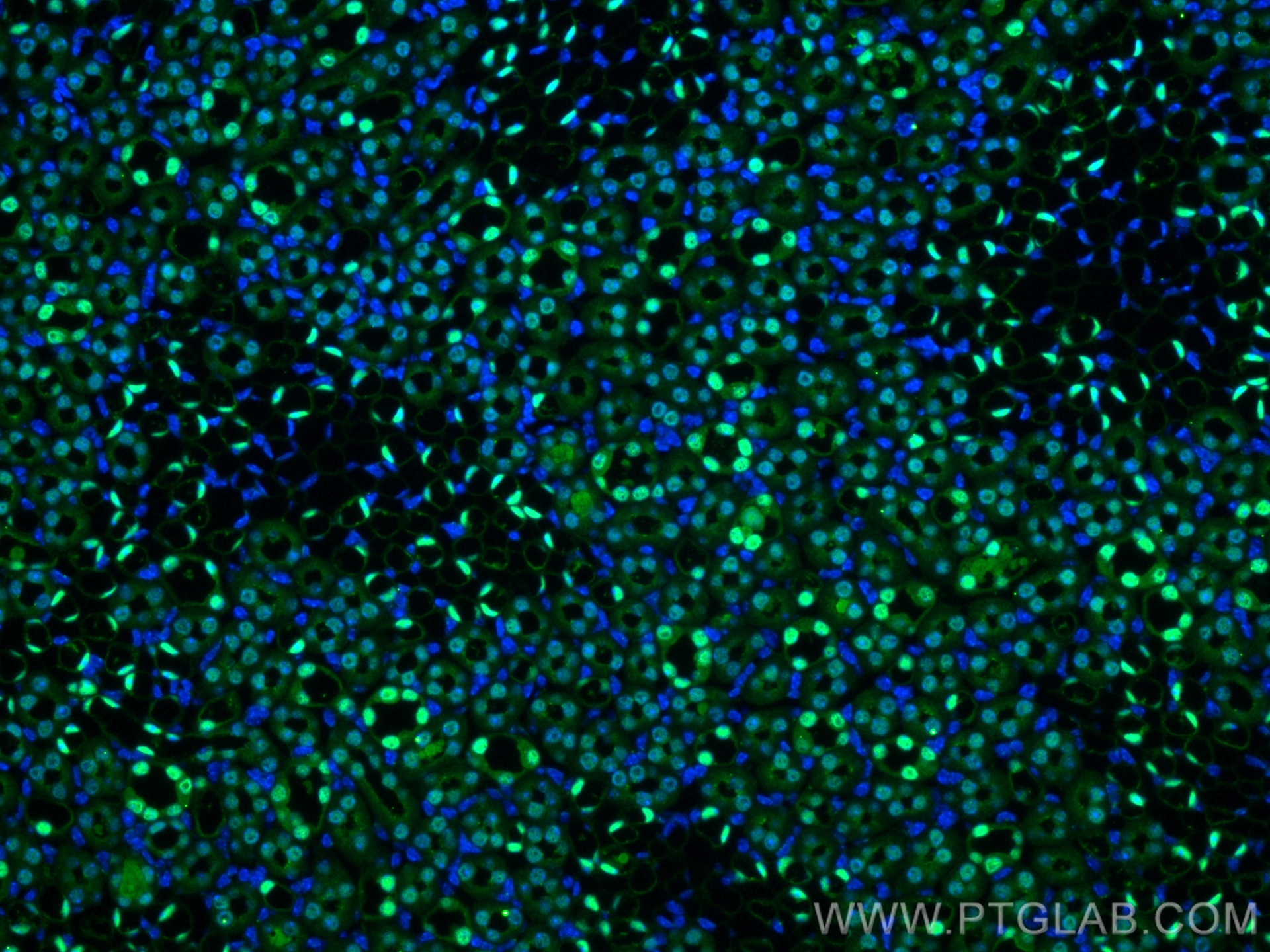
| Positive IF-P detected in | mouse kidney tissue |
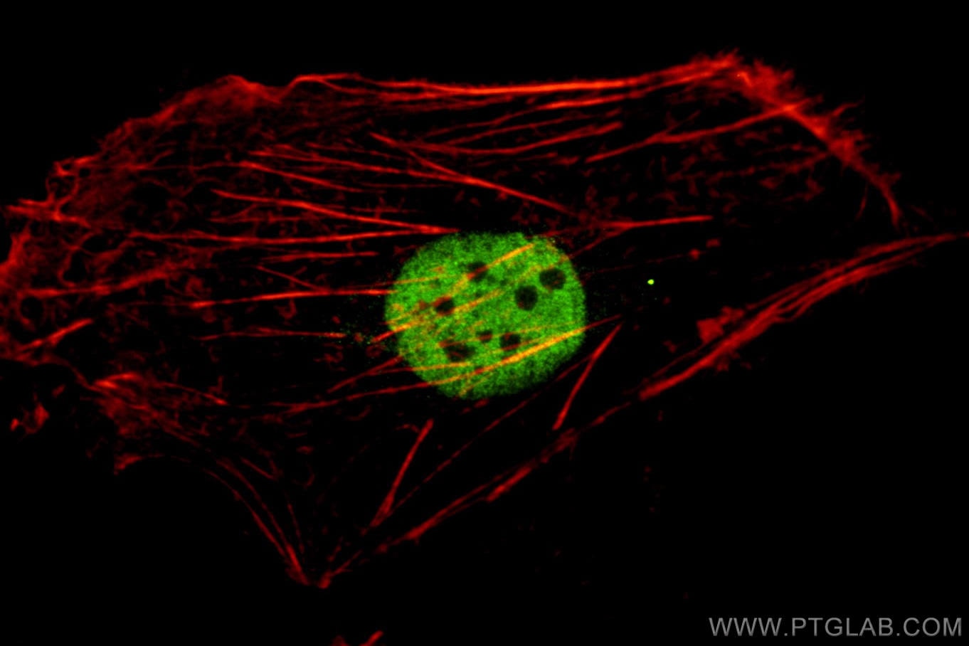
| Positive IF/ICC detected in | SKOV-3 cells |
Recommended dilution
| Application | Dilution |
|---|---|
| Western Blot (WB) | WB : 1:1000-1:4000 |
| Immunohistochemistry (IHC) | IHC : 1:5000-1:20000 |
| Immunofluorescence (IF)-P | IF-P : 1:200-1:800 |
| Immunofluorescence (IF)/ICC | IF/ICC : 1:50-1:500 |
| It is recommended that this reagent should be titrated in each testing system to obtain optimal results. | |
| Sample-dependent, Check data in validation data gallery. | |
Published Applications
| WB | See 1 publications below |
| IHC | See 6 publications below |
| IF | See 3 publications below |
Product Information
60145-4-Ig targets PAX8 in WB, IHC, IF/ICC, IF-P, ELISA applications and shows reactivity with human, mouse, rat samples.
| Tested Reactivity | human, mouse, rat |
| Cited Reactivity | human, mouse |
| Host / Isotype | Mouse / IgG2b |
| Class | Monoclonal |
| Type | Antibody |
| Immunogen |
CatNo: Ag0306 Product name: Recombinant human PAX8 protein Source: e coli.-derived, PGEX-4T Tag: GST Domain: 1-212 aa of BC001060 Sequence: MPHNSIRSGHGGLNQLGGAFVNGRPLPEVVRQRIVDLAHQGVRPCDISRQLRVSHGCVSKILGRYYETGSIRPGVIGGSKPKVATPKVVEKIGDYKRQNPTMFAWEIRDRLLAEGVCDNDTVPSVSSINRIIRTKVQQPFNLPMDSCVATKSLSPGHTLIPSSAVTPPESPQSDSLGSTYSINGLLGIAQPGSDKRKMDDSDQDSCRLSIDS Predict reactive species |
| Full Name | paired box 8 |
| Calculated Molecular Weight | 48 kDa |
| Observed Molecular Weight | 48 kDa |
| GenBank Accession Number | BC001060 |
| Gene Symbol | PAX8 |
| Gene ID (NCBI) | 7849 |
| RRID | AB_10643528 |
| Conjugate | Unconjugated |
| Form | Liquid |
| Purification Method | Protein A purification |
| UNIPROT ID | Q06710 |
| Storage Buffer | PBS with 0.02% sodium azide and 50% glycerol, pH 7.3. |
| Storage Conditions | Store at -20°C. Stable for one year after shipment. Aliquoting is unnecessary for -20oC storage. 20ul sizes contain 0.1% BSA. |
Background Information
PAX8 is a member of the paired box (PAX) family of transcription factors, typically containing a paired box domain and a paired-type homeodomain. It is expressed during organogenesis of the thyroid gland, kidney and Mullerian system. It is thought to regulate the expression of Wilms tumor suppressor (WT1) gene and mutations in PAX8 have been associated with Wilms tumor cells, thyroid and ovarian carcinomas. PAX8 is a useful marker in distinguishing ovarian carcinomas from mammary carcinomas (PMID: 18724243). PAX8 is expressed in a high percentage of ovarian serous, endometrioid, and clear cell carcinomas, but only rarely in primary ovarian mucinous adenocarcinomas.
PAX8 has 5 isoforms with calculated MW 31kd, 34kd, 41kd, 43kd and 48kd. And there is a 60kd band corresponding to the electrophoretic mobility of PAX8 (PMID:15650356).
Protocols
| Product Specific Protocols | |
|---|---|
| IF protocol for PAX8 antibody 60145-4-Ig | Download protocol |
| IHC protocol for PAX8 antibody 60145-4-Ig | Download protocol |
| WB protocol for PAX8 antibody 60145-4-Ig | Download protocol |
| Standard Protocols | |
|---|---|
| Click here to view our Standard Protocols |
Publications
| Species | Application | Title |
|---|---|---|
Dev Cell Identification of a core transcriptional program driving the human renal mesenchymal-to-epithelial transition | ||
Mod Pathol Xp11.2 translocation renal cell carcinoma with NONO-TFE3 gene fusion: morphology, prognosis, and potential pitfall in detecting TFE3 gene rearrangement. | ||
Stem Cell Res Ther Genetic correction of induced pluripotent stem cells from a DFNA36 patient results in morphologic and functional recovery of derived hair cell-like cells | ||
Front Cell Dev Biol Lgr4 Regulates Oviductal Epithelial Secretion Through the WNT Signaling Pathway | ||
Hum Pathol Clinicopathological and Molecular Characterization of Biphasic Hyalinizing Psammomatous Renal Cell Carcinoma (BHP RCC): Further Support for the Newly Proposed Entity. |
Reviews
The reviews below have been submitted by verified Proteintech customers who received an incentive for providing their feedback.
FH Kenzo (Verified Customer) (03-28-2024) | It worked well for immunolabeling in mouse retina tissues
|
FH Juan (Verified Customer) (04-18-2019) | It works well in Western Blot experiment.
|

