Green fluorescent protein (GFP) in plant research
Why does GFP work so well in plants?
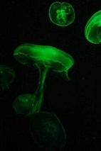 Although the jellyfish Aequorea Victoria Green fluorescent protein (GFP) was already discovered in the 1960s, it took three more decades until it was eventually cloned and could be utilized as a marker protein in E.coli and C. elegans. Since then it has developed into one of the most widely studied and exploited proteins in life sciences. Correspondingly, the importance of GFP was recognized in 2008 when the Nobel Committee awarded Osamu Shimomura, Marty Chalfie, and Roger Tsien the Chemistry Nobel Prize "for the discovery and development of the green fluorescent protein, GFP."
Why does GFP work so well in plants?
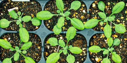
Next to its application in bacteria and animals, GFP was also established as a versatile tool for plant research. It became one of the most powerful reporter proteins in all kinds of plants because of its outstanding properties:
- small size of just 26.9 kD, which can tolerate N- and C-terminal protein fusions
- direct, visible fluorescence without the need for additional substrates or co-factors and relatively low photobleaching
- stable in a wide pH and redox potential range, especially important when GFP is located and analyzed in different organelles of the plant cell
- maintains fluorescence even after prolonged incubation in denaturing agents, which are required for sample preparation, e.g. 6 M guanidine HCl, 8 M urea or 1% SDS
- extremely thermostable
- good expression in a multitude of plants from different families and genus, independent of cell type and location
- stable expression with little degradation after 24 h and mild toxicity to the plant cells
Over time, a plethora of GFP variants and other fluorescent proteins have been discovered and developed as research tools.
For what applications is GFP used in plant research?
GFP is increasingly being used as a universal reporter in plant biology from the cellular to whole plant level:
- in vivo reporter to assess frequency of transient and stable transformation
- monitoring trafficking and subcellular localization of proteins
- macroscopic visualization of GFP in whole leaves and plants
- monitor virus movement in and among whole plants
- monitor transgene movement and transgenic plants in the field
Since 2008 the ChromoTek’s GFP-Trap contributes to plant research: It has already been used in more than 250 publications for qualitative and quantitative analysis of plant-derived GFP-fusion proteins and their binding partners by immunoprecipitation (IP).
Why is GFP-Trap beneficial for plant research?
- Just 25 µL of GFP-Trap slurry are sufficient for large cell extract volumes.
In general, recombinant proteins including GFP-tagged proteins can only be expressed to low levels in plants. Therefore, several grams of plant material are needed resulting in large cell lysate volumes of about 1-10 mL. Due to GFP-Trap’s high affinity the IP reaction can still be performed with only 25 µL of bead slurry.
- Low-expressed proteins are captured efficiently.
Because the expression levels of GFP-fusion proteins in plant cells are frequently poor, the GFP-fusion protein’s concentration in the cell lysate may be very low, and sometimes even below the detection limit of a Western Blot (WB). However, GFP-Trap can effectively bind to even smallest amounts (<1 ng) of the GFP-tagged protein and enriches the GFP-fusion protein to enable WB detection.
- Harsh lysis buffer ingredients don’t affect the IP.
During the preparation of plant cell extract the lysis buffer is often supplemented with glycerol, reducing agents, and detergents to keep the GFP-fusion protein in solution. GFP-Trap tolerates even elevated concentrations of such harsh buffer ingredients without sacrificing its binding capacity or efficiency.
- Stringent washing conditions minimize background.
Highly concentrated plant cell extracts are prone to bind non-specifically to all kind of affinity resins including the resin or GFP Nanobody framework of GFP-Trap. However, because of the extraordinary stability of the GFP:GFP Nanobody complex, multiple washing steps can be performed and harsh wash buffers, supplemented with e.g. detergents or high salt, can be used. The GFP-fusion protein remains bound to the beads while non-specific background from plant proteins is effectively minimized.
What Antibody is recommended for Western blotting of plant protein samples?
For WB analysis of GFP-tagged proteins from plants we recommend the GFP antibody [3H9] (rat monoclonal) because of its extraordinary low background.
Application Note: GFP immunoprecipitation of A. thaliana plant samples
ChromoTek provides an application note for immunoprecipitation (IP) of GFP-tagged proteins from Arabidopsis thaliana. This application note comprises a step-by-step protocol and is based on more than 70 peer reviewed publications available. This protocol may also work as starting point for other plants e.g. Nicotiana, Oryza, Solanum, Petunia, etc..
You can download the new applications note “How to conduct a GFP-fusion protein immunoprecipitation of Arabidopsis thaliana plant samples with ChromoTek’s GFP-Trap” here:
Application note
GFP immunoprecipitation of A. thaliana plant samples
Need a quick explanation about GFP-Trap? Watch this:
Thank you to Sid Balachandran for the fluorescent jellyfish picture at unsplash
References
DeBlasio SL, Sylvester AW, Jackson D. Illuminating plant biology: using fluorescent proteins for high-throughput analysis of protein localization and function in plants. Brief Funct Genomics. 2010;9(2):129–138.
Hanson MR, Köhler RH. GFP imaging: methodology and application to investigate cellular compartmentation in plants. J Exp Bot. 2001;52(356):529–539.
Leffel SM, Mabon SA, Stewart CN Jr. Applications of green fluorescent protein in plants. Biotechniques. 1997;23(5):912–918.
Sheen J, Hwang S, Niwa Y, Kobayashi H, Galbraith DW. Green-fluorescent protein as a new vital marker in plant cells. Plant J. 1995;8(5):777–784.
Tsien RY. The green fluorescent protein. Annu Rev Biochem. 1998;67:509–544